H Pylori Open Access
Copyright ©The Author(s) 2003. Published by Baishideng Publishing Group Inc. All rights reserved.
World J Gastroenterol. Sep 15, 2003; 9(9): 2021-2024
Published online Sep 15, 2003. doi: 10.3748/wjg.v9.i9.2021
Stability of randomly amplified polymorphic DNA fingerprinting in genotyping clinical isolates of Helicobacter pylori
Feng-Chan Han, Han-Chong Ng, Bow Ho, Department of Microbiology, Faculty of Medicine, National University of Singapore, 5 Science Drive 2, Singapore 117595, Republic of Singapore
Author contributions: All authors contributed equally to the work.
Supported by the National Medical Research Council (NMRC) Sponsored Project, No. R-182-000-037-213
Correspondence to: Feng-Chan Han, Institute of Genetic Diagnosis, Fourth Military Medical University, 169 Changle West Road, Xi’an710033, Shaanxi Province, China. biohanfc@hotmail.com
Telephone: +86-29-3374772 Fax: +86-29-3285729
Received: March 2, 2003
Revised: April 23, 2003
Accepted: May 11, 2003
Published online: September 15, 2003

Abstract

AIM: H pylori genomes are highly diversified. This project was designed to genotype H pylori isolates by the polymerase chain reaction (PCR)-based randomly amplified polymorphic DNA (RAPD) fingerprinting technique and to verify its stability by Southern blotting and DNA sequencing.

METHODS: Clinical isolates of H pylori were cultured from gastric antra and cardia of 73 individuals, and genomic DNA was prepared for each isolate. RAPD was carried out under optimized conditions. 23S rDNA was regarded as an internal control, and a 361 bp rDNA fragment (RDF) was used as a probe to screen the RAPD products by Southern blotting. Ten RDFs from different clinical isolates and the flanking regions (both upstream and downstream) of four RDFs were amplified and sequenced.

RESULTS: H pylori isolates from different individuals had different RAPD profiles, but the profiles for isolates cultured from different gastric sites of a given individual were identical in all but one case. Isolates from 27 individuals were RDF positive by Southern blotting. Sequences of the RDFs and their flanking regions were almost the same between the RDF positive and negative isolates as determined by Southern blotting. There was no binding site for random PCR primer inside the sequences.

CONCLUSION: RAPD is very useful in genotyping H pylori grossly on a large scale. However, it seems unstable in amplification of low yield fragments, especially those that do not appear as visible bands on the agarose gel stained with EB, since the primer is partially matched to the template.


INTRODUCTION

H pylori is a spiral, Gram-negative bacterium, which chronically infects more than half of population worldwide, and is implicated in gastritis, peptic ulcer and gastric cancer[1-12]. One of its features is high genome diversity[13-15]. Clinical isolates of H pylori from different individuals show enormous variation in their genomic fingerprints[16-18]. Studies based on the two sequenced genomes of H pylori strains (i.e. 26695 and J99) have revealed that among the H pylori genes, 22% are dispensable and about 6% are unique[14,15]. Sequence variation of certain genes or regions, such as the cag pathogenicity island[19,20], vacA[21], babA and babB[22], drug resistant genes[23-26] and restriction-modification genes[27-29], is also remarkable. For a given individual, the genes of the colonized strain even change over the course of colonization[17,30]. Genotyping of the bacterium is important for the epidemiological and pathogenic investigation, and genes that are present in one strain but absent or substantially different in others can be of great interest biologically.

The polymerase chain reaction (PCR)-based randomly amplified polymorphic DNA (RAPD) method has widely applied to the distinction of H pylori isolates due to its sensitivity, efficiency and promptness[16,17]. However, problems with its stability or reproducibility arise. In this study, we obtained RAPD fingerprints of clinical isolates of H pylori under optimized conditions, and then determined the stability of RAPD products by Southern blotting and DNA sequencing techniques.

MATERIALS AND METHODS
H pylori culture

Clinical isolates of H pylori cultured from gastric antra and cardia of 73 patients, 12 with gastric ulcer (GU), 18 with duodenum ulcer (DU), 8 with both GU and DU, and 25 with non-ulcer dyspepsia (NUD), were selected for this study. Each isolate preserved in the brain heart infusion broth (BHI, Gibco, Edinburgh, UK) supplemented with 100 mL/L horse serum and 200 mL/L glycerol was inoculated onto H pylori selective chocolate blood agar containing 40 g/L blood agar base No. 2 (Oxoid, Basingstoke, UK) and 50 mL/L horse blood (Gibco, Edinburgh, UK). Antibiotics (Sigma, St. Louis, MO, USA) were used at the following concentrations: 3 mg/L vancomycin, 5 mg/L trimethoprim, 10 mg/L nalidixic acid and 2 mg/L amphotericin B. The plates were incubated in a microaerobic atmosphere (50 mL/L CO2) in a CO2 incubator (Forma Scientific, Marietta, OH, USA) at 37 °C for up to 5 days.

Extraction of genomic DNA

H pylori cells on the plates were harvested by using a sterile swab, and transferred into an Eppendorf tube containing 1.5 mL TE buffer (10 mmol/L Tris-HCl, pH8.0; 1 mmol/L EDTA, pH8.0). The suspension was centrifuged at 8000 g for 10 min and washed once with TE buffer. The pellet was then suspended in 800 μL TE buffer was incubated with 10 μL of 100 g/L lysozyme (Sigma, St. Louis, MO, USA) at 37 °C for 30 min. The suspension was then lysed with 100 μL of 100 g/L sodium dodecyl sulfate (SDS) by incubating at 37 °C for another 30 min. Then 5 μL of proteinase K (10 g/L) (Boehninger, Mannheim, Germany) was added into the mixture and incubated at 56 °C for 1 h. Afterward, H pylori DNA was purified by extracting twice with equal volume of phenol and once with equal volume of chloroform, followed by centrifugation at 12000 g for 10 min each time. The supernatant was transferred into a new Eppendorf tube, and precipitated with 2 volume of absolute ethanol and 20 μL of 3 M sodium acetate at -20 °C overnight. The DNA preparation was centrifuged at 12000 g for 20 min and washed once with 700 mL/L ethanol. The pellet was then vacuum-dried and suspended in 50 μL distilled water. RNase A was added at the final concentration of 20 mg/L. After incubated at room temperature fo r 30 min, DNA concentration was determined spectrophotometrically.

RAPD for genomic DNA

A universal primer was chosen for the PCR-based RAPD, according to the paper of Akopyanz, et al[31] to fingerprint the genomic DNA. The primer was 5-AAGAGCCCGT-3. The volume of the PCR mixture was 25 μL containing 50 ng of H pylori genomic DNA, 20 pmol primer, 1 unit of Taq DNA polymerase, 0.25 mmol/L dNTPs, 10 mmol/L Tris-HCl, 50 mmol/L KCl, 2 mmol/L MgCl2 and 0.1 g/L gelatin (Promega, Madison WI, USA). PCR was performed with a thermal cycler (Perkin-Elmer 2400, Boston, MA, USA) consisting of an initial step at 94 °C for 5 min. This was followed by 39 cycles of denaturation at 94 °C for 1 min, annealing at 36 °C for 1 min and extension at 72 °C for 1 min. Ten microlitres of the PCR products were electrophoresed in 10 g/L horizontal agarose gel in TBE buffer at 68 V for 2 h. The gels were stained with ethidium bromide (EB, 1 mg/L) and photographed.

DNA fragment cloning and sequencing

At first, two primers were designed: RDF1-CGCAAGCTTCCACACAGAACCAC, and RDF2-CGCGGATCCGGACCTTTACTACAAC. A 23S rDNA fragment (RDF) was amplified according to the gene sequence reported[32]. The PCR mixture was 40 μL in volume containing 4 μL of 10 × polymerase buffer, 2 μL of genomic DNA (20 ng) from the reference strain NCTC11637, 1 μL (2 units) of Taq DNA polymerase, 4 μL of 2.5 mmol/L dNTPs, and 4 μL of 10 mmol/L primers. The sample was subjected to denaturation at 94 °C for 3 min. The PCR program was 30 cycles at 94 °C for 30 s, at 55 °C for 45 s, and at 72 °C for 1 min, followed by an extra extension at 72 °C for 5min. The PCR product (10 μL) was then electrophoresed in a 15 g/L agarose gel. DNA was recovered by using QIAEXÐ agarose gel extract kits (Qiagen, Santa Clarita, CA, USA). The purified PCR product was then cloned into the pT-Adv vector (Clontech, Palo Alto, CA, USA) and transformed into E. coli Top10, which was then inoculated on the selective plates containing 20 µL of 50 g/L ampicillin, 35 µL of 100 mmol/L IPTG and 40 µL of 20 g/L X-gal. White colonies were randomly picked up, suspended in 5 mL of Luria-Bertani medium containing ampicillin (50 mg/L) and cultured at 37 °C overnight. The recombinant plasmid was extracted by using Qiaprep spin miniprep kit (Qiagen, Santa Clarita, CA, USA). In the same way, RDFs from different H pylori isolates were amplified and cloned into the pGEM-T-Easy Vector (Promega, Madison, WI, USA). Then, A 23S rDNA fragment (1050 bp) containing the RDF in the middle was also amplified from different H pylori isolates by using the primers DMSR1-TAAGTTCGCGATAAGGTGTGC and DMSR1-GGTTCCTGCTTAGATGCTTTC. The inserts in the recombinant plasmids and the RDF flanking regions were sequenced by using the Big dye terminator DNA sequencing kit (Perkin-Elmer, Boston, MA, USA) and ABI automated sequencer (GMI, Minnesota, USA).

Southern blotting

Southern blotting was carried out following the protocol provided in the Enhanced chemiluminescence (ECL) direct nucleic acid labeling and detection system (Amersham Pharmacia Biotech, Buckinghamshire, UK). In brief, after agarose gel electrophoresis of the RAPD products, the DNA was submitted to denaturation by immersing the gel (200 cm2) in 200 ml buffer containing 0.5 mol/L NaOH and 1.5 mol/L NaCl for 30 min. Then the gel was neutralized in 200 ml of 0.5 mol/L Tris-HCl and 1.5 mol/L NaCl (pH7.5) for 30 min. Afterward, the DNA was transferred to the Hybond N+ membrane (Amersham Pharmacia Biotech, Buckinghamshire, UK) by using a capillary blotting apparatus. DNA fixation was carried out by exposing the membrane to an UV transilluminator (Vilber Lourmat, Marne la Vallee Cedex 1, France) for 5 min. RDF sequenced and digested by EcoR I from pT-Adv vector was used as a probe. The pre-hybridization and hybridization were performed in a buffer containing 0.5 mol/L NaCl and 50 g/L blocking agent in a hybridization oven at 42 °C for 1 h and 5 h respectively. After stringency washing (twice for 20 min in 6 mol/L urea, 4 g/L SDS and 0.1 × standard saline citrate, SSC; twice for 5 min in 2 × SSC), chemiluminenscence signals were detected by exposing the hyperfilm to the membrane for 5 to 30 min in a dark room.

RESULTS
H pylori gene profiles identified by RAPD

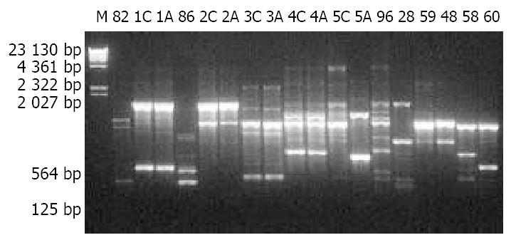
For all H pylori isolates from the 73 individuals, bacterial growth, genomic DNA extraction and RAPD were carried out under the same conditions. RAPD products were analyzed by 10 g/L agarose gel electrophoresis (Figure 1 and Figure 2), which showed that H pylori from gastric antra of different individuals had different RAPD profiles whereas the isolates from antrum and cardia of the same individual had almost identical RAPD profiles with only one exception (lanes 5C and 5A in Figure 1).

Figure 1
Figure 1 10 g/L agarose gel electrophoresis of RAPD products of H pylori isolates. M: λDNA Hind III markers; 82, 86, 96, 28, 59, 48, 59 and 60: Designations of H pylori isolates; A: Antrum; C: Cardia.
Figure 2
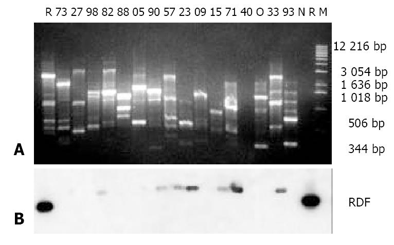
Figure 2 Results of agarose gel electrophoresis of RAPD products (A) and Southern blotting (B). A: 10 g/L agarose gel elec-trophoresis of RAPD products of H pylori isolates; M: 1 Kb DNA ladder; 73, 27, 98, 82, 88, 05, 90, 57, 23, 09, 15, 71, 40, 33 and 93: Designations of the H pylori isolates; N: H pylori reference strain NCTC11637; R: 1 ng RDF loaded in the well; O: Template blank control; B: Southern blotting results using the RDF as a probe, the dark blots on either side of Picture B corresponding to the 1 ng RDF in the agarose gel in Picture A.
Probe preparation

The sequence of RDF from the reference strain NCTC11637 was 99% identical to that reported in the GenBank. It was a 361 bp fragment of the 23S rDNA (from 2133 to 2493), which is just inside the loop (2134-2684) to form peptidyltransferase center in the ribosome. The fragment was then used as a probe in Southern blotting to investigate if the random PCR primer would pick up the same fragment with the same amount from genomes of different clinical isolates of H pylori.

Southern blotting

Firstly, the sensitivity of Southern blotting, in which the RDF was used as both target and probe, was determined by using the ECL system. The results showed that the sensitive limitation of this system was 4 pg. Then, the probe was used to screen the RAPD products of different H pylori isolates. Parts of the results are shown in Figure 2. H pylori isolates from 27 of the 73 individuals were RDF positive, and the darkness of the blots was quite different from one isolate to another. As the size of the RAPD fragment hybridized with the RDF was bigger (about 400 bp) than that of the RDF, at least one primer binding site would be outside the RDF region.

Sequencing RDFs and their flanking regions

In order to know if there were sequence differences that might facilitate the binding of the random primer, the RDFs and their flanking regions from different H pylori isolates were sequenced. Firstly, the RDFs from 10 different H pylori isolates were cloned for sequencing. The isolates with designated numbers of 15, 22, 38, 58, 90 and 93 were RDF positive and the isolates 20, 41, 84 and 90 were RDF negative in Southern blotting. Blast analysis of the sequenced results exhibited that the RDFs from the 10 isolates were quite similar.

The rates of identity were 98% to 100%. There was no random primer binding siteinside the RDFs. Next, the RDF flanking regions were sequenced by using the PCR products directly. Four isolates were used. Sequences of the upstream regions or the downstream regions of RDFs were also quite similar (98% to 99% identity) between the RDF positive isolates (numbers 38 and 93) and the RDF negative isolates (numbers 84 and 90) in Southern blotting, and there was no random primer binding site inside the upstream regions or the downstream regions.

DISCUSSION

It is known that DNA fingerprints of clinical H pyori isolates are highly diversified[16,17] and the fingerprinting profiles are changeable even for the isolates from different parts of the stomach in a single host[33,34]. The results in this study were basically in according with those obtained in the previous studies. The gene diversity of H pylori is associated with certain diseases[20,35,36], and clinical background and geographic origin of the isolates may be responsible for the changes[37].

It is always considered that the gene of rRNA is relatively conservative, and two copies of 23S rDNA exist in H pylori genome[32,38]. Theoretically, when the RDF probe is used to screen the RAPD products of different clinical isolates, Southern blotting profiles should be the same. However, our results showed that the Southern blotting profiles of 23S rDNA from different H pylori isolates were also polymorphic. Since genomes of different H pylori isolates contain almost identical RDF as well as the flanking regions and the same amount of genomic DNA was used as the templates in this study, instability of using RAPD to amplify this gene fragment at the pg level might be responsible for the results.

The random primer was first proposed for fingerprinting of H pylori by Akopyanz et al[31]. They recommended the primer since it had a relative high CG content and could be used to distinguish different H pylori isolates. In fact, when one employs this primer, there will be numerous potentially amplifiable fragments and only a few of them are visible on agarose gels after EB staining. This depends on the matching extent of the primer to the genomic DNA. For the bright or visible bands, the primer may be completely or mostly matched, but for the light or invisible bands, the primer is probably partially matched.

In conventionally used PCR, the specificity depends mainly on the properties of the primers and the annealing temperature. However, in PCR-based RAPD, the random primer is only 10 mers and the annealing temperature is 36 °C. Therefore, specificity of amplification is relatively low. Since we could not find any primer binding site inside or outside the RDF region in this experiment, we propose that, for amplification of the RDF, the primer be partially matched to the genomic DNA, and thus, the profiles of RAPD and Southern blotting are diversified.

From our experience, RAPD depends highly on the quality and quantity of the template. If we make a comparison between two RAPD profiles of a given H pylori isolate, the concentration, purity and even the integrity of the DNA templates should be considered. The RAPD results will be quite different if the genomic DNA is prepared using two different methods. Sometimes different PCR machines might also give different results. So, when we employed this method, we had tried to treat every sample under the same conditions, and handled the DNA preparation carefully. Even so, RAPD seems to be unstable in amplification of low yield fragments, especially those that do not appear as visible binds in agarose gel electrophoresis.

RAPD has reasonable discriminatory power[39] and is effective for H pylori genotyping grossly on large scale. But stability of this method should be taken into consideration.

Although we found many strain specific RAPD profiles in this study, using the RAPD-based methods to screen H pylori strain specific genes are not recommended. To identify strain specific genes, the PCR sequencing techniques are more reliable and reproducible.

ACKNOWLEDGEMENTS

We are grateful to Ms. Min Gong and Yan-Wing Ho, and also Mr. Mun-Fai Loke, Department of Microbiology, National University of Singapore, for their help in the preparation of H pylori isolates.

Footnotes

Edited by Xia HHX

References
1.  Israel DA, Peek RM. pathogenesis of Helicobacter pylori-induced gastric inflammation. Aliment Pharmacol Ther. 2001;15:1271-1290.  [PubMed]  [DOI]  [Cited in This Article: ]  [Cited by in Crossref: 112]  [Cited by in F6Publishing: 101]  [Article Influence: 4.2]  [Reference Citation Analysis (0)]
2.  Sanders MK, Peura DA. Helicobacter pylori-Associated Diseases. Curr Gastroenterol Rep. 2002;4:448-454.  [PubMed]  [DOI]  [Cited in This Article: ]  [Cited by in Crossref: 46]  [Cited by in F6Publishing: 47]  [Article Influence: 2.0]  [Reference Citation Analysis (0)]
3.  Dawsey SM, Mark SD, Taylor PR, Limburg PJ. Gastric cancer and H pylori. Gut. 2002;51:457-458.  [PubMed]  [DOI]  [Cited in This Article: ]  [Cited by in Crossref: 37]  [Cited by in F6Publishing: 40]  [Article Influence: 1.7]  [Reference Citation Analysis (0)]
4.  Blaser MJ. Linking Helicobacter pylori to gastric cancer. Nat Med. 2000;6:376-377.  [PubMed]  [DOI]  [Cited in This Article: ]  [Cited by in Crossref: 47]  [Cited by in F6Publishing: 45]  [Article Influence: 1.8]  [Reference Citation Analysis (0)]
5.  Blaser MJ, Berg DE. Helicobacter pylori genetic diversity and risk of human disease. J Clin Invest. 2001;107:767-773.  [PubMed]  [DOI]  [Cited in This Article: ]  [Cited by in Crossref: 239]  [Cited by in F6Publishing: 245]  [Article Influence: 10.2]  [Reference Citation Analysis (0)]
6.  Wang RT, Wang T, Chen K, Wang JY, Zhang JP, Lin SR, Zhu YM, Zhang WM, Cao YX, Zhu CW. Helicobacter pylori infection and gastric cancer: evidence from a retrospective cohort study and nested case-control study in China. World J Gastroenterol. 2002;8:1103-1107.  [PubMed]  [DOI]  [Cited in This Article: ]
7.  Meining A, Riedl B, Stolte M. Features of gastritis predisposing to gastric adenoma and early gastric cancer. J Clin Pathol. 2002;55:770-773.  [PubMed]  [DOI]  [Cited in This Article: ]  [Cited by in Crossref: 35]  [Cited by in F6Publishing: 39]  [Article Influence: 1.7]  [Reference Citation Analysis (0)]
8.  Han SR, Zschausch HC, Meyer HG, Schneider T, Loos M, Bhakdi S, Maeurer MJ. Helicobacter pylori: clonal population structure and restricted transmission within families revealed by molecular typing. J Clin Microbiol. 2000;38:3646-3651.  [PubMed]  [DOI]  [Cited in This Article: ]
9.  Shibata A, Parsonnet J, Longacre TA, Garcia MI, Puligandla B, Davis RE, Vogelman JH, Orentreich N, Habel LA. CagA status of Helicobacter pylori infection and p53 gene mutations in gastric adenocarcinoma. Carcinogenesis. 2002;23:419-424.  [PubMed]  [DOI]  [Cited in This Article: ]  [Cited by in Crossref: 54]  [Cited by in F6Publishing: 56]  [Article Influence: 2.4]  [Reference Citation Analysis (0)]
10.  Lan J, Xiong YY, Lin YX, Wang BC, Gong LL, Xu HS, Guo GS. Helicobacter pylori infection generated gastric cancer through p53-Rb tumor-suppressor system mutation and telomerase reactivation. World J Gastroenterol. 2003;9:54-58.  [PubMed]  [DOI]  [Cited in This Article: ]
11.  Cai L, Yu SZ, Zhang ZF. Helicobacter pylori infection and risk of gastric cancer in Changle County,Fujian Province,China. World J Gastroenterol. 2000;6:374-376.  [PubMed]  [DOI]  [Cited in This Article: ]
12.  Liu HF, Liu WW, Fang DC, Wang GA, Teng XC. Relationship between Helicobacter pylori infection and gastric precancerous lesions: a follow-up study. Shijie Huaren Xiaohua Zazhi. 2002;10:912-915.  [PubMed]  [DOI]  [Cited in This Article: ]
13.  Israel DA, Salama N, Arnold CN, Moss SF, Ando T, Wirth HP, Tham KT, Camorlinga M, Blaser MJ, Falkow S. Helicobacter pylori strain-specific differences in genetic content, identified by microarray, influence host inflammatory responses. J Clin Invest. 2001;107:611-620.  [PubMed]  [DOI]  [Cited in This Article: ]  [Cited by in Crossref: 251]  [Cited by in F6Publishing: 232]  [Article Influence: 9.7]  [Reference Citation Analysis (0)]
14.  Salama N, Guillemin K, McDaniel TK, Sherlock G, Tompkins L, Falkow S. A whole-genome microarray reveals genetic diversity among Helicobacter pylori strains. Proc Natl Acad Sci USA. 2000;97:14668-14673.  [PubMed]  [DOI]  [Cited in This Article: ]  [Cited by in Crossref: 427]  [Cited by in F6Publishing: 407]  [Article Influence: 16.3]  [Reference Citation Analysis (0)]
15.  Björkholm BM, Oh JD, Falk PG, Engstrand LG, Gordon JI. Genomics and proteomics converge on Helicobacter pylori. Curr Opin Microbiol. 2001;4:237-245.  [PubMed]  [DOI]  [Cited in This Article: ]  [Cited by in Crossref: 13]  [Cited by in F6Publishing: 13]  [Article Influence: 0.5]  [Reference Citation Analysis (0)]
16.  Yakoob J, Hu GL, Fan XG, Yang HX, Liu SH, Tan DM, Li TG, Zhang Z. Diversity of Helicobacter pylori among Chinese persons with H. pylori infection. APMIS. 2000;108:482-486.  [PubMed]  [DOI]  [Cited in This Article: ]  [Cited by in Crossref: 12]  [Cited by in F6Publishing: 12]  [Article Influence: 0.5]  [Reference Citation Analysis (0)]
17.  Israel DA, Salama N, Krishna U, Rieger UM, Atherton JC, Falkow S, Peek RM. Helicobacter pylori genetic diversity within the gastric niche of a single human host. Proc Natl Acad Sci USA. 2001;98:14625-14630.  [PubMed]  [DOI]  [Cited in This Article: ]  [Cited by in Crossref: 255]  [Cited by in F6Publishing: 251]  [Article Influence: 10.5]  [Reference Citation Analysis (0)]
18.  Sillakivi T, Aro H, Ustav M, Peetsalu M, Peetsalu A, Mikelsaar M. Diversity of Helicobacter pylori genotypes among Estonian and Russian patients with perforated peptic ulcer, living in Southern Estonia. FEMS Microbiol Lett. 2001;195:29-33.  [PubMed]  [DOI]  [Cited in This Article: ]  [Cited by in Crossref: 11]  [Cited by in F6Publishing: 12]  [Article Influence: 0.5]  [Reference Citation Analysis (0)]
19.  Peters TM, Owen RJ, Slater E, Varea R, Teare EL, Saverymuttu S. Genetic diversity in the Helicobacter pylori cag pathogenicity island and effect on expression of anti-CagA serum antibody in UK patients with dyspepsia. J Clin Pathol. 2001;54:219-223.  [PubMed]  [DOI]  [Cited in This Article: ]  [Cited by in Crossref: 31]  [Cited by in F6Publishing: 31]  [Article Influence: 1.3]  [Reference Citation Analysis (0)]
20.  Dong Q, O'Sullivan M, Hall W, Herra C, Kean C, O'Morain C, Buckley M. Identification of a new segment involved in cagA 3' region variation of Helicobacter pylori. FEMS Immunol Med Microbiol. 2002;33:51-55.  [PubMed]  [DOI]  [Cited in This Article: ]  [Cited by in Crossref: 2]  [Cited by in F6Publishing: 3]  [Article Influence: 0.1]  [Reference Citation Analysis (0)]
21.  Catalano M, Matteo M, Barbolla RE, Jimenez Vega DE, Crespo O, Leanza AG, Toppor J, Antelo P. Helicobacter pylori vacA genotypes, cagA status and ureA-B polymorphism in isolates recovered from an Argentine population. Diagn Microbiol Infect Dis. 2001;41:205-210.  [PubMed]  [DOI]  [Cited in This Article: ]  [Cited by in Crossref: 14]  [Cited by in F6Publishing: 16]  [Article Influence: 0.7]  [Reference Citation Analysis (0)]
22.  Pride DT, Meinersmann RJ, Blaser MJ. Allelic Variation within Helicobacter pylori babA and babB. Infect Immun. 2001;69:1160-1171.  [PubMed]  [DOI]  [Cited in This Article: ]  [Cited by in Crossref: 92]  [Cited by in F6Publishing: 101]  [Article Influence: 4.2]  [Reference Citation Analysis (0)]
23.  Yakoob J, Fan X, Hu G, Liu L, Zhang Z. Antibiotic susceptibility of Helicobacter pylori in the Chinese population. J Gastroenterol Hepatol. 2001;16:981-985.  [PubMed]  [DOI]  [Cited in This Article: ]  [Cited by in Crossref: 15]  [Cited by in F6Publishing: 19]  [Article Influence: 0.8]  [Reference Citation Analysis (0)]
24.  Piana A, Are BM, Maida I, Dore MP, Sotgiu G, Realdi G, Mura I. Genotypic characterization of clarithromycin-resistant Helicobacter pylori strains. New Microbiol. 2002;25:123-130.  [PubMed]  [DOI]  [Cited in This Article: ]
25.  Gerrits MM, de Zoete MR, Arents NL, Kuipers EJ, Kusters JG. 16S rRNA mutation-mediated tetracycline resistance in Helicobacter pylori. Antimicrob Agents Chemother. 2002;46:2996-3000.  [PubMed]  [DOI]  [Cited in This Article: ]  [Cited by in Crossref: 84]  [Cited by in F6Publishing: 83]  [Article Influence: 3.6]  [Reference Citation Analysis (0)]
26.  Jeong JY, Mukhopadhyay AK, Dailidiene D, Wang Y, Velapatiño B, Gilman RH, Parkinson AJ, Nair GB, Wong BC, Lam SK. Sequential inactivation of rdxA (HP0954) and frxA (HP0642) nitroreductase genes causes moderate and high-level metronidazole resistance in Helicobacter pylori. J Bacteriol. 2000;182:5082-5090.  [PubMed]  [DOI]  [Cited in This Article: ]  [Cited by in Crossref: 97]  [Cited by in F6Publishing: 97]  [Article Influence: 3.9]  [Reference Citation Analysis (0)]
27.  Nobusato A, Uchiyama I, Kobayashi I. Diversity of restriction-modification gene homologues in Helicobacter pylori. Gene. 2000;259:89-98.  [PubMed]  [DOI]  [Cited in This Article: ]  [Cited by in Crossref: 54]  [Cited by in F6Publishing: 64]  [Article Influence: 2.6]  [Reference Citation Analysis (0)]
28.  Lin LF, Posfai J, Roberts RJ, Kong H. Comparative genomics of the restriction-modification systems in Helicobacter pylori. Proc Natl Acad Sci USA. 2001;98:2740-2745.  [PubMed]  [DOI]  [Cited in This Article: ]  [Cited by in Crossref: 116]  [Cited by in F6Publishing: 123]  [Article Influence: 5.1]  [Reference Citation Analysis (0)]
29.  Takata T, Aras R, Tavakoli D, Ando T, Olivares AZ, Blaser MJ. Phenotypic and genotypic variation in methylases involved in type II restriction-modification systems in Helicobacter pylori. Nucleic Acids Res. 2002;30:2444-2452.  [PubMed]  [DOI]  [Cited in This Article: ]  [Cited by in Crossref: 25]  [Cited by in F6Publishing: 26]  [Article Influence: 1.1]  [Reference Citation Analysis (0)]
30.  Kuipers EJ, Israel DA, Kusters JG, Gerrits MM, Weel J, van Der Ende A, van Der Hulst RW, Wirth HP, Höök-Nikanne J, Thompson SA. Quasispecies development of Helicobacter pylori observed in paired isolates obtained years apart from the same host. J Infect Dis. 2000;181:273-282.  [PubMed]  [DOI]  [Cited in This Article: ]  [Cited by in Crossref: 127]  [Cited by in F6Publishing: 131]  [Article Influence: 5.2]  [Reference Citation Analysis (0)]
31.  Akopyanz N, Bukanov NO, Westblom TU, Kresovich S, Berg DE. DNA diversity among clinical isolates of Helicobacter pylori detected by PCR-based RAPD fingerprinting. Nucleic Acids Res. 1992;20:5137-5142.  [PubMed]  [DOI]  [Cited in This Article: ]  [Cited by in Crossref: 565]  [Cited by in F6Publishing: 595]  [Article Influence: 18.0]  [Reference Citation Analysis (0)]
32.  Taylor DE, Ge Z, Purych D, Lo T, Hiratsuka K. Cloning and sequence analysis of two copies of a 23S rRNA gene from Helicobacter pylori and association of clarithromycin resistance with 23S rRNA mutations. Antimicrob Agents Chemother. 1997;41:2621-2628.  [PubMed]  [DOI]  [Cited in This Article: ]
33.  Wong BC, Wang WH, Berg DE, Fung FM, Wong KW, Wong WM, Lai KC, Cho CH, Hui WM, Lam SK. High prevalence of mixed infections by Helicobacter pylori in Hong Kong: metronidazole sensitivity and overall genotype. Aliment Pharmacol Ther. 2001;15:493-503.  [PubMed]  [DOI]  [Cited in This Article: ]  [Cited by in Crossref: 38]  [Cited by in F6Publishing: 39]  [Article Influence: 1.6]  [Reference Citation Analysis (0)]
34.  Thoreson AC, Hosseini N, Svennerholm AM, Bölin I. Different Helicobacter pylori strains colonize the antral and duodenal mucosa of duodenal ulcer patients. Helicobacter. 2000;5:69-78.  [PubMed]  [DOI]  [Cited in This Article: ]  [Cited by in Crossref: 13]  [Cited by in F6Publishing: 14]  [Article Influence: 0.6]  [Reference Citation Analysis (0)]
35.  Azuma T, Yamakawa A, Yamazaki S, Fukuta K, Ohtani M, Ito Y, Dojo M, Yamazaki Y, Kuriyama M. Correlation between variation of the 3' region of the cagA gene in Helicobacter pylori and disease outcome in Japan. J Infect Dis. 2002;186:1621-1630.  [PubMed]  [DOI]  [Cited in This Article: ]  [Cited by in Crossref: 93]  [Cited by in F6Publishing: 104]  [Article Influence: 4.5]  [Reference Citation Analysis (0)]
36.  Arents NL, van Zwet AA, Thijs JC, Kooistra-Smid AM, van Slochteren KR, Degener JE, Kleibeuker JH, van Doorn LJ. The importance of vacA, cagA, and iceA genotypes of Helicobacter pylori infection in peptic ulcer disease and gastroesophageal reflux disease. Am J Gastroenterol. 2001;96:2603-2608.  [PubMed]  [DOI]  [Cited in This Article: ]  [Cited by in Crossref: 42]  [Cited by in F6Publishing: 48]  [Article Influence: 2.0]  [Reference Citation Analysis (0)]
37.  Göttke MU, Fallone CA, Barkun AN, Vogt K, Loo V, Trautmann M, Tong JZ, Nguyen TN, Fainsilber T, Hahn HH. Genetic variability determinants of Helicobacter pylori: influence of clinical background and geographic origin of isolates. J Infect Dis. 2000;181:1674-1681.  [PubMed]  [DOI]  [Cited in This Article: ]  [Cited by in Crossref: 15]  [Cited by in F6Publishing: 16]  [Article Influence: 0.6]  [Reference Citation Analysis (0)]
38.  van Doorn LJ, Glupczynski Y, Kusters JG, Mégraud F, Midolo P, Maggi-Solcà N, Queiroz DM, Nouhan N, Stet E, Quint WG. Accurate prediction of macrolide resistance in Helicobacter pylori by a PCR line probe assay for detection of mutations in the 23S rRNA gene: multicenter validation study. Antimicrob Agents Chemother. 2001;45:1500-1504.  [PubMed]  [DOI]  [Cited in This Article: ]  [Cited by in Crossref: 111]  [Cited by in F6Publishing: 118]  [Article Influence: 4.9]  [Reference Citation Analysis (0)]
39.  Burucoa C, Lhomme V, Fauchere JL. Performance criteria of DNA fingerprinting methods for typing of Helicobacter pylori isolates: experimental results and meta-analysis. J Clin Microbiol. 1999;37:4071-4080.  [PubMed]  [DOI]  [Cited in This Article: ]