Published online Apr 15, 2000. doi: 10.3748/wjg.v6.i2.275
Revised: July 6, 1999
Accepted: July 14, 1999
Published online: April 15, 2000
- Citation: Cheng J, Zhong YW, Liu Y, Dong J, Yang JZ, Chen JM. Cloning and sequence analysis of human genomic DNA of augmenter of liver regeneration. World J Gastroenterol 2000; 6(2): 275-277
- URL: https://www.wjgnet.com/1007-9327/full/v6/i2/275.htm
- DOI: https://dx.doi.org/10.3748/wjg.v6.i2.275
The liver is one of the organs, which have potential regenerative capability in mammalian animal[1]. The study of the canine model indicated that the liver could regenerate to original size after 70% hepatectomy in only two weeks[2]. So it is a hot research topic for the cellular and molecular mechanism of liver regeneration. Accumulated results demonstrated that the hepatocyte growth factor (HGF)[3], insulin-like growth factor I and II (IGF-I, II)[4], epidermal growth factor (EGF), transforming growth factor alpha (TG F alpha)[5] and insulin[6] are among the most important growth factors for liver regenerative regulation. In recent years, a heat-stable protein in the serum of the patients with various liver diseases has been noted for its potential stimulation effects on the liver regeneration, and this growth factor is called hepatocyte-stimulatory substance (HSS). Gradient purification and sequence analysis of HSS protein indicated that the HSS protein itself is the augmenter of liver regeneration (ALR)[7], or called hepatopoietin (HPO)[8]. The immunohistochemical staining indicated that the expression of the ALR mainly existed in platelets and the sperm cells in testes, and ALR also could be found in the liver and the spleen which contain many platelets[9]. The analysis of the protein structure of the human and mouse ALR indicated that the primary protein structure of ALR does not contain a typical signal peptide sequence, and it is unknown if a specific receptor is necessary for the effect of the ALR. Therefore, it is important to clone the genomic DNA sequence of the ALR and it is also very helpful for the analysis of the structure of the ALR genomic DNA and regulation at the transcriptional and post-transcriptional levels.
Using human ALR cDNA sequence as a reference, and BLAST search path as a tool, the GenBank established by National Center for Biological Information (NCBI), USA, has been searched for the homologous sequences.
According to the Breathnath-Chambon rule and the human ALR cDNA coding sequence, the intron-exon structure of human genomic DNA was defined.
The homology of human and mouse ALR genomic DNA sequences was analyzed for their 5′-UTR, intron-exon structure and 3′-UTR sequences.
Using human ALR cDNA (AF124604, human HPO2 mRNA, complete coding sequence) sequence as a reference, and BLAST path as a search tool, homologous DNA sequence was searched on GenBank. It was found that 5 cDNA and DNA fragments were homologous to human ALR cDNA sequence, including mouse ALR genomic DNA, rat ALR cDNA, human HPO1 cDNA partial sequence, human ERV1 cDNA and DNA sequence of human genomic DNA P1 clone derived from human chromosome 16 (Table 1).
| GenBank No. | Name | Character |
| U31176 | Human ERV1 mRNA | Complete coding sequence |
| AF124604 | Human HPO2 mRNA | Complete coding sequence |
| AC005606 | Human genomic DNA seuence | Chromosome 16 |
| AF124603 | Human HPO1 mRNA | Partial coding sequence |
| U40494 | Mouse ALR genomic DNA | Complete coding sequence |
| D30735 | Rat ALR mRNA | Complete coding sequence |
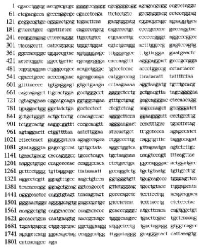
From Table 1, it is clear that human ALR cDNA has a high homology to P1 clone 109.8C (LANL) which is 16 (GenBank No. AC005606). Further analysis of human HPO2 cDNA complete sequence and HPO1 cDNA partial sequence showed that human ALR genomic DNA was between 44742-46554 nt of P1 clone 109-8C (LANL). Human ALR genomic DNA consisted of 1813 bp (Figure 1).
Human genomic DNA consists of introns and exons. According to the Breathnath-Chambon rule of intron-exon junction structure, in conjunction with the coding sequence of human ALR cDNA, we found that human ALR genomic DNA has 3 exons and 2 introns. The 3 exons were located between 158 nt-175 nt, 446 nt-642 nt and 1565 nt-1727 nt of P1 clone 109-8C (LANL) of human chromosome 16, respectively.
To compare human and mouse ALR genomic DNA sequences, we found that the 3 exons were similar in length, but different in their 5′-UTR, introns and 3′-UTR regions in length. The 3 exons for both human and mouse ALR were 18 nt, 197 nt and 163 nt, respectively. The comparative results are shown in Table 2.
| Human | Mouse | |
| 5′-UTR | 157 nt | 252 nt |
| Exon 1 | 18 nt | 18 nt |
| Intron 1 | 270 nt | 398 nt |
| Exon 2 | 197 nt | 197 nt |
| Intron 2 | 922 nt | 483 nt |
| Exon 3 | 163 nt | 163 nt |
| 3′-UTR | 86 nt | 535 nt |
The human ALR genomic DNA was homologous to a genomic DNA fragment derived from P1 clone 109-8C (LANL) of human chromosome 16p13.3, so human ALR genomic DNA should be assigned to human chromosome 16p13.3.
Augmenter of liver regeneration (ALR) plays a very important role in the regulation of liver regeneration. The expression sites were mainly located in platelets and sperm cells of testes. But the mechanism of triggering the expression, transportation and secretion of ALR from platelets and testes remained unknown. It is not clear if the secreted ALR function as a liver tropic factor via specific receptor on the hepatocyte membrane. Molecular cloning of human cDNA has been completed, but the transcription and post transcriptional regulation based genomic structure of ALR is still unclear. So it is very important to know the structure of human ALR genomic DNA. The regulation of human gene expression occurred at multiple levels, but there is no doubt that the transcription and post-transcriptional regulation is among the most important steps of their expressive regulations. In this study, we conducted DNA sequence homology search on the World Wide Web (WWW) in an attempt to find the homologous DNA sequence to human ALR cDNA in GenBank using BLAST as a tool, and found that human ALR genomic DNA consisted of 1813 nt (GenBank accession number: AF146394). According to the Breathnath-Chambon rule and ALR cDNA coding sequence, we defined 3 exons and 2 introns in the genomic DNA sequence. Human ALR gene was also highly conserved, indicating that ALR plays a very important role in the whole evolution process.
Human genome project (HGP) has been planned to complete before the year of 2005. But in recent years, along with more scientists involved in this project and large investment into this project, there is strong evidence to predict that this HGP will be finished soon. The conduction of HGP will result in a big database of human genomic DNA nucleotide sequence, and will define the final restriction map for human whole genome. The GenBank is a good and important information resource for both analysis and functional DNA cloning.
Wells et al[10] used conserved motif sequence of chemokine as a reference, searched on the GenBank and obtained a gene coding for a new chemokine. This is the first example to clone a new gene only from GenBank database homology DNA sequence search. In the research of apoptosis, the CED-3 gene in C. elegans was demonstrated as a dead gene. Miura et al[11] used this sequence as a reference to search homology DNA sequence to CED-3 gene in GenBank and found that interleukin-1 beta converting enzyme (ICE) is homologous gene to CED-3 gene in C. elegans. Later studies demonstrated that the gene transduction of ICE expressive vector could induce apoptosis in the NIH 3T3 murine fibroblast cell line. As a result, the gene homologous analysis is a good means to define the new functional gene. We also used the principle of gene homology and cloned a parasite surface protein amastin coding DNA for Leishmania major parasites[12]. So the GenBank is not only a accumulated data bank of cloned nucleotide sequence, but a good channel to define new gene and new functional gene. After the HGP was completed, the post-HGP works will need the GenBank to identify new genes, and this will be a good alternative for the molecular biological studies.
The human ALR genomic DNA sequence described in this paper was accepted by GenBank, GenBank accession number for it is AF146394.
Edited by Ma JY
| 1. | Francavilla A, Polimeno L, Barone M, Azzarone A, Starzl TE. Hepatic regeneration and growth factors. J Surg Oncol Suppl. 1993;3:1-7. [RCA] [PubMed] [DOI] [Full Text] [Cited by in Crossref: 20] [Cited by in RCA: 24] [Article Influence: 0.7] [Reference Citation Analysis (0)] |
| 2. | Francavilla A, Hagiya M, Porter KA, Polimeno L, Ihara I, Starzl TE. Augmenter of liver regeneration: its place in the universe of hepatic growth factors. Hepatology. 1994;20:747-757. [RCA] [PubMed] [DOI] [Full Text] [Cited by in Crossref: 91] [Cited by in RCA: 89] [Article Influence: 2.8] [Reference Citation Analysis (0)] |
| 3. | Burr AW, Toole K, Chapman C, Hines JE, Burt AD. Anti-hepatocyte growth factor antibody inhibits hepatocyte proliferation during liver regeneration. J Pathol. 1998;185:298-302. [RCA] [PubMed] [DOI] [Full Text] [Cited by in RCA: 3] [Reference Citation Analysis (0)] |
| 4. | Bae MH, Lee MJ, Bae SK, Lee OH, Lee YM, Park BC, Kim KW. Insulin-like growth factor II (IGF-II) secreted from HepG2 human hepatocellular carcinoma cells shows angiogenic activity. Cancer Lett. 1998;128:41-46. [RCA] [PubMed] [DOI] [Full Text] [Cited by in Crossref: 33] [Cited by in RCA: 34] [Article Influence: 1.2] [Reference Citation Analysis (0)] |
| 5. | Francavilla A, Starzl TE, Porter K, Foglieni CS, Michalopoulos GK, Carrieri G, Trejo J, Azzarone A, Barone M, Zeng QH. Screening for candidate hepatic growth factors by selective portal infusion after canine Eck's fistula. Hepatology. 1991;14:665-670. [RCA] [PubMed] [DOI] [Full Text] [Cited by in Crossref: 65] [Cited by in RCA: 53] [Article Influence: 1.5] [Reference Citation Analysis (0)] |
| 6. | Kogut KA, Nifong LW, Witt MJ, Krusch DA. Hepatic insulin extraction after major hepatectomy. Surgery. 1998;123:415-420. [RCA] [PubMed] [DOI] [Full Text] [Cited by in Crossref: 2] [Cited by in RCA: 4] [Article Influence: 0.1] [Reference Citation Analysis (0)] |
| 7. | Hagiya M, Francavilla A, Polimeno L, Ihara I, Sakai H, Seki T, Shimonishi M, Porter KA, Starzl TE. Cloning and sequence analysis of the rat augmenter of liver regeneration (ALR) gene: expression of biologically active recombinant ALR and demonstration of tissue distribution. Proc Natl Acad Sci USA. 1994;91:8142-8146. [RCA] [PubMed] [DOI] [Full Text] [Cited by in Crossref: 137] [Cited by in RCA: 143] [Article Influence: 4.5] [Reference Citation Analysis (0)] |
| 8. | He F, Wu C, Tu Q, Xing G. Human hepatic stimulator substance: a product of gene expression of human fetal liver tissue. Hepatology. 1993;17:225-229. [RCA] [PubMed] [DOI] [Full Text] [Cited by in Crossref: 26] [Cited by in RCA: 26] [Article Influence: 0.8] [Reference Citation Analysis (0)] |
| 9. | Newton E, Chyung Y, Kang P, Bucher N, Andrews D, Kurnick J, DeLeo A, Rao A, Trucco M, Starzl TE. The immunofluoresence visualization of ALR (augmenter of liver regeneration) reveals its presence in platelets and male germ cells. J Histochem Cytochem. 1999;in press. |
| 10. | Wells TN, Peitsch MC. The chemokine information source: identification and characterization of novel chemokines using the WorldWideWeb and expressed sequence tag databases. J Leukoc Biol. 1997;61:545-550. [PubMed] |
| 11. | Miura M, Zhu H, Rotello R, Hartwieg EA, Yuan J. Induction of apoptosis in fibroblasts by IL-1 beta-converting enzyme, a mammalian homolog of the C. elegans cell death gene ced-3. Cell. 1993;75:653-660. [RCA] [PubMed] [DOI] [Full Text] [Cited by in Crossref: 1002] [Cited by in RCA: 1012] [Article Influence: 30.7] [Reference Citation Analysis (0)] |


