Published online Dec 28, 2020. doi: 10.3748/wjg.v26.i48.7619
Peer-review started: September 28, 2020
First decision: November 8, 2020
Revised: November 19, 2020
Accepted: December 6, 2020
Article in press: December 6, 2020
Published online: December 28, 2020
Processing time: 88 Days and 6.7 Hours
Stress-related gastric mucosal damage or ulcer remains an unsolved issue for critically ill patients. Stress ulcer prophylaxis has been part of routine intensive care, but uncertainty and controversy still exist. Co-secreted with mucins, intestinal trefoil factor (ITF) is reported to promote restitution and regeneration of intestinal mucosal epithelium, although the mechanism remains unknown.
To elucidate the protective effects of ITF on gastric mucosa and explore the possible mechanisms.
We used a rat model of gastric mucosal damage induced by water immersion restraint stress and lipopolysaccharide-treated human gastric epithelial cell line to investigate the potential effects of ITF on damaged gastric mucosa both in vivo and in vitro.
ITF promoted the proliferation and migration and inhibited necrosis of gastric mucosal epithelia in vitro. It also preserved the integrity of gastric mucosa by upregulating expressions of occludin and zonula occludens-1. In the rat model, pretreatment with ITF ameliorated the gastric mucosal epithelial damage and facilitated mucosal repair. The protective effects of ITF were confirmed to be exerted via activation of Akt signaling, and the specific inhibitor of Akt signaling LY249002 reversed the protective effects.
ITF might be a promising candidate for prevention and treatment of stress-induced gastric mucosal damage, and further studies should be undertaken to verify its clinical feasibility.
Core Tip: Stress-related gastric mucosal damage remains an issue for critical care patients. As an endogenous peptide, intestinal trefoil factor was found to alleviate both macroscopic and microscopic gastric mucosal damage in vivo induced by acute stress stimulation and promote mucosal epithelial cell survival, accelerate wound closure, and preserve mucosal integrity in vitro. Akt signaling pathway could play an essential role. Therefore, intestinal trefoil factor is a promising candidate for prevention and treatment of stress-induced gastric mucosal damage.
- Citation: Huang Y, Wang MM, Yang ZZ, Ren Y, Zhang W, Sun ZR, Nie SN. Pretreatment with intestinal trefoil factor alleviates stress-induced gastric mucosal damage via Akt signaling. World J Gastroenterol 2020; 26(48): 7619-7632
- URL: https://www.wjgnet.com/1007-9327/full/v26/i48/7619.htm
- DOI: https://dx.doi.org/10.3748/wjg.v26.i48.7619
Stress-related gastric mucosal damage is one of the most common complications for critically ill patients in the intensive care unit (ICU), and it may evolve to ulceration and bleeding[1]. It can be induced by strong stimulation or chronic stress, including severe trauma, shock, infection, burns, surgery, or other etiological factors that involve complex pathophysiological changes. Most ICU patients are believed to develop gastrointestinal ulcers, and most of these ulcers are superficial and asymptomatic. Only a small proportion (2%-5%) progress to upper gastrointestinal bleeding, leading to increased morbidity or mortality[2,3]. However, the underlying mechanisms of the typically acute gastric mucosal lesions remain incompletely understood, as decreased blood flow, local tissue hypoxia, oxidative stress, ischemia, and reperfusion injury may contribute to the pathophysiology. Therefore, prevention and intervention of gastric mucosal injury has been the most common concern from both clinical and basic research perspectives.
Stress ulcer prophylaxis has been regarded as part of routine critical care for ICU patients[4]. However, pharmacotherapy of such gastric mucosal lesions is not consistent, and recommendations in some guidelines are conflicting. Recently, some new clinical trials and updated systematic reviews on the benefits and harms of the two most commonly prescribed agents, proton pump inhibitors and H2-receptor antagonists, have been reported, but the quality of evidence has limited their clinical decision-making value[5]. Therefore, further clinical trials, in-depth mechanism studies, and novel strategies are needed for gastric mucosal damage.
In the gastrointestinal tract, mucus plays an important role in protecting epithelial cells against mechanical damage, infection, or other stimuli and maintaining stability of the luminal microenvironment[6]. Co-secreted with mucins, trefoil factor family (TFF) peptides (TFF1, TFF2, and TFF3) are recognized as integral constituents of the mucus barrier. TFF3, also known as intestinal trefoil factor (ITF), is a small protease-resistant protein (59 amino acids) that is predominantly expressed in mucin-secreting goblet cells of the small intestine and colon[7]. Previous studies have demonstrated that ITF plays pivotal roles in maintaining epithelial integrity of the intestines through regulation of restitution and regeneration of the intestinal epithelium, which involves regulation of E-cadherin function in epithelial cells, epidermal growth factor receptor signaling, and the extracellular signal-regulated kinase and janus kinase/signal transducer and activator of transcription 3 pathways[8,9]. Additionally, ITF is upregulated in various pathological conditions such as infection and carcinogenesis. It may promote malignant progression by activating the epithelial–mesenchymal transition process in colorectal cancer and facilitate other tumors, including prostate and breast cancer[10-12]. Intriguingly, in normal gastric mucosa, expression of ITF is too low to detect, but recent studies have indicated that serum ITF levels could be an independent prognostic factor in patients with gastric cancer, and ITF downregulation could inhibit both proliferation and invasion of gastric cancer cells in vitro[13].
Our previous study demonstrated that ITF can protect gastric epithelial cells (GES-1) from non-steroidal anti-inflammatory drugs in vitro, leaving the therapeutic potential and mechanisms to be elucidated[14]. Here, we aimed to investigate the effects of ITF on gastric mucosal lesions induced by stress both in vivo and in vitro, and possible mechanisms have also been preliminarily examined.
Male Sprague–Dawley rats (8-10 wk old, 180–200 g) and their formula diet were obtained from the Model Animal Research Center of Nanjing University (Nanjing, China). Rats were housed individual cages in a room with controlled conditions (22 ± 2 °C, relative humidity of 50% ± 5%), a 12 h light/12 h dark cycle, and free access to food and drinking water. Water immersion restraint stress (WIRS) model was adopted in the present study as previously described, with some modifications[15]. Briefly, in the stress exposure session, each rat was restrained individually in a plastic cage and immersed up to its xiphoid in temperature-controlled water (23 °C) for 16 h.
The animal study was approved by the Institution Animal Committee of Jinling Hospital, Medical School of Nanjing University, and the rats were maintained in accordance with the guidelines for the care and use of laboratory animals.
Sprague–Dawley rats were randomly divided into four groups (n = 10), and all rats were fasted for 24 h with free access to water before modelling. (1) Control group: The rats received intraperitoneal injection of saline without stress exposure; (2) WIRS group: The rats received intraperitoneal injection of saline with stress exposure; (3) WIRS + ITF group: The rats received intraperitoneal injection of ITF (0.1 mg/kg, PeproTech, Rocky Hill, NJ, United States) for 3 d before stress exposure; (4) WIRS + ITF + LY group: The rats received intraperitoneal injection of ITF (0.1 mg/kg) for 3 d before stress exposure and received additional injection of LY294002 (20 mg/kg, Cell Signaling Technology, Danvers, MA, United States) prior to stress exposure.
All the rats were anesthetized with intraperitoneal sodium pentobarbital (50 mg/kg, Sigma Aldrich, St. Louis, MO, United States) and sacrificed by cervical dislocation 24 h after the stress exposure session. Stomach samples of each rat were fixed in 4% buffered paraformaldehyde and embedded in paraffin. Paraffin sections were then cut to a thickness of 5 μm and stained with hematoxylin and eosin for histological examination according to standard procedures. The histological damage was assessed and scored [gastric lesion index (GLI)] by two experienced pathologists as described previously[16].
Human gastric mucosal epithelial cell line GES-1 were obtained from American Type Culture Collection (ATCC, Rockville, MD, United States). Cells were cultured as an adherent monolayer in cell culture flasks (25 cm2, Cat. No. 430639, Corning, Corning, NY, United States) using high glucose-Dulbecco’s modified Eagle medium (Thermo Scientific, Waltham, MA, United States) with 10% fetal bovine serum (Gibco, Gaithersburg, MD, United States) at 37 °C in a humidified incubator with 5% CO2. When GES-1 cells reached 80% confluence, they were routinely passaged using 0.25% trypsin and were diluted 1:2 at each passage. Cells treated with optimum concentration of ITF (100 ng/mL), LY294002 (15 μmol/L), and lipopolysaccharides (LPS, 10 μg/mL, Sigma Aldrich) were used in the following experiments as reported in our previous study[17].
Cells were placed on a 6-well culture plate at 2 × 10⁶ cells/mL in 2 mL culture medium, then incubated at 37 °C in an atmosphere of 95% air and 5% CO2 for 24 h, 48 h, and 72 h. After treated at different times, cells in each group were plated on a 96-well culture plate at 2 × 10⁴ cells/well in 100 μL culture medium; after incubated for 12 h, the culture medium was removed, and 100 μL free-serum medium was added with 10 μL Cell Counting Kit-8 solution (CCK-8, Cat. No. CK04-11, Dojindo Laboratories, Kumamoto, Japan) to each well of culture plate. After incubated for 4 h, absorbance (optical density) was measured at 450 nm with a multi- detection micro plate reader (VersaMax, Molecular Devices, San Jose, CA, United States).
The migration ability of GES-1 cells was determined using Transwell and wound healing migration assay. Briefly, the former was performed in Transwell chambers (8 μm pore size, Cat. No. 3422, Corning) according to the manufacturer’s instructions. Cells were suspended in serum-free culture medium at a concentration of 4 × 10⁵ cells/mL and then added to the upper chamber (at 4 × 10⁴ cells/well). Simultaneously, 0.5 mL of culture medium with 10% fetal bovine serum containing ITF (100 ng/mL) or LY294002 (15 mol/L) was added to the lower compartment. The cells were allowed to migrate in a humidified CO2 incubator at 37 °C for 12 h. After incubation, cells that had entered the lower surface of the filter membrane were fixed with 90% ethanol for 30 min at room temperature, washed three times with distilled water, and stained with 0.1% Crystal Violet in 0.1 mol/L borate and 2% ethanol for 30 min at room temperature. Cells remaining on the upper surface of the filter membrane were gently scraped off with a cotton swab. Images of penetrated cells were captured by a photomicroscope (BX51, Olympus, Tokyo, Japan). Cell migration ability was quantified in a blinded manner by counting the number of penetrated cells on the lower surface of the membrane with five fields (100 × magnification) per chamber. During wound healing migration assay, the confluent monolayers of cells were wounded by scratching lines with a sterile micropipette tip. After removing the cellular debris with phosphate buffered saline (PBS), cells were cultured for 72 h with serum-free medium. The cells migrated to the wounded region were observed by inverted microscope (CK-2 L, Olympus) and photographed (100 × magnification), and the percentages of wound closure were calculated.
Proteins were obtained from tissue specimens or GES-1 cells, and western blot analysis was performed as previously described. Briefly, the isolated protein samples (30 μg) were loaded on 12% sodium dodecyl sulfate-polyacrylamide gel to perform electrophoresis. The separated proteins were then transferred to polyvinylidene fluoride membranes using standard procedures. For immunoblotting, the membranes were incubated at 37 °C for 1 h in blocking buffer [0.1% Tween 20, 1% bovine serum albumin (BSA), and 5% non-fat milk in PBS]. The primary antibodies were added to the membranes and incubated at 4 °C overnight. After three-times washing with PBS, the membranes were incubated with 1:10000 diluted secondary antibodies (horseradish peroxidase-conjugated goat anti-rabbit/mouse IgG, Boster, Wuhan, China) at 37 °C for 1 h. After additional washing with tris-buffered saline containing 0.1% Tween 20 detergent, the target proteins on the blot membrane were visualized using the enhanced chemiluminescence system (Cat. No. 345818, Merck, Darmstadt, Germany). The Odyssey Scanning System (LI-COR, Lincoln, NE, United States) was used for image capture. Equal loading of proteins was confirmed by visualization of β-actin. Band intensities were quantified by densitometry using Image J Software (version 1.41). The primary antibodies were employed as follows: Rabbit anti-Akt, rabbit anti-pAkt (Cell Signaling Technology), rabbit anti-occludin, rabbit anti-zonula occludens (ZO)-1, and mouse anti-β-actin (Abcam, Cambridge, United Kingdom).
For studying the protective effect of ITF on maintaining integrity of GES-1 cells and investigating the regulation of Akt signaling pathway, 10 μg/mL LPS was added into cultured GES-1 cells. After 4 h, 100 ng/mL ITF was added, and then treated GES-1 cells were cultured in this condition for 48 h. Immunofluorescence analysis of GES-1 cells was performed as described previously. Briefly, cells were first fixed with 4% paraformaldehyde for 10 min. To block unspecific binding sites, the cells were incubated with PBS containing 2% BSA for 1 h at 37 °C. After blocking of the non-specific staining, the cells were incubated with the primary antibodies rabbit anti-occludin and rabbit anti-ZO-1 (Abcam) at a dilution of 1:200 at 4 °C overnight. After three washes with PBS with 0.2% Triton X-100, GES-1 cells were incubated with a secondary antibody (goat anti-rabbit/mouse Alexa Fluor 594 or 488, Life Technologies, Carlsbad, CA, United States) at a 1:400 dilution in 2% BSA for 1 h at 37 °C in dark. Then cells were stained with 10 μg/mL 4´, 6´-diamidino-2-phenylindole (Biyuntian, Nantong, China) for 10 min to identify cellular nuclei. The images were captured using a confocal fluorescence microscope (FV3000, Olympus).
The integrity of the cell membrane was detected using fluorescein diacetate and propidium iodide staining. Cells were placed on a 6-well culture plate at 1 × 10⁶ cells/mL in 2 mL culture medium. After 12 h, cells were treated with LPS (10 μg/mL), ITF (100 ng/mL), and LY294002 (15 μmol/L) for 24 h, and then plated at a density of 2 × 10⁵ cells/well onto 96-well plates, stained with 5 μg/mL propidium iodide and 4 μg/mL fluorescein diacetate, and observed under a fluorescent microscope (Nikon ECLIPSE TE2000-S, Tokyo, Japan).
All data were analyzed using SPSS 21.0 software (Armonk, NY, United States). Experimental results are expressed as mean ± standard deviation (m ± SD) and were compared by one-way analysis of variance followed by SNK-Q test. P < 0.05 was defined as indicating a statistical significance.
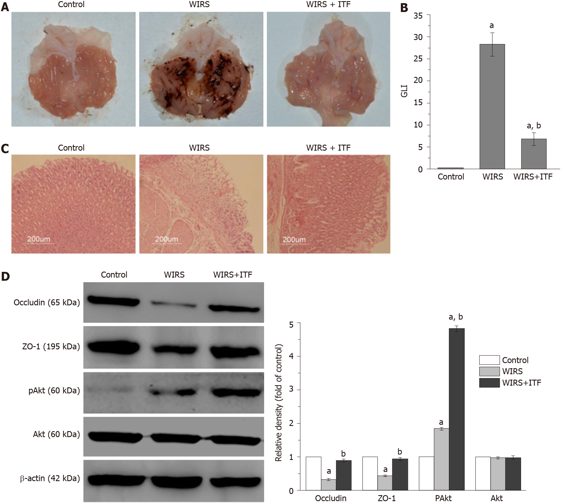
The stomachs of the WIRS group presented with severe gastric mucosal lesions, and part of the lesion area developed severe edema, bleeding, and ulceration (Figure 1A). Compared with the WIRS group, the gastric lesions in the WIRS + ITF group were almost negligible, and the GLI was significantly decreased (7.50 ± 2.10 vs 28.50 ± 3.20, P < 0.01) (Figure 1B), although the GLI was still higher than that of the control group (without stress exposure). More-detailed histological examination showed that the WIRS group displayed extensive destruction of the gastric mucosa. The pathological changes, including epithelial necrosis, congestion, bleeding, inflammatory cell infiltration, dilating and congesting vessels, and edema, were also present in the submucosa. Minimal damage was observed in the WIRS + ITF group (Figure 1C). Integrity of the mucosa is the basis of its barrier function, and we investigated expression of epithelial tight junction markers occludin and ZO-1. Occludin and ZO-1 were significantly downregulated in the stomach of the WIRS group (Figure 1D). The potential mechanisms that may be involved in this wound healing process were also preliminarily investigated. Akt/protein kinase B is a widely recognized vital regulatory factor responsible for maintaining cell viability and survival. After treatment with ITF, the Akt signaling pathway was activated and expression of pAkt in the gastric specimens was increased in the WIRS + ITF group (Figure 1D).
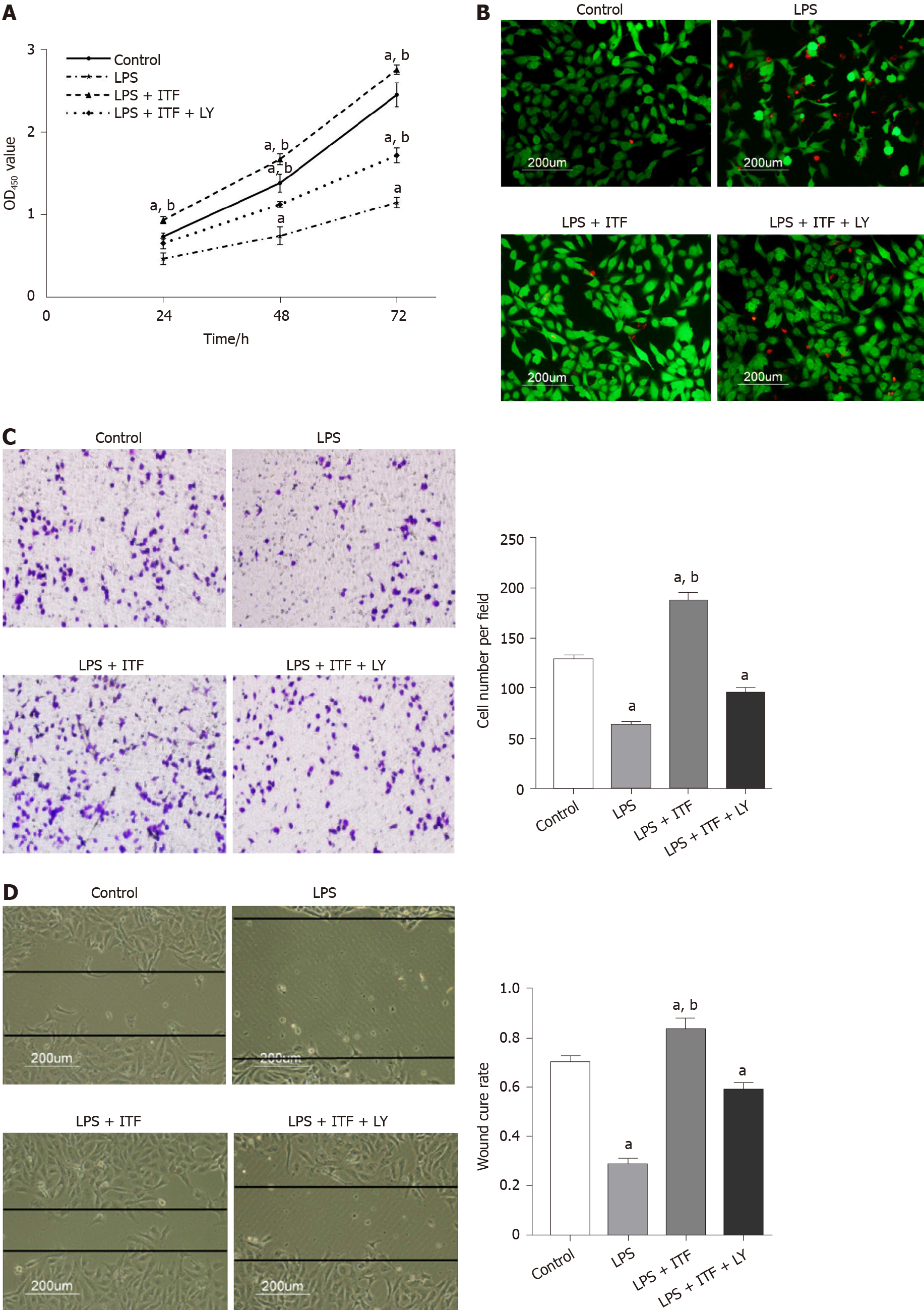
When treated with LY294002, cell viability was inhibited compared with the LPS + ITF group but was still significantly increased at 48 and 72 h compared with the LPS group (P < 0.01) (Figure 2A). Necrosis was induced in GES-1 cells by LPS and was attenuated by ITF, while the number of necrotic cells was increased when treated with LY294002 (Figure 2B). Epithelial cell migration and wound closure were promoted by ITF but hindered by LPS (Figure 2C and D). Inhibition of the Akt pathway by LY294002 suppressed ITF-induced cell migration.
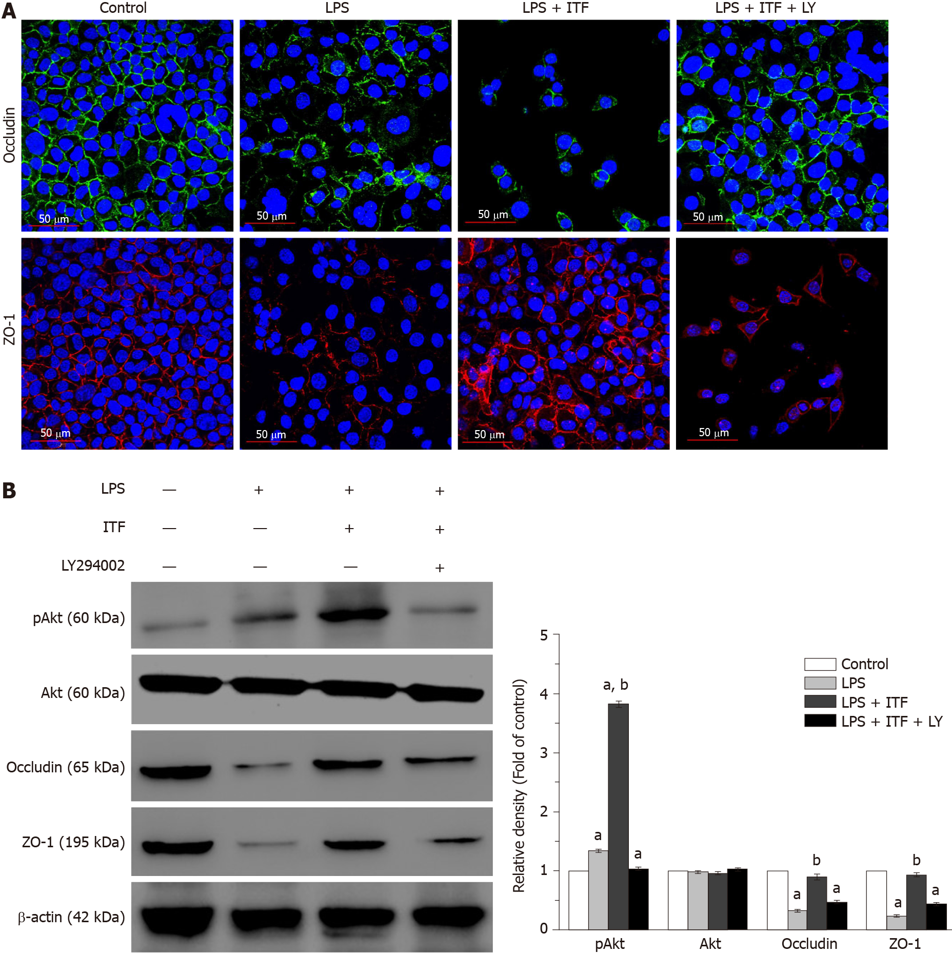
To explore the effects of ITF on gastric mucosal epithelial integrity, expression of tight junction makers was detected. Immunofluorescence and western blotting demonstrated that LPS induced epithelial tight junction damage and decreased expression of occludin and ZO-1, whereas ITF reversed the decreased expression of occludin and ZO-1 (Figure 3A and B). Expression of Akt phosphorylation in GES-1 cells was increased when treated with ITF (Figure 3B), but LY294002 inhibited activation of the Akt signaling pathway induced by ITF and suppressed the protective effects of ITF on epithelial integrity (Figure 3A and B). These results indicated that ITF had protective effects on gastric mucosal epithelial integrity by activating Akt signaling, while the inhibitor of Akt signaling pathway, LY294002, eliminated the protective effects of ITF.
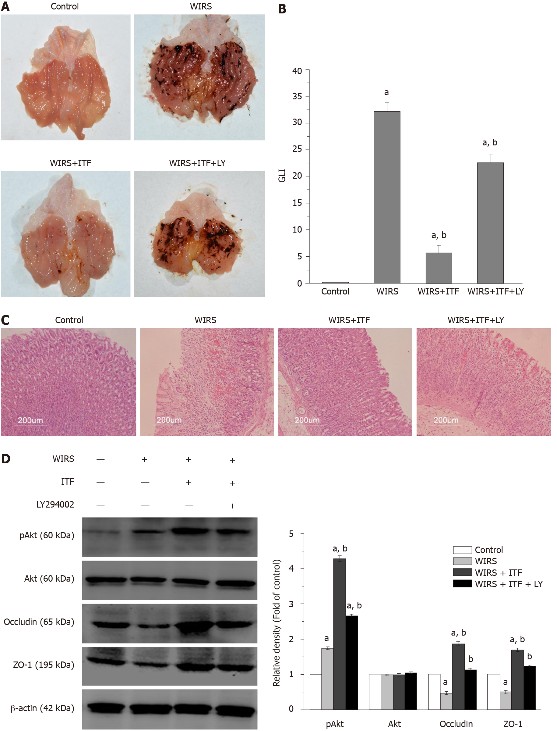
To confirm the gastroprotective effects of ITF and involvement of Akt signaling in this process, another group of rats (WIRS + ITF + LY group) was enrolled and treated with Akt signaling pathway inhibitor LY294002. Stress exposure caused formation of gastric lesions in all groups except for the control group with intact mucosa (Figure 4A). Rats from the WIRS and WIRS + ITF + LY groups developed severe gastric mucosal lesions and the typical macroscopic signs including hyperemia, hemorrhage and edema. On the contrary, only minimal morphological lesions and reduced areas of gastric ulcer formation were observed in the WIRS + ITF group. Correspondingly, the GLI in the WIRS + ITF group was markedly decreased compared with that in the WIRS group (5.50 ± 1.20 vs 32.0 ± 2.50, P < 0.01) (Figure 4B), but this trend was reversed by LY294002 (GLI: 22.0 ± 1.50). Microscopic examination showed that ITF prevented stress-induced histological changes in the gastric mucosa with inflammation infiltration, congestion, and hemorrhage. Administration of LY294002 interfered with the protective effects of ITF, and the mucosal damage was slight compared with that in the WIRS group (Figure 4C).
Expression of occludin, ZO-1, Akt, and pAkt in gastric mucosa was determined. Expression of occludin and ZO-1 in the gastric tissues was significantly enhanced by ITF, and the effects were partially inhibited by LY294002 (Figure 4D). Akt phosphorylation was upregulated when challenged by stress exposure and was further increased by ITF. However, the inhibitor of the Akt pathway reduced phosphorylation in the WIRS + ITF + LY group compared with the WIRS + ITF group (P < 0.05) (Figure 4D).
The gastrointestinal epithelial barrier is crucial for the maintenance of homeostasis, and it is also constantly exposed to various stimuli and susceptible to those threats. It is not surprising that critically ill patients are often confronted with the risk of mucosal damage, alterations in epithelial barrier function, and various complications. Despite the increased risk of myocardial ischemia, Clostridium difficile enteritis and hospital-acquired pneumonia, prophylactic medication for stress ulcers is still widely adopted in the ICU, along with treating the primary diseases[18]. Novel approaches have been explored to control and prevent mucosal damage or promote epithelial restitution.
As an endogenous peptide and a key constituent of mucus barrier, ITF is a potential choice for mucosal damage prophylaxis and treatment. In recent decades, changes in ITF expression level have been recognized in various diseases, especially in gastrointestinal disorders. Serum levels of ITF are significantly increased after skeletal trauma in humans, which could enhance motility and migration of mesenchymal progenitor cells and promote skeletal repair[19]. For critically ill children, where the body is in a stressed state, serum ITF concentration is associated with gastrointestinal failure and prognosis[20]. Another study has suggested that urinary ITF can help diagnose and predict the disease course in necrotizing enterocolitis at the early stage in newborns[21]. Previous studies have also paid close attention to the application of ITF to inflammatory bowel disease, showing that ITF could be used as a biomarker to predict disease activity and assess mucosal healing in ulcerative colitis[22,23]. All these reports suggest the therapeutic potential of ITF in gastrointestinal mucosal damage, but there is an absence of direct evidence.
However, few studies have explored the roles of ITF in stress-induced gastric mucosal injury. Previously, we have reported the ITF-mediated protection of gastric epithelial mucosa cells from NSAIDs in vitro without mechanistic explanation and further investigation in vivo[14]. In the current study, we initially found that pretreatment with ITF attenuated gastric lesions and alleviated local inflammation in the WIRS rat model, and the altered expression of epithelium tight junction markers indicated the ability of ITF to maintain or restore epithelial integrity. Preliminary results also suggested the activation of Akt kinase. In the following experiments, the cytoprotective effects of ITF were verified using LPS to induce GES-1 cell injury, and ITF promoted cell proliferation and migration, inhibited LPS-induced necrosis, and preserved the intercellular tight junction in vitro without signaling. Intriguingly, when treated with LY294002, a specific inhibitor of the Akt signaling pathway, these protective effects of ITF were attenuated significantly in vitro. The results with the GES-1 cell line suggested that Akt signaling was essential for ITF to function biologically, and this was validated in another set of WIRS rat models.
The Akt signaling pathway is reported to play essential roles in the process of cell proliferation, differentiation, apoptosis, and migration. It has also been shown to preserve epithelial integrity during inflammation, which was confirmed in the current study[24]. Our results in vitro and in vivo demonstrated that ITF was a rapid responder to stress stimuli. Although mucosal restitution is an intrinsic function of gastrointestinal epithelial cells, ITF still exerted promising wound healing effects.
Several issues involved in the subtle regulatory networks still need to be discussed. Whether ITF responsiveness requires a receptor is unclear. Belle and co-workers found that the leucine rich repeat receptor and nogo-interacting protein 2 (LINGO2) is essential for ITF-mediated functions, and ITF–LINGO2 interactions derepress inhibitory LINGO2–epidermal growth factor receptor complexes, allowing ITF to drive wound healing and immunity[25]. ITF has also been reported to interact with other receptors including chemokine CXC receptor 4 and 7, protease-activated receptors, and classic signaling pathways[26-28].
The histological examination in our study indicated that ITF could exert anti-inflammatory effects in vivo. These effects were also demonstrated when the microglial cells were cultured in the presence of ITF, and subsequent reduced expression and secretion of pro-inflammatory cytokines after LPS stimulation were detected[29]. ITF derived from human breast milk can downregulate proinflammatory cytokines and upregulate human β-defensin expression via regulating intracellular Ca2+ activity, and the significance of ITF as an immunomodulator should be given more emphasis[27]. Recombinant human ITF is reported to protect mucosal barrier function in rats with nonalcoholic steatohepatitis and reduce inflammatory injury by reducing expression of Toll-like receptor 4 and nuclear factor-κB[30]. Supplementation of ITF also rescued Toll-like receptor 2-deficient mice from increased inflammatory-stress-induced mucosal damage linked to innate immune protection[31]. Additionally, the TFFs, including ITF, are reported to share a divalent lectin activity that recognizes the N-acetylglucosamine-α-1, 4-galactose disaccharide and interact with mucosal glycoproteins relying on the glycosylation state. How their lectin activities might promote cell migration to achieve epithelial restitution remains unclear[32]. Accumulating evidence has implicated glycosylation as an underappreciated post-translational modification, and glycan alterations have an important impact on the mucus layer, glycan–lectin interactions, and mucosal immunity[32-34].
With regard to clinical implications of ITF in adult critically ill patients, the latest research shows that plasma ITF levels are sustained elevated in abdominal sepsis patients, and higher ITF levels are associated with shock and multiple organ (≥ three) failure. However, elevated ITF level is not an independent risk factor for 30 d mortality[35]. These results suggest that gastrointestinal injury contributes to the pathogenesis of critical illness, and ITF could be a potential therapeutic agent. ITF has been utilized for enema in patients with mild-to-moderate left-sided ulcerative colitis in a clinical trial, but this well-tolerated regimen did not provide any benefit above that of adding 5-aminosalicylic acid alone[36]. A novel delivery method, use of the systemic route, and adjustment of medication duration should be taken into consideration in subsequent trials. The latest study found that protein disulfide isomerase A1 can directly catalyze dimerization of ITF, and changes in the modification protein disulfide isomerase A1 reduce its activity, resulting in a corresponding decrease in ITF dimerization and delayed intestinal mucosal repair during sepsis. This work suggests novel mechanisms for the inhibition of mucosal repair and promising targets for the prevention and treatment of sepsis[37]. The aforementioned potential adverse effects of conventional antiulcer agents such as proton pump inhibitors and H2-receptor antagonists and the current results support the prospect of clinical translation of ITF, especially for critically ill patients who are in a of stress.
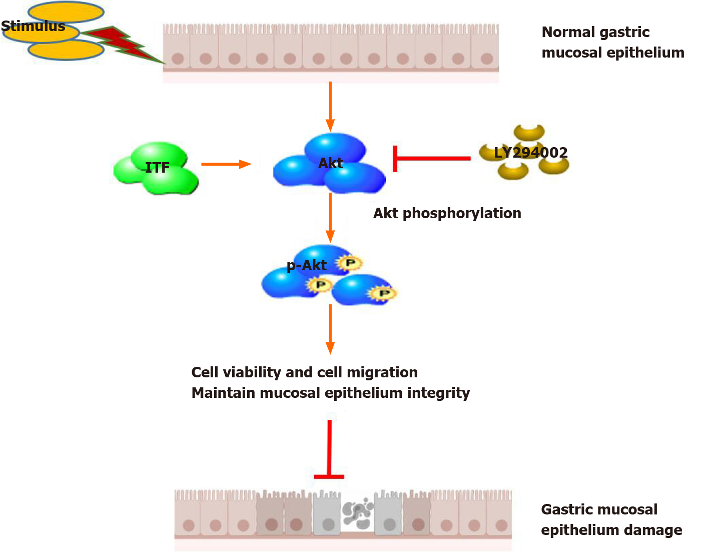
In conclusion, ITF can alleviate both macroscopic and microscopic gastric mucosal damage in vivo induced by acute stress stimulation and promote mucosal epithelial cell survival, accelerate wound closure, and preserve mucosal integrity in vitro. Akt signaling pathway could play essential roles in this process (Figure 5). Further studies should be implemented to explore the feasibility of clinical application of ITF for prevention and treatment of stress ulcer and gastric mucosal damage caused by other factors.
Stress-related gastric mucosal damage is a prevalent complication in critically ill patients in the intensive care unit, and it may evolve to ulceration and bleeding. Stress ulcer prophylaxis has been common in routine intensive care, but with controversy. Co-secreted with mucins, intestinal trefoil factor (ITF) is reported to promote the restitution and regeneration of intestinal mucosal epithelium, but the mechanism is unknown.
As an endogenous peptide, ITF harbors innate advantages over conventional anti-ulcer agents, and might be a new candidate for stress ulcer prophylaxis.
To investigate the protective effects of ITF on gastric mucosa and explore the underlying mechanisms.
We utilized water immersion restraint stress-induced gastric mucosal damage rat model and lipopolysaccharide-induced gastric epithelium cell damage model to investigate the potential functions of ITF on damaged gastric mucosa both in vivo and in vitro.
We found that ITF promoted proliferation and migration and inhibited necrosis of gastric epithelium cells and preserved the integrity of gastric mucosa by increasing expression of occludin and zonula occludens-1. Additionally, pretreatment with ITF ameliorated the gastric mucosal epithelial damage and promoted mucosal repair in vivo. We found that the protective effects of ITF were exerted by activation of Akt signaling, and the specific inhibitor of this pathway, LY249002, abolished the protective effects.
Pretreatment with ITF alleviated stress-induced gastric mucosal damage by activation of Akt signaling.
This study provides insight into the translational potential of ITF as a promising candidate for prevention and treatment of stress-induced gastric mucosal damage.
| 1. | Alhazzani W, Guyatt G, Alshahrani M, Deane AM, Marshall JC, Hall R, Muscedere J, English SW, Lauzier F, Thabane L, Arabi YM, Karachi T, Rochwerg B, Finfer S, Daneman N, Alshamsi F, Zytaruk N, Heel-Ansdell D, Cook D; Canadian Critical Care Trials Group. Withholding Pantoprazole for Stress Ulcer Prophylaxis in Critically Ill Patients: A Pilot Randomized Clinical Trial and Meta-Analysis. Crit Care Med. 2017;45:1121-1129. [RCA] [PubMed] [DOI] [Full Text] [Cited by in Crossref: 55] [Cited by in RCA: 79] [Article Influence: 8.8] [Reference Citation Analysis (0)] |
| 2. | Krag M, Perner A, Møller MH. Stress ulcer prophylaxis in the intensive care unit. Curr Opin Crit Care. 2016;22:186-190. [RCA] [PubMed] [DOI] [Full Text] [Cited by in Crossref: 5] [Cited by in RCA: 11] [Article Influence: 1.1] [Reference Citation Analysis (0)] |
| 3. | Marker S, Krag M, Møller MH. What's new with stress ulcer prophylaxis in the ICU? Intensive Care Med. 2017;43:1132-1134. [RCA] [PubMed] [DOI] [Full Text] [Cited by in Crossref: 14] [Cited by in RCA: 12] [Article Influence: 1.3] [Reference Citation Analysis (0)] |
| 4. | Ye Z, Reintam Blaser A, Lytvyn L, Wang Y, Guyatt GH, Mikita JS, Roberts J, Agoritsas T, Bertschy S, Boroli F, Camsooksai J, Du B, Heen AF, Lu J, Mella JM, Vandvik PO, Wise R, Zheng Y, Liu L, Siemieniuk RAC. Gastrointestinal bleeding prophylaxis for critically ill patients: a clinical practice guideline. BMJ. 2020;368:l6722. [RCA] [PubMed] [DOI] [Full Text] [Cited by in Crossref: 104] [Cited by in RCA: 89] [Article Influence: 14.8] [Reference Citation Analysis (0)] |
| 5. | Rice TW, Kripalani S, Lindsell CJ. Proton Pump Inhibitors vs Histamine-2 Receptor Blockers for Stress Ulcer Prophylaxis in Critically Ill Patients: Issues of Interpretability in Pragmatic Trials. JAMA. 2020;323:611-613. [RCA] [PubMed] [DOI] [Full Text] [Cited by in Crossref: 4] [Cited by in RCA: 8] [Article Influence: 1.3] [Reference Citation Analysis (0)] |
| 6. | Pelaseyed T, Bergström JH, Gustafsson JK, Ermund A, Birchenough GM, Schütte A, van der Post S, Svensson F, Rodríguez-Piñeiro AM, Nyström EE, Wising C, Johansson ME, Hansson GC. The mucus and mucins of the goblet cells and enterocytes provide the first defense line of the gastrointestinal tract and interact with the immune system. Immunol Rev. 2014;260:8-20. [RCA] [PubMed] [DOI] [Full Text] [Cited by in Crossref: 646] [Cited by in RCA: 974] [Article Influence: 88.5] [Reference Citation Analysis (0)] |
| 7. | Thim L. Trefoil peptides: from structure to function. Cell Mol Life Sci. 1997;53:888-903. [RCA] [PubMed] [DOI] [Full Text] [Cited by in Crossref: 176] [Cited by in RCA: 170] [Article Influence: 5.9] [Reference Citation Analysis (0)] |
| 8. | Aihara E, Engevik KA, Montrose MH. Trefoil Factor Peptides and Gastrointestinal Function. Annu Rev Physiol. 2017;79:357-380. [RCA] [PubMed] [DOI] [Full Text] [Cited by in Crossref: 85] [Cited by in RCA: 136] [Article Influence: 13.6] [Reference Citation Analysis (0)] |
| 9. | Le J, Zhang DY, Zhao Y, Qiu W, Wang P, Sun Y. ITF promotes migration of intestinal epithelial cells through crosstalk between the ERK and JAK/STAT3 pathways. Sci Rep. 2016;6:33014. [RCA] [PubMed] [DOI] [Full Text] [Full Text (PDF)] [Cited by in Crossref: 22] [Cited by in RCA: 26] [Article Influence: 2.6] [Reference Citation Analysis (0)] |
| 10. | Yusufu A, Shayimu P, Tuerdi R, Fang C, Wang F, Wang H. TFF3 and TFF1 expression levels are elevated in colorectal cancer and promote the malignant behavior of colon cancer by activating the EMT process. Int J Oncol. 2019;55:789-804. [RCA] [PubMed] [DOI] [Full Text] [Full Text (PDF)] [Cited by in Crossref: 6] [Cited by in RCA: 27] [Article Influence: 3.9] [Reference Citation Analysis (0)] |
| 11. | Al-Salam S, Sudhadevi M, Awwad A, Al Bashir M. Trefoil factors peptide-3 is associated with residual invasive breast carcinoma following neoadjuvant chemotherapy. BMC Cancer. 2019;19:135. [RCA] [PubMed] [DOI] [Full Text] [Full Text (PDF)] [Cited by in Crossref: 8] [Cited by in RCA: 14] [Article Influence: 2.0] [Reference Citation Analysis (0)] |
| 12. | Perera O, Evans A, Pertziger M, MacDonald C, Chen H, Liu DX, Lobie PE, Perry JK. Trefoil factor 3 (TFF3) enhances the oncogenic characteristics of prostate carcinoma cells and reduces sensitivity to ionising radiation. Cancer Lett. 2015;361:104-111. [RCA] [PubMed] [DOI] [Full Text] [Cited by in Crossref: 26] [Cited by in RCA: 30] [Article Influence: 2.7] [Reference Citation Analysis (0)] |
| 13. | Taniguchi Y, Kurokawa Y, Takahashi T, Mikami J, Miyazaki Y, Tanaka K, Makino T, Yamasaki M, Nakajima K, Mori M, Doki Y. Prognostic Value of Trefoil Factor 3 Expression in Patients with Gastric Cancer. World J Surg. 2018;42:3997-4004. [RCA] [PubMed] [DOI] [Full Text] [Cited by in Crossref: 6] [Cited by in RCA: 11] [Article Influence: 1.6] [Reference Citation Analysis (0)] |
| 14. | Lin J, Sun Z, Zhang W, Liu H, Shao D, Ren Y, Wen Y, Cao L, Wolfram J, Yang Z, Nie S. Protective effects of intestinal trefoil factor (ITF) on gastric mucosal epithelium through activation of extracellular signal-regulated kinase 1/2 (ERK1/2). Mol Cell Biochem. 2015;404:263-270. [RCA] [PubMed] [DOI] [Full Text] [Cited by in Crossref: 8] [Cited by in RCA: 11] [Article Influence: 1.0] [Reference Citation Analysis (0)] |
| 15. | Shigeshiro M, Tanabe S, Suzuki T. Repeated exposure to water immersion stress reduces the Muc2 gene level in the rat colon via two distinct mechanisms. Brain Behav Immun. 2012;26:1061-1065. [RCA] [PubMed] [DOI] [Full Text] [Cited by in Crossref: 15] [Cited by in RCA: 19] [Article Influence: 1.4] [Reference Citation Analysis (0)] |
| 16. | Huang P, Zhou Z, Wang H, Wei Q, Zhang L, Zhou X, Hutz RJ, Shi F. Effect of the IGF-1/PTEN/Akt/FoxO signaling pathway on the development and healing of water immersion and restraint stress-induced gastric ulcers in rats. Int J Mol Med. 2012;30:650-658. [RCA] [PubMed] [DOI] [Full Text] [Cited by in Crossref: 17] [Cited by in RCA: 26] [Article Influence: 1.9] [Reference Citation Analysis (0)] |
| 17. | Sun Z, Liu H, Yang Z, Shao D, Zhang W, Ren Y, Sun B, Lin J, Xu M, Nie S. Intestinal trefoil factor activates the PI3K/Akt signaling pathway to protect gastric mucosal epithelium from damage. Int J Oncol. 2014;45:1123-1132. [RCA] [PubMed] [DOI] [Full Text] [Cited by in Crossref: 42] [Cited by in RCA: 53] [Article Influence: 4.4] [Reference Citation Analysis (0)] |
| 18. | Barbateskovic M, Marker S, Granholm A, Anthon CT, Krag M, Jakobsen JC, Perner A, Wetterslev J, Møller MH. Stress ulcer prophylaxis with proton pump inhibitors or histamin-2 receptor antagonists in adult intensive care patients: a systematic review with meta-analysis and trial sequential analysis. Intensive Care Med. 2019;45:143-158. [RCA] [PubMed] [DOI] [Full Text] [Cited by in Crossref: 34] [Cited by in RCA: 44] [Article Influence: 6.3] [Reference Citation Analysis (0)] |
| 19. | Krüger K, Schmid S, Paulsen F, Ignatius A, Klinger P, Hotfiel T, Swoboda B, Gelse K. Trefoil Factor 3 (TFF3) Is Involved in Cell Migration for Skeletal Repair. Int J Mol Sci. 2019;20. [RCA] [PubMed] [DOI] [Full Text] [Full Text (PDF)] [Cited by in Crossref: 3] [Cited by in RCA: 9] [Article Influence: 1.3] [Reference Citation Analysis (0)] |
| 20. | Ma MJ, Han B, Xu SQ. Trefoil factor 3 related to gastrointestinal failure in pediatric critical illness. Arch Pediatr. 2016;23:681-684. [RCA] [PubMed] [DOI] [Full Text] [Cited by in Crossref: 2] [Cited by in RCA: 2] [Article Influence: 0.2] [Reference Citation Analysis (0)] |
| 21. | Coufal S, Kokesova A, Tlaskalova-Hogenova H, Frybova B, Snajdauf J, Rygl M, Kverka M. Urinary I-FABP, L-FABP, TFF-3, and SAA Can Diagnose and Predict the Disease Course in Necrotizing Enterocolitis at the Early Stage of Disease. J Immunol Res. 2020;2020:3074313. [RCA] [PubMed] [DOI] [Full Text] [Full Text (PDF)] [Cited by in Crossref: 16] [Cited by in RCA: 36] [Article Influence: 6.0] [Reference Citation Analysis (0)] |
| 22. | Srivastava S, Kedia S, Kumar S, Pratap Mouli V, Dhingra R, Sachdev V, Tiwari V, Kurrey L, Pradhan R, Ahuja V. Serum human trefoil factor 3 is a biomarker for mucosal healing in ulcerative colitis patients with minimal disease activity. J Crohns Colitis. 2015;9:575-579. [RCA] [PubMed] [DOI] [Full Text] [Cited by in Crossref: 24] [Cited by in RCA: 27] [Article Influence: 2.5] [Reference Citation Analysis (0)] |
| 23. | Nakov R, Velikova T, Nakov V, Ianiro G, Gerova V, Tankova L. Serum trefoil factor 3 predicts disease activity in patients with ulcerative colitis. Eur Rev Med Pharmacol Sci. 2019;23:788-794. [RCA] [PubMed] [DOI] [Full Text] [Cited by in RCA: 9] [Reference Citation Analysis (0)] |
| 24. | Fruman DA, Chiu H, Hopkins BD, Bagrodia S, Cantley LC, Abraham RT. The PI3K Pathway in Human Disease. Cell. 2017;170:605-635. [RCA] [PubMed] [DOI] [Full Text] [Cited by in Crossref: 1679] [Cited by in RCA: 2068] [Article Influence: 229.8] [Reference Citation Analysis (0)] |
| 25. | Belle NM, Ji Y, Herbine K, Wei Y, Park J, Zullo K, Hung LY, Srivatsa S, Young T, Oniskey T, Pastore C, Nieves W, Somsouk M, Herbert DR. TFF3 interacts with LINGO2 to regulate EGFR activation for protection against colitis and gastrointestinal helminths. Nat Commun. 2019;10:4408. [RCA] [PubMed] [DOI] [Full Text] [Full Text (PDF)] [Cited by in Crossref: 46] [Cited by in RCA: 72] [Article Influence: 10.3] [Reference Citation Analysis (0)] |
| 26. | Dieckow J, Brandt W, Hattermann K, Schob S, Schulze U, Mentlein R, Ackermann P, Sel S, Paulsen FP. CXCR4 and CXCR7 Mediate TFF3-Induced Cell Migration Independently From the ERK1/2 Signaling Pathway. Invest Ophthalmol Vis Sci. 2016;57:56-65. [RCA] [PubMed] [DOI] [Full Text] [Cited by in Crossref: 31] [Cited by in RCA: 39] [Article Influence: 3.9] [Reference Citation Analysis (0)] |
| 27. | Barrera GJ, Tortolero GS. Trefoil factor 3 (TFF3) from human breast milk activates PAR-2 receptors, of the intestinal epithelial cells HT-29, regulating cytokines and defensins. Bratisl Lek Listy. 2016;117:332-339. [RCA] [PubMed] [DOI] [Full Text] [Cited by in Crossref: 9] [Cited by in RCA: 10] [Article Influence: 1.1] [Reference Citation Analysis (0)] |
| 28. | Braga Emidio N, Hoffmann W, Brierley SM, Muttenthaler M. Trefoil Factor Family: Unresolved Questions and Clinical Perspectives. Trends Biochem Sci. 2019;44:387-390. [RCA] [PubMed] [DOI] [Full Text] [Cited by in Crossref: 38] [Cited by in RCA: 49] [Article Influence: 7.0] [Reference Citation Analysis (0)] |
| 29. | Arnold P, Rickert U, Helmers AK, Spreu J, Schneppenheim J, Lucius R. Trefoil factor 3 shows anti-inflammatory effects on activated microglia. Cell Tissue Res. 2016;365:3-11. [RCA] [PubMed] [DOI] [Full Text] [Cited by in Crossref: 15] [Cited by in RCA: 19] [Article Influence: 1.9] [Reference Citation Analysis (0)] |
| 30. | Wang Y, Liang K, Kong W. Intestinal Trefoil Factor 3 Alleviates the Intestinal Barrier Function Through Reducing the Expression of TLR4 in Rats with Nonalcoholic Steatohepatitis. Arch Med Res. 2019;50:2-9. [RCA] [PubMed] [DOI] [Full Text] [Cited by in Crossref: 7] [Cited by in RCA: 13] [Article Influence: 1.9] [Reference Citation Analysis (0)] |
| 31. | Podolsky DK, Gerken G, Eyking A, Cario E. Colitis-associated variant of TLR2 causes impaired mucosal repair because of TFF3 deficiency. Gastroenterology. 2009;137:209-220. [RCA] [PubMed] [DOI] [Full Text] [Full Text (PDF)] [Cited by in Crossref: 192] [Cited by in RCA: 191] [Article Influence: 11.2] [Reference Citation Analysis (0)] |
| 32. | Järvå MA, Lingford JP, John A, Soler NM, Scott NE, Goddard-Borger ED. Trefoil factors share a lectin activity that defines their role in mucus. Nat Commun. 2020;11:2265. [RCA] [PubMed] [DOI] [Full Text] [Full Text (PDF)] [Cited by in Crossref: 19] [Cited by in RCA: 36] [Article Influence: 6.0] [Reference Citation Analysis (0)] |
| 33. | Kudelka MR, Stowell SR, Cummings RD, Neish AS. Intestinal epithelial glycosylation in homeostasis and gut microbiota interactions in IBD. Nat Rev Gastroenterol Hepatol. 2020;17:597-617. [RCA] [PubMed] [DOI] [Full Text] [Cited by in Crossref: 139] [Cited by in RCA: 241] [Article Influence: 40.2] [Reference Citation Analysis (0)] |
| 34. | Hoffmann W. Trefoil Factor Family (TFF) Peptides and Their Diverse Molecular Functions in Mucus Barrier Protection and More: Changing the Paradigm. Int J Mol Sci. 2020;21. [RCA] [PubMed] [DOI] [Full Text] [Full Text (PDF)] [Cited by in Crossref: 27] [Cited by in RCA: 70] [Article Influence: 11.7] [Reference Citation Analysis (0)] |
| 35. | Meijer MT, Uhel F, Cremer OL, Scicluna BP, Schultz MJ, van der Poll T; MARS consortium. Elevated trefoil factor 3 plasma levels in critically ill patients with abdominal sepsis or non-infectious abdominal illness. Cytokine. 2020;133:155181. [RCA] [PubMed] [DOI] [Full Text] [Cited by in Crossref: 2] [Cited by in RCA: 4] [Article Influence: 0.7] [Reference Citation Analysis (0)] |
| 36. | Mahmood A, Melley L, Fitzgerald AJ, Ghosh S, Playford RJ. Trial of trefoil factor 3 enemas, in combination with oral 5-aminosalicylic acid, for the treatment of mild-to-moderate left-sided ulcerative colitis. Aliment Pharmacol Ther. 2005;21:1357-1364. [RCA] [PubMed] [DOI] [Full Text] [Cited by in Crossref: 37] [Cited by in RCA: 38] [Article Influence: 1.8] [Reference Citation Analysis (0)] |
| 37. | Shi Y, Wang C, Wu D, Zhu Y, Wang ZE, Peng X. Mechanistic study of PDIA1-catalyzed TFF3 dimerization during sepsis. Life Sci. 2020;255:117841. [RCA] [PubMed] [DOI] [Full Text] [Cited by in Crossref: 1] [Cited by in RCA: 1] [Article Influence: 0.2] [Reference Citation Analysis (0)] |
Open-Access: This article is an open-access article that was selected by an in-house editor and fully peer-reviewed by external reviewers. It is distributed in accordance with the Creative Commons Attribution NonCommercial (CC BY-NC 4.0) license, which permits others to distribute, remix, adapt, build upon this work non-commercially, and license their derivative works on different terms, provided the original work is properly cited and the use is non-commercial. See: http://creativecommons.org/Licenses/by-nc/4.0/
Manuscript source: Unsolicited manuscript
Specialty type: Gastroenterology and hepatology
Country/Territory of origin: China
Peer-review report’s scientific quality classification
Grade A (Excellent): 0
Grade B (Very good): B
Grade C (Good): C
Grade D (Fair): D
Grade E (Poor): 0
P-Reviewer: Enosawa S, Matsukawa J S-Editor: Zhang L L-Editor: Filipodia P-Editor: Liu JH


