Published online Sep 7, 2020. doi: 10.3748/wjg.v26.i33.4945
Peer-review started: May 17, 2020
First decision: June 18, 2020
Revised: June 27, 2020
Accepted: August 12, 2020
Article in press: August 12, 2020
Published online: September 7, 2020
Processing time: 109 Days and 22 Hours
Intestinal mucosal barrier dysfunction plays an important role in the pathogenesis of ulcerative colitis (UC). Recent studies have revealed that impaired autophagy is associated with intestinal mucosal dysfunction in the mucosa of colitis mice. Resveratrol exerts anti-inflammatory functions by regulating autophagy.
To investigate the effect and mechanism of resveratrol on protecting the integrity of the intestinal mucosal barrier and anti-inflammation in dextran sulfate sodium (DSS)-induced ulcerative colitis mice.
Male C57BL/6 mice were divided into four groups: negative control group, DSS model group, DSS + resveratrol group, and DSS + 5-aminosalicylic acid group. The severity of colitis was assessed by the disease activity index, serum inflammatory cytokines were detected by enzyme-linked immunosorbent assay. Colon tissues were stained with haematoxylin and eosin, and mucosal damage was evaluated by mean histological score. The expression of occludin and ZO-1 in colon tissue was evaluated using immunohistochemical analysis. In addition, the expression of autophagy-related genes was determined using reverse transcription-polymerase chain reaction and Western-blot, and morphology of autophagy was observed by transmission electron microscopy.
The resveratrol treatment group showed a 1.72-fold decrease in disease activity index scores and 1.42, 3.81, and 1.65-fold decrease in the production of the inflammatory cytokine tumor necrosis factor-α, interleukin-6 and interleukin-1β, respectively, in DSS-induced colitis mice compared with DSS group (P < 0.05). The expressions of the tight junction proteins occludin and ZO-1 in DSS model group were decreased, and were increased in resveratrol-treated colitis group. Resveratrol also increased the levels of LC3B (by 1.39-fold compared with DSS group) and Beclin-1 (by 1.49-fold compared with DSS group) (P < 0.05), as well as the number of autophagosomes, which implies that the resveratrol may alleviate intestinal mucosal barrier dysfunction in DSS-induced UC mice by enhancing autophagy.
Resveratrol treatment decreased the expression of inflammatory factors, increased the expression of tight junction proteins and alleviated UC intestinal mucosal barrier dysfunction; this effect may be achieved by enhancing autophagy in intestinal epithelial cells.
Core tip: We established a chronic colitis model successfully via administration of dextran sulfate sodium (DSS), and we found that resveratrol ameliorates the production of the inflammatory cytokines in DSS-induced colitis mice. Meanwhile, resveratrol treatment alleviated intestinal mucosal barrier dysfunction in DSS-induced colitis and increased the expression of the tight junction proteins occludin and ZO-1. Further studies showed that resveratrol treatment increased the levels of LC3B and Beclin-1 in the colons of colitis mice, as well as the number of autophagosomes, which may via enhancing autophagy.
- Citation: Pan HH, Zhou XX, Ma YY, Pan WS, Zhao F, Yu MS, Liu JQ. Resveratrol alleviates intestinal mucosal barrier dysfunction in dextran sulfate sodium-induced colitis mice by enhancing autophagy. World J Gastroenterol 2020; 26(33): 4945-4959
- URL: https://www.wjgnet.com/1007-9327/full/v26/i33/4945.htm
- DOI: https://dx.doi.org/10.3748/wjg.v26.i33.4945
Inflammatory bowel disease (IBD) is a chronic intestinal inflammatory condition involving the gastrointestinal tract, comprising ulcerative colitis (UC) and Crohn’s disease. UC is characterized by abdominal pain, diarrhoea, and haematochezia. The incidence of UC is rising with changes in dietary habits and the rhythm of life[1,2]. UC is often persistent or recurrent in patients and increases the risk of colorectal cancer[3]. Currently, drugs such as 5-aminosalicylic acid (5-ASA), corticosteroids and immunosuppressants are used, but the effect of these drugs is poor, and the long-term use of these drugs may cause severe adverse reactions, resulting in great psychological and economic burdens on the patients[4,5]. Therefore, effective and safe drugs against UC are urgently needed.
The initiating event and pathogenesis of UC currently remain elusive. Recent work has highlighted the importance of intestinal mucosal barrier dysfunction in disease pathophysiology[6]. Many studies have shown that the destruction of the intestinal mucosal barrier plays an important role in the deterioration of IBD, especially in UC patients[7-9]. Protecting the integrity of the intestinal mucosal barrier is thought to be a potential clinical treatment approach for UC.
Resveratrol (3,4′,5-Trihydroxy-trans-stilbene), a natural plant polyphenol found in grapes, is the principal biologically active component in red wine. Resveratrol has anti-inflammatory and immunoregulatory functions, with the advantages of low price and few side effects[10-12]. So far, very few clinical studies have indicated that resveratrol can alleviate clinical colitis activity and improve quality of life and in patients with active UC[13,14]. Although these limited studies show that resveratrol may ameliorate inflammation in UC, the mechanism remains unclear. Intestinal epithelial cells (IECs) are reported to act as the first line of defence in the intestinal mucosal barrier. The IECs tight junctions may play an important role in intestinal mucosal barrier. Occludin and ZO-1 are important tight junction proteins that play a significant role in maintaining the integrity of the intestinal mucosal barrier. Thus, we focused on whether resveratrol can alleviate intestinal mucosal barrier injury and inflammation. In addition, the mechanism underlying the anti-inflammatory effect of resveratrol in colitis was explored.
Autophagy is a cellular recycling process involving self-degradation and the reconstruction of damaged organelles and proteins[15]. Recent studies have shown that impaired autophagy is related to intestinal mucosal damage in mice with colitis, and stimulation of autophagy can prevent intestinal mucosal inflammation and ameliorates murine colitis in mice[16]. Furthermore, the regulatory effect of resveratrol on autophagy in inflammatory diseases has gradually attracted attention[17,18]. In the present study, we evaluated the effect of resveratrol on dextran sulfate sodium (DSS)-induced colitis in mice and explore the mechanism of resveratrol on protecting the integrity of the intestinal mucosal barrier and anti-inflammation.
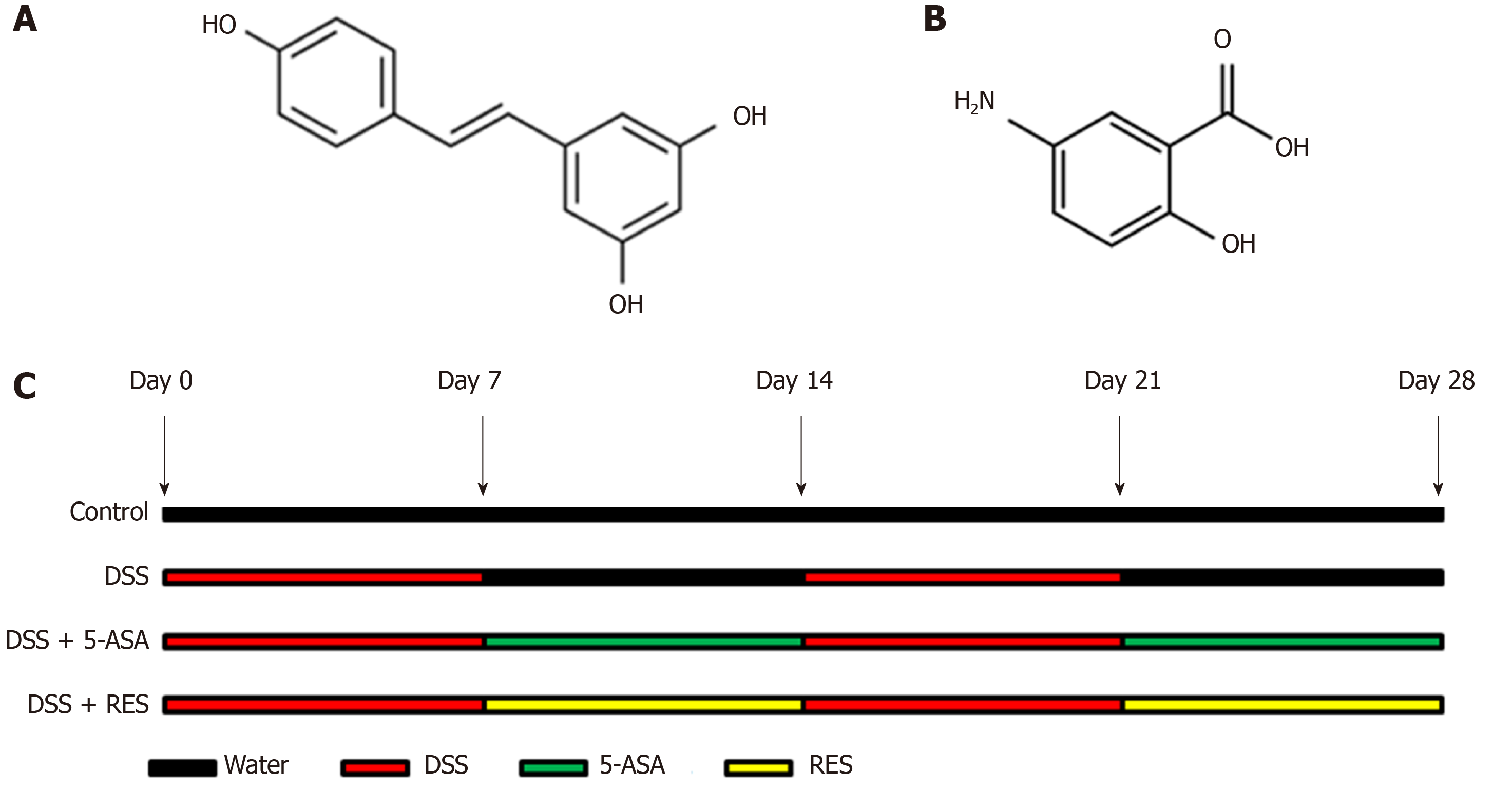
Resveratrol (3,4′,5-Trihydroxy-trans-stilbene; C14H12O3; Molecular Weight: 228.24 g/moL; Figure 1A) and 5-Aminosalicylic acid (5-ASA; C7H7NO3; Molecular Weight: 153.14 g/moL; Figure 1B) were purchased from Sigma Chemical (United States). The purity was checked via HPLC analysis and exceeded 99%. Dextran Sulfate Sodium (DSS, Molecular Weight: 36000-50000) was purchased from MP Biomedicals (United States).
Male C57BL/6 mice aged 5 wk and weighing 17-19 g were purchased from Shanghai SLAC Laboratory Animal Co., Ltd. Animals were housed in mouse cages at a controlled temperature (21 ± 2 °C) with a pre-set 12-h light/dark cycle. The mice were fed a standard laboratory diet and normal drinking water. The normal diet included crude proteins ≥ 20%, crude fat ≥ 6%, crude fibre ≤ 5%, crude ash ≤ 8%, lysine 1.32%, calcium 1%-1.8%, and phosphorus 0.6%-1.2%, respectively. The animals were acclimated to the experimental conditions for one week before the experiment. According to the guidelines for the care and use of laboratory animals, all the procedures for the treatment and execution of mice were approved by the Animal Care and Use Committee of Zhejiang Chinese Medical University.
DSS-induced colitis is a widely used model that morphologically and symptomatically resembles UC in humans. In this study, we established DSS-induced chronic colitis model to study UC. Mice were randomly divided into four experimental groups (n = 12 mice/group): Negative control group, DSS model group, resveratrol-treated group and 5-ASA positive-control group. Mice in the negative control group received normal drinking water for 28 d. In the DSS model group, chronic colitis was induced by two cycles of giving drinking water containing 3% DSS for 7 d and normal drinking water for 7 d. Resveratrol treatment group (DSS + RES) mice received two cycles of DSS by drinking water, and treatment of resveratrol 100 mg/kg per day by gavage for two cycles followed by DSS at the same time (Figure 1C). 5-ASA, the first-line therapy for UC, was used as a positive control to evaluate the efficacy of resveratrol. Mice in the positive-control group (DSS + 5-ASA) were treated with two cycles of DSS, and 5-ASA 200 mg/kg per day for 28 d at the same time. Body mass was detected every 5 d, and body mass loss was calculated as (detected body mass-initial mass) /initial mass. All animals were euthanized with carbon dioxide and sacrificed at humane end points.
To assess the severity of colitis, the disease activity index (DAI) was calculated daily based on weight loss, stool consistency and rectal bleeding (Table 1)[19]. Stool consistency (0, normal; 2, loose stool; 4, watery diarrhea); bloody stools (0, normal; 2, slight bleeding; 4, gross bleeding); and body weight loss (0, < 2%; 1, decreased ≥ 2 - < 5%; 2, decreased ≥ 5 - < 10%; 3, decreased ≥ 10 - < 15%; 4, decreased ≥ 15). These values were assessed for each animal, and the sum of the 3 values constituted the DAI. For each parameter, the scores ranged from 0 to 4, resulting in a total DAI score ranging from 0 (unaffected) to 12 (severe colitis).
| Body mass loss (%) | Stool consistency | Bleeding | Score |
| < 2% | Normal | No bleeding | 0 |
| ≥ 2 - < 5% | - | - | 1 |
| ≥ 5 - < 10% | Loose stools | Slight bleeding | 2 |
| ≥ 10 - < 15% | - | - | 3 |
| ≥ 15 | Watery diarrhea | Gross bleeding | 4 |
Mice were sacrificed, and blood samples (1.0 mL) were collected from the heart. The expression levels of tumour necrosis factor-α (TNF-α, MTA00B, R&D Systems Europe Ltd., Abingdon, England), interleukin (IL)-6 (M6000B, R&D Systems Europe Ltd., Abingdon, England ), and IL-1β (MLB00C, RD Systems Europe Ltd., Abingdon, England) in the plasma were detected using the enzyme-linked immunosorbent assay kits according to the manufacturer’s instructions. The concentration of the cytokines was expressed as pg/mL per mg protein.
Colon length can be used as an indirect marker of inflammation. Thus, when mice were sacrificed, the length of colons from the caecum to the anus was measured. Then, the colon was fixed in 10% formalin, dehydrated at gradient concentrations of ethanol, and embedded in paraffin. Tissue sections (8 µm) were prepared and stained with haematoxylin and eosin (HE), and mucosal damage was evaluated. To evaluate the colonic mucosal damage severity, 9 fields were randomly selected and observed under an inverted fluorescence microscope by two pathologists who were blinded to the experimental groups. Colonic mucosal damage was confirmed by the mean histological score (Table 2)[20]. Each histological score, such as inflammation (0-3), extent (0-3), regeneration (0-4) and crypt damage (0-4), was multiplied by the percentage of compromised tissue (1 point for 25%, 2 points for 26%-50%, 3 points for 51%-75%, and 4 points for 76%-100%). Therefore, the scores of inflammation and extent range from 0 to 12, and the scores of regeneration and crypt damage range from 0 to 16, which reached a maximum score of 56.
| Features | Grade | Description |
| Inflammation | 0 | None |
| 1 | Slight | |
| 2 | Moderate | |
| 3 | Severe | |
| Extent | 0 | None |
| 1 | Mucosa | |
| 2 | Mucosa and submucosa | |
| 3 | Transmural | |
| Regeneration | 4 | No tissue repair |
| 3 | Surface epithelium not intact | |
| 2 | Regeneration with crypt depletion | |
| 1 | Almost complete regeneration | |
| 0 | Complete regeneration or normal tissue | |
| Crypt damage | 0 | None |
| 1 | Basal 1/3 damaged | |
| 2 | Basal 2/3 damaged | |
| 3 | Only surface epithelium intact | |
| 4 | Entire crypt and epithelium lost | |
| Percent involvement | 1 | 1%-25% |
| 2 | 26%-50% | |
| 3 | 51%-75% | |
| 4 | 76%-100% |
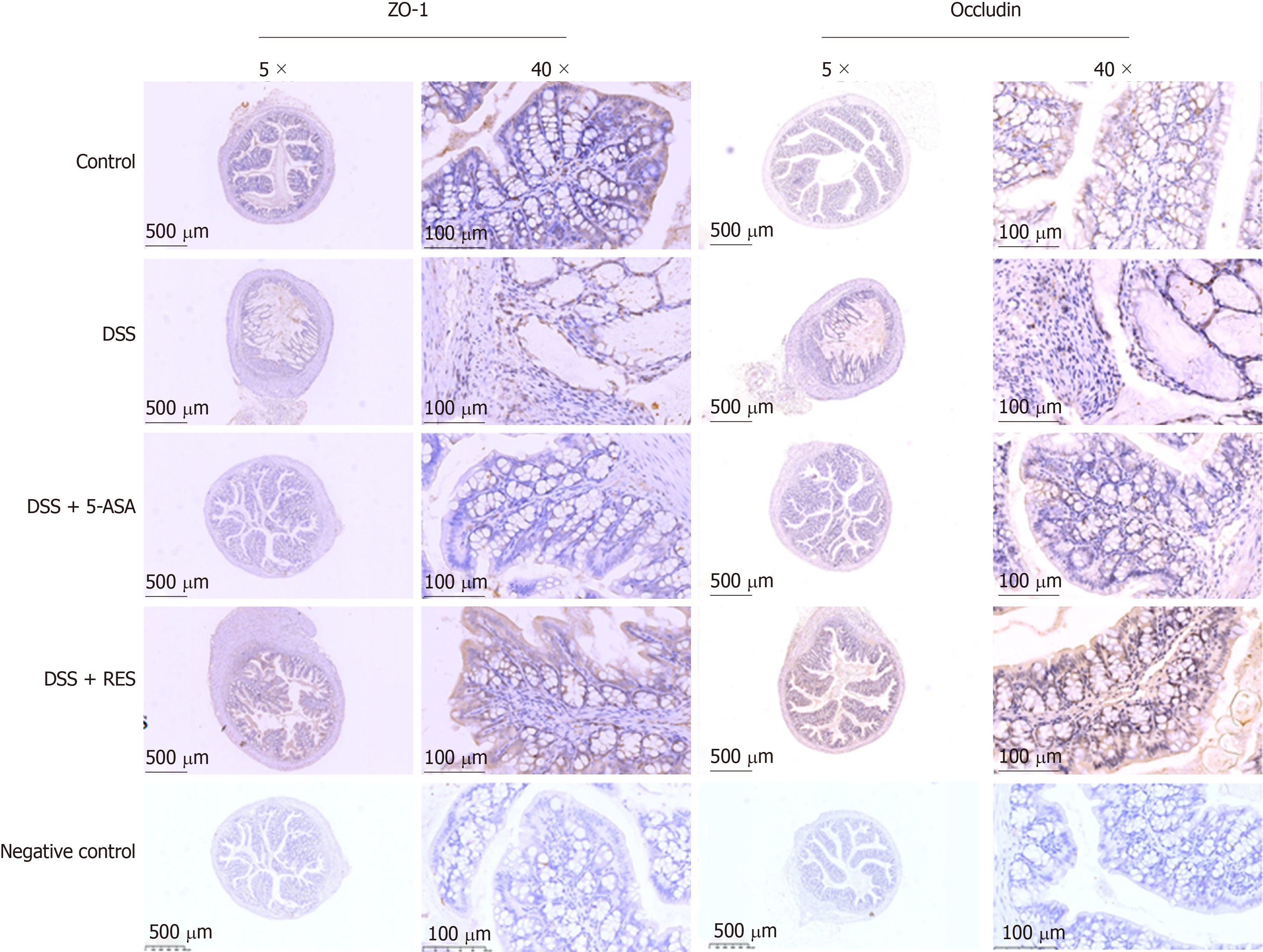
The expression of occludin and ZO-1 was detected by immunohistochemical analysis. Briefly, all blocks of colonic tissue were sectioned (3-5 µm). Then, sections were deparaffinized, rehydrated at graded ethanol concentrations and incubated with fresh 3% hydrogen peroxide for 10 min. After being rinsed with phosphate buffer saline (PBS), each tissue section was subjected to antigen retrieval by a suitable antigen retrieval method (boiling in 0.01 M citrate buffer). After endogenous peroxidases were blocked, sections were incubated with anti-occludin antibody (ab222691, 1:200 dilution in PBS, Abcam, United States) and a monoclonal ZO-1 antibody (ab221546, 1:300, Abcam, United States) overnight at 4°C. Subsequently, the sections were incubated for 20 min at room temperature with biotin-labelled secondary antibodies, stained with 3,3-diaminobenzidine, counterstained with haematoxylin, dehydrated and mounted. Primary antibody was replaced by PBS as the negative control. The following criterion was used for semi-quantitative staining: 0 (no staining), 1 (weak staining, light yellow), 2 (moderate staining, yellow brown), and 3 (strong staining, brown). The proportion of stained tumor cells was scored according to the proportion of positively stained tumor cells as follows: 0 (< 5%); 1 (6-25%); 2 (26-50%); and 3 (> 51%). A composite score was obtained by the staining intensity score multiplied the proportion scores. The total score ≤ 4 was defined as low expression, and > 5 was regarded as high expression.
Briefly, proteins were extracted using radioimmunoprecipitation assay buffer, separated by sodium dodecyl sulphate-polyacrylamide gel (5%-10%) electrophoresis, and then transferred to polyvinylidene difluoride membranes (Millipore, Billerica, MA, United States). These membranes were washed with PBS and blocked with Tris-buffered saline-Tween with 5% skim milk (232100, BD, Bioscience, United States). Primary β-actin (3700S, 1:1000, Cell Signal Technology, United States), LC3B (3868T, 1:1000, Cell Signal Technology, United States) and Beclin-1 (3495T, 1:1000, Cell Signal Technology, United States) antibodies were applied, followed by horseradish peroxidase-conjugated secondary antibodies. Then, an enhanced chemiluminescence detection system (Thermo Fisher Scientific, United States) was used to detect antibody binding. Meanwhile, β-actin was used as a negative control.
Total ribonucleic acid was extracted from tissue specimens using TRIzol reagent, and messenger ribonucleic acid (mRNA) was reverse transcribed to complementary deoxyribonucleic acid with a QuantiTect reverse transcription kit (205311, Qiagen, German) according to the manufacturer’s instructions. Quantitative polymerase chain reaction (qPCR) was performed using a SuperScript III Platinum SYBR green one-step qPCR kit (11732088, Thermo Fisher). The qPCR primer sequences were as follows: actin: 5’-CAT CCG TAA AGA CCT CTA TGC CAA C-3’ (forward) and 5’-ATG GAG CCA CCG ATC CAC A-3’ (reverse); Beclin: 5’-ATG GAG GGG TCT AAG GCG TC-3’ (forward) and 5’-TGG GCT GTG GTA AGT AAT GGA-3’ (reverse); LC3B: 5’-TCA AGT CCA ACT ACC GAG TCC-3’ (forward) and 5’-TCA GAG GTT TCC CAT CCA AG-3’ (reverse). Relative messenger ribonucleic acid expression levels were calculated based on the computerized tomography values and normalized using actin expression. All experiments were performed in triplicate on a Roche LightCycler 480 platform.
First, colon tissues were fixed with glutaraldehyde and osmium teroxid, then dehydrated in ethanol, passed through propylene oxide, and embedded in Spurr resin. Fifty nanometres thick sections were cut by an ultramicrotome, and the sections were subsequently post-stained with 4% uranyl acetate for 10 min and Reynold's lead citrate for 1.5 min. The structure of the IECs and autolysosomes were observed by transmission electron microscopy (HITACHI H-7650).
All statistical analyses were performed using Statistical Package for the Social Sciences software (version 13.0; Statistic Package for Social Science Inc., Chicago, IL, United States). Continuous variables were presented as the mean and standard deviation and analysed using one-way Anova or Kruskal-Wallis test. All values were expressed as the mean ± standard deviation. All statistical analyses were conducted with α = 0.05, and P < 0.05 was considered significant.
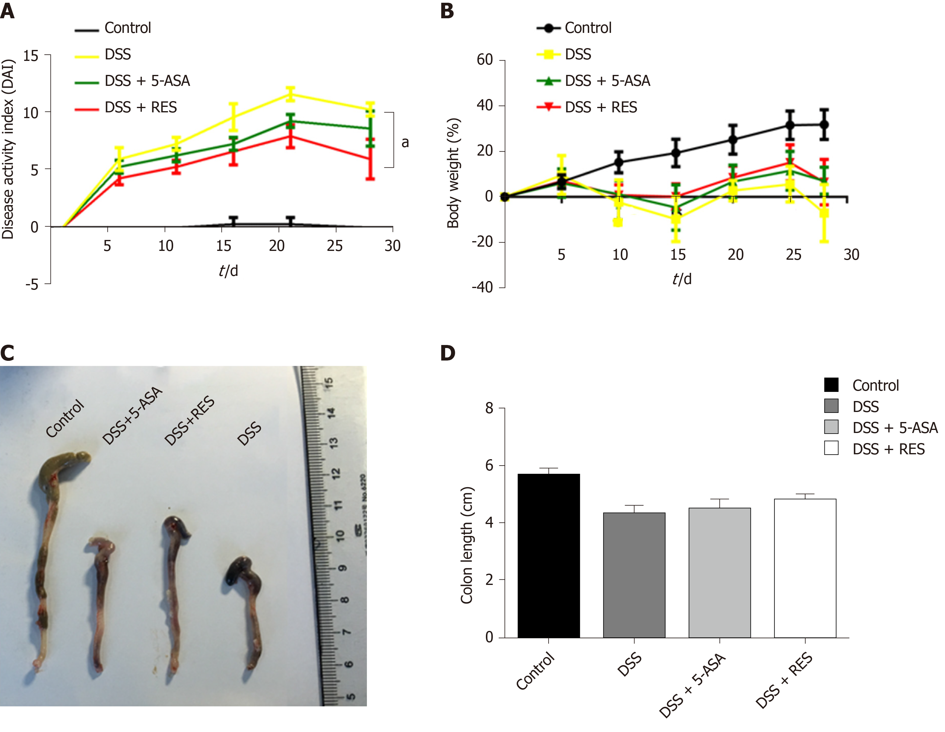
DSS-induced colitis was evaluated, and the clinical course was monitored and scored for the presence of bloody stool, watery diarrhoea and weight loss. During this experiment, three mice in the DSS group died, and one of the mice died on the 13th day, and the other two died on the 14th day; one mouse in the DSS + 5-ASA group died on the 13th day, and no mice in the DSS + RES group died. In comparison with those in the negative control group, mice in the DSS group showed progressively increased DAI score. However, the DSS + RES group showed 1.72-fold decrease in DAI score compared with the DSS group (P < 0.05, Figure 2A), which indicated that resveratrol may have a favourable effect on colitis.
Body mass could be decreased by DSS-induced inflammation; therefore, we studied the effect of resveratrol on body mass in DSS-induced colitis mice. The body mass was measured every 5 d, and the results showed that DSS-treated mice exhibited a statistically significant decrease. Resveratrol and 5-ASA treatment increased the body mass of DSS-induced colitis mice, but the difference was not significant (Figure 2B). We also measured the colon length of DSS-induced colitis mice and found that the colon length of the resveratrol-treated group was longer than that of the DSS group, but the difference was also not significant (Figure 2C).
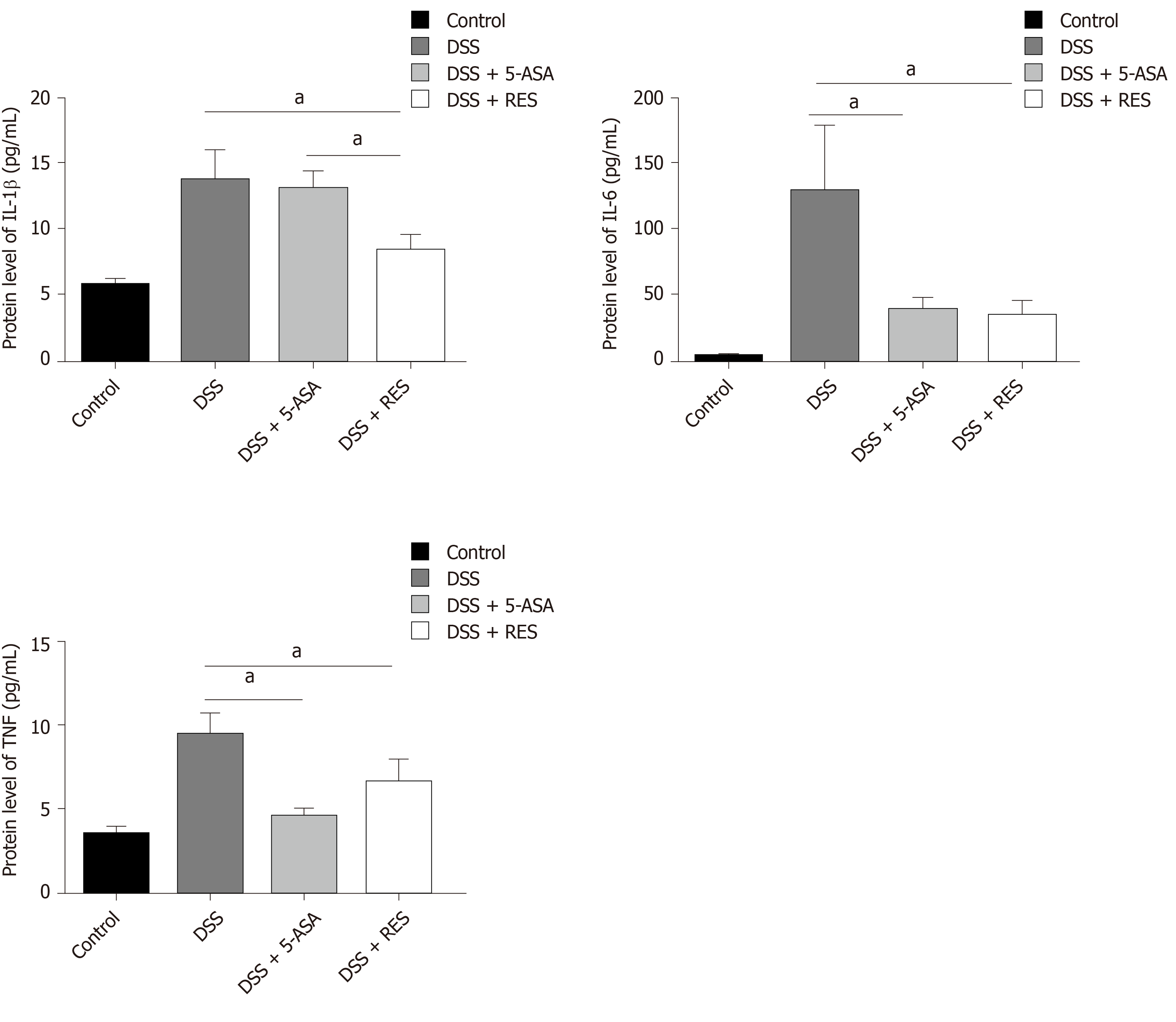
The enzyme-linked immunosorbent assay results demonstrated that the protein expression levels of the inflammatory cytokines TNF-α, IL-6, and IL-1β were higher in the DSS-induced colitis group than in the control group (P < 0.05, Figure 3). Furthermore, the levels of TNF-α and IL-6 in the 5-ASA-treatment group were lower than those in the DSS-induced colitis group (P < 0.05). However, the levels of TNF-α, IL-6 and IL-1β showed 1.42, 3.81, and 1.65-fold decrease in the resveratrol group compared with the DSS group (P < 0.05), and level of IL-1β also showed a 1.57-fold decrease in the resveratrol group compared with that in the DSS + 5-ASA group (P < 0.05).
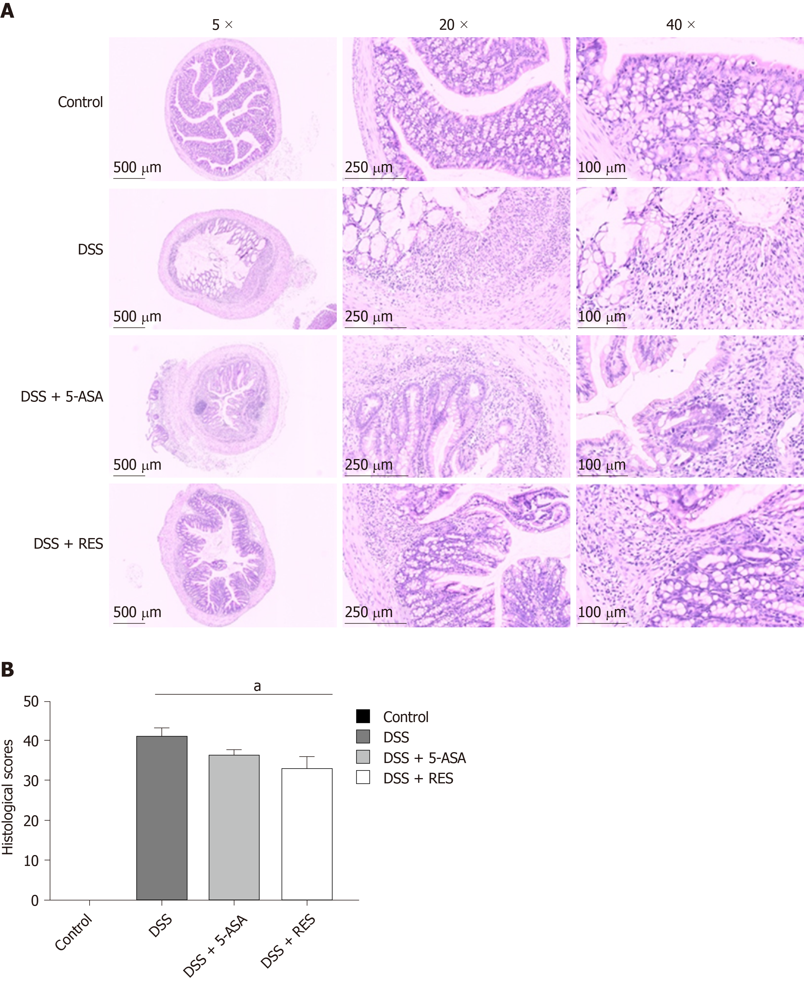
We also assessed colon damage by histologic examination. DSS group showed significant colonic mucosal damages, crypt depletion, infiltration of inflammatory cells into the mucosa and submucosa, loss of epithelial barrier. The colon damage and intestinal inflammation in mice treated with resveratrol and 5-ASA were less severe than that in DSS-induced colitis mice (Figure 4A). Consistent with the histologic damage, the histological scores of colitis mice treated with resveratrol were significantly lower than those of DSS-induced colitis mice (P < 0.05, Figure 4B), while the histological scores of 5-ASA-treated colitis mice were not significantly different from those of mice in the DSS group. These results indicate that resveratrol may alleviate intestinal mucosal barrier dysfunction in DSS-induced colitis mice more effectively than 5-ASA.
The tight junction proteins occludin and ZO-1 have been reported to play an important role in maintaining the integrity of the intestinal mucosal epithelial barrier. In the present study, we found that the expression levels of occludin and ZO-1 were higher in the resveratrol-treated DSS group than in the 5-ASA and DSS groups (Figure 5 and Table 3).
| Group | ZO-1 expression | χ2/P value | Occludin expression | χ2/P value | ||
| Low (%) | High (%) | Low (%) | High (%) | |||
| Control | 6 (50.0%) | 6 (50.0%) | 8.812/0.029 | 7 (58.3%) | 5 (41.7%) | 10.987/0.010 |
| DSS | 7 (77.8%) | 2 (22.2%) | 8 (88.9%) | 1 (11.1%) | ||
| DSS + 5-ASA | 7 (63.6%) | 4 (36.4%) | 9 (81.8%) | 2 (18.2%) | ||
| DSS + RES | 2 (16.7%) | 10 (83.3%) | 3 (25.0%) | 9 (75.0%) | ||
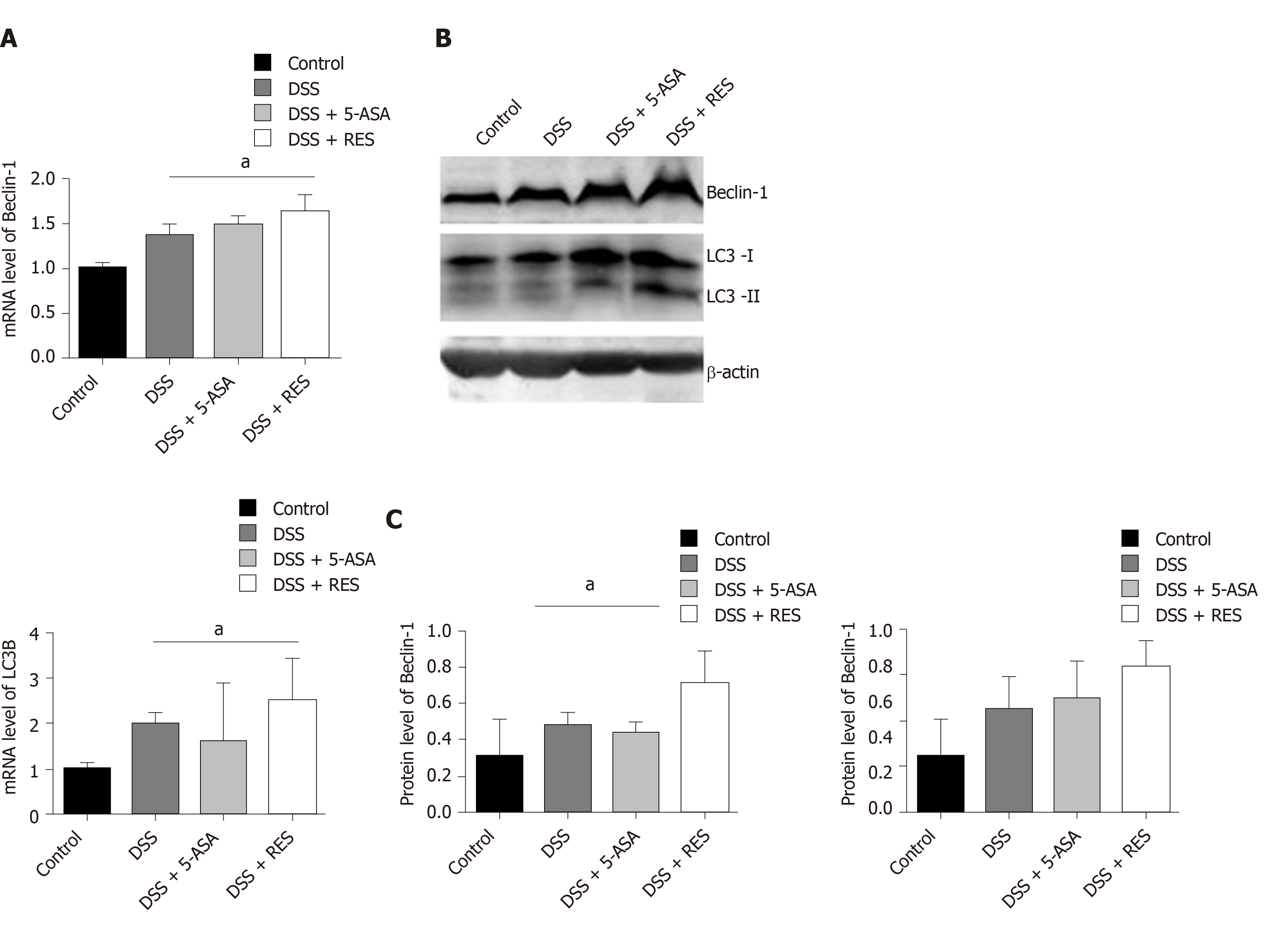
To explore whether the inhibition of colitis by resveratrol treatment is mediated by autophagy activation, we examined the mRNA and protein levels of LC3B and Beclin-1. As determined by real-time PCR, a substantial increase in the mRNA expression level of LC3B and Beclin-1 was observed in the DSS + RES group compared with the DSS group, suggesting that resveratrol increases the mRNA levels of LC3B and Beclin-1 in the colons of DSS-treated mice (P < 0.05, Figure 6A). Consistent with the mRNA level analysis, the western blot results also showed that resveratrol treatment induced significant increases in the LC3-II/I ratio and Beclin-1 level in DSS-induced colitis mice (P < 0.05, Figure 6B and C). However, the difference in the mRNA levels of LC3B and Beclin-1 was not significant between the 5-ASA-treated colitis group and the DSS group (P > 0.05).
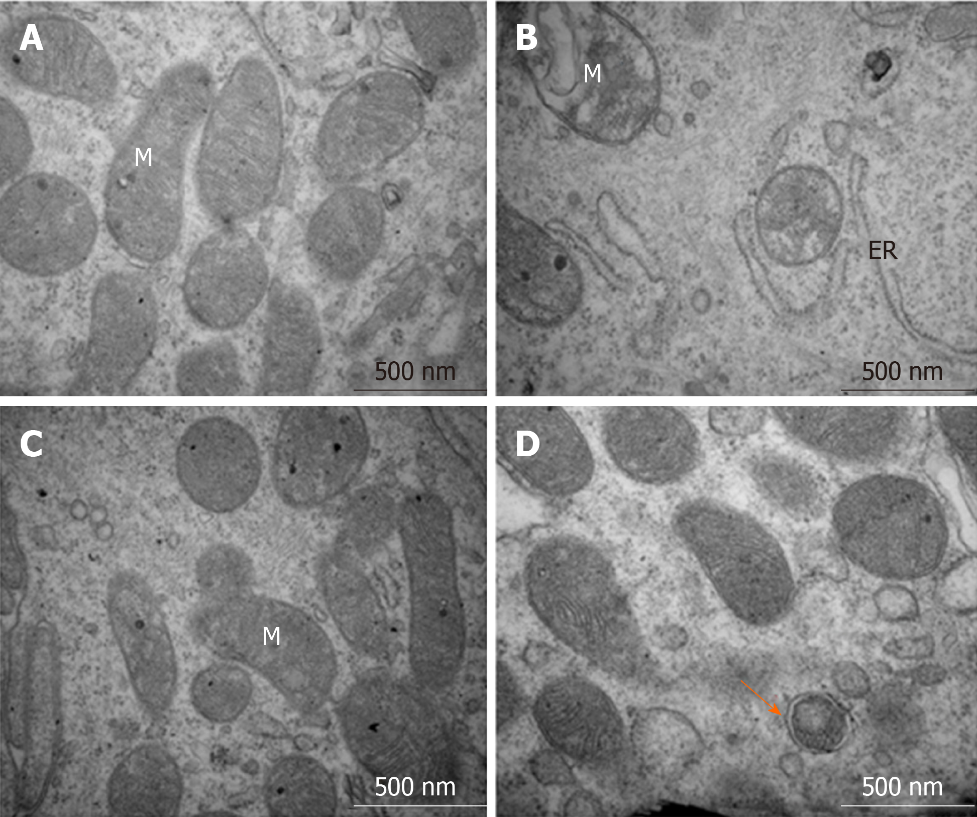
The transmission electron microscopy results showed that IECs in the control group had normal organelle structure with a normal endoplasmic reticulum and mitochondria (Figure 7A). However, in the DSS-induced colitis group, the organelle structure was markedly damaged, with swollen mitochondria, disrupted mitochondrial inner ridges and dilated rough endoplasmic reticulum (Figure 7B). Inflammation induced by the 5-ASA treatment and autophagosomes were rare (Figure 7C). Additionally, resveratrol administration increased the number of autophagosomes and improved the condition of the endoplasmic reticulum and mitochondria, which indicated that resveratrol promoted the progression of autophagy (Figure 7D).
UC is a recurrent chronic inflammation of the colon. DSS-induced colitis models, including acute and chronic colitis models, are most commonly used in UC. Acute colitis model is usually induced by administering DSS for 7 d. In this study, a chronic colitis model was successfully established by the administration of two cycles of DSS treatment. Each cycle involved the administration of 3% DSS for 7 d followed by normal drinking water for 7 d[21]. Mice in the chronic colitis group developed clinical symptoms, such as bloody stool and weight loss; the mice also showed pathological features similar to UC patients, such as shortened colon length, intestinal mucosa congestion, oedema and erosion, ulceration, crypt damage, and the infiltration of inflammatory cells into the mucosa and submucosa; the histological scores of colitis and serum inflammation indices (TNF-α, IL-6, IL-1β) in the DSS group increased. The chronic colitis model induced by administering repeated cycles of DSS more closely resembled human UC than the acute colitis model did.
In recent years, the role of intestinal mucosal barrier dysfunction in the pathogenesis of UC has attracted wide attention. IECs are the first-line defence in the intestinal mucosal barrier[22]. The IECs tight junctions are key epithelial intracellular junctions[23,24]. Occludin and ZO-1 are important tight junction proteins that play a significant role in maintaining the integrity of the intestinal mucosal barrier. The integrity of the intestinal mucosal barrier prevents and defends against the invasion of inflammatory factors and bacteria and further mitigates intestinal inflammation. Resveratrol is a well-known phytophenol with pleiotropic properties and has anti-oxidative and anti-inflammatory activity. Recently, resveratrol has also been shown to alleviate intestinal inflammation[25-27]. Consistent with these studies, our results also showed that resveratrol can reduce intestinal inflammation and alleviate intestinal mucosal barrier dysfunction in DSS-induced colitis mice. Resveratrol significantly increased levels of tight junction proteins (occludin and ZO-1) and reduced histological colonic mucosa injury scores, even than 5-ASA did. Resveratrol also decreased serum inflammation indices (TNF-α, IL-6, and IL-1β), although there was no significant difference between resveratrol and 5-ASA treatment. These results showed that both resveratrol and 5-ASA alleviated intestinal inflammation, and resveratrol performed better at maintaining the integrity of the intestinal mucosal epithelial barrier than 5-ASA.
Autophagy is a cellular self-protective mechanism stimulated by both internal and external adverse environmental factors. In this process, degenerated proteins, aged and damaged organelles, even pathogenic microorganisms, are recycled via autophagic degradation to maintain cellular metabolic balance and homeostasis[15,28,29]. Autophagy can be activated when an organ is stimulated by adverse factors. Saito M and others found a reduced LC3-II/I ratio, expression of adhesion proteins and IECs adhesion, and a damaged mucosal epithelial barrier when IECs were exposed to TNF-α and treated with autophagy inhibitors[30]. In this study, we found that resveratrol alleviated the intestinal mucosal barrier dysfunction and reduced the intestinal inflammatory response by enhancing autophagy in experimental chronic colitis(two cycles of DSS treatment for 28 d), as the expression of LC3B and Beclin-1, and the number of autophagosomes in resveratrol-treated group were increased when compared to that in the chronic colitis model group and the negative control group.
A previous study conducted by Zhang et al[31] reported that DSS-inducing increased the autophagy in acute colitis models(DSS treatment for 7 d), and curcumin and resveratrol could protect against colitis by reducing autophagy rather than enhancing autophagy[31]. We speculated that this inconsistency may be caused by the different colitis models used in the two studies. The acute colitis model resembles the early stage of human UC, while the chronic colitis model resembles the advanced stage of UC. Our previous study showed that autophagy was dynamic in the progression of colitis. It was activated in IECs by inflammatory factors in the early stage of a rat model of sepsis-induced acute colitis,and decreased as the disease progressed, thereby reducing the removal of oxidative stress products and the damaged organelles and other substances in cells, which lead to the destruction of the intestinal mucosal barrier and the aggravation of intestinal inflammation[32]. We believe that the chronic colitis mouse model is a better tool to explore the pathogenesis and drug effects of ulcerative colitis.
However, there are still many limitations in the present study. The detailed autophagy involved in resveratrol-induced protection of intestinal mucosal barrier was unclear as we did not study the effect of autophagy inhibitors on colitis treated by resveratrol. On the other hand, we only investigated the effect of resveratrol on animals, therefore, further research is still needed for clinical application.
Intestinal mucosal barrier disorder plays a very important role in the pathogenesis of ulcerative colitis (UC). Recent studies have revealed that impaired autophagy is associated with intestinal mucosal dysfunction in the mucosa of colitis mice.
Resveratrol can regulate autophagy in the treatment of a few inflammatory diseases. Recently, few studies have indicated that resveratrol can alleviate clinical colitis activity in patients with active UC, while the mechanism for its anti-inflammatory effect remains elusive.
The aim of the study was to explore the effect and mechanism of resveratrol on protecting the integrity of the intestinal mucosal barrier and anti-inflammation in dextran sulfate sodium (DSS)-induced ulcerative colitis.
DSS-induced ulcerative colitis was induced by DSS, then the disease activity index was used to assess the severity of colitis. Inflammatory cytokines were detected by enzyme-linked immunosorbent assay. Tissue sections were stained with haematoxylin and eosin, and mucosal damage was evaluated by mean histological score. The expression of occludin and ZO-1 was detected by immunohistochemical analysis. Reverse transcription-polymerase chain reaction and Western-blot were used to analyze autophagy-related gene expression, and morphology of autophagy was observed by transmission electron microscopy.
The resveratrol treatment group showed a 1.72-fold decrease in disease activity index scores and 1.42-, 3.81-, and 1.65-fold decrease in the production of the inflammatory cytokines tumor necrosis factor-α, interleukin-6 and interleukin-1β, respectively, in DSS-induced colitis mice compared with DSS group (P < 0.05). The expression of the tight junction proteins occludin and ZO-1 in DSS model group was reduced, while in resveratrol-treated colitis group was increased. Resveratrol also increased the levels of LC3B (1.39-fold compared with DSS group) and Beclin-1 (1.49-fold compared with DSS group) (P < 0.05), as well as the number of autophagosomes, which implies that the resveratrol may alleviate intestinal mucosal barrier dysfunction in DSS-induced UC mice by enhancing autophagy.
Resveratrol treatment reduces the expression of inflammatory factors, increases the expression of tight junction proteins and alleviates UC intestinal mucosal barrier dysfunction; this effect is achieved via the regulation of autophagy in intestinal epithelial cells.
This work suggests that resveratrol may be useful as a new approach to treat UC by enhancing autophagy. Further study of the functionary mechanism will help us to understand the role of resveratrol in colitis and provide a theoretical basis for future clinical application.
| 1. | Sairenji T, Collins KL, Evans DV. An Update on Inflammatory Bowel Disease. Prim Care. 2017;44:673-692. [RCA] [PubMed] [DOI] [Full Text] [Cited by in Crossref: 217] [Cited by in RCA: 365] [Article Influence: 40.6] [Reference Citation Analysis (13)] |
| 2. | Yu YR, Rodriguez JR. Clinical presentation of Crohn's, ulcerative colitis, and indeterminate colitis: Symptoms, extraintestinal manifestations, and disease phenotypes. Semin Pediatr Surg. 2017;26:349-355. [RCA] [PubMed] [DOI] [Full Text] [Cited by in Crossref: 135] [Cited by in RCA: 252] [Article Influence: 28.0] [Reference Citation Analysis (0)] |
| 3. | Ananthakrishnan AN. Epidemiology and risk factors for IBD. Nat Rev Gastroenterol Hepatol. 2015;12:205-217. [RCA] [PubMed] [DOI] [Full Text] [Cited by in Crossref: 930] [Cited by in RCA: 1309] [Article Influence: 119.0] [Reference Citation Analysis (0)] |
| 4. | Ungaro R, Mehandru S, Allen PB, Peyrin-Biroulet L, Colombel JF. Ulcerative colitis. Lancet. 2017;389:1756-1770. [RCA] [PubMed] [DOI] [Full Text] [Cited by in Crossref: 2199] [Cited by in RCA: 2714] [Article Influence: 301.6] [Reference Citation Analysis (2)] |
| 5. | Garcia-Planella E, Mañosa M, Van Domselaar M, Gordillo J, Zabana Y, Cabré E, López San Román A, Domènech E. Long-term outcome of ulcerative colitis in patients who achieve clinical remission with a first course of corticosteroids. Dig Liver Dis. 2012;44:206-210. [RCA] [PubMed] [DOI] [Full Text] [Cited by in Crossref: 35] [Cited by in RCA: 43] [Article Influence: 3.1] [Reference Citation Analysis (0)] |
| 6. | Vindigni SM, Zisman TL, Suskind DL, Damman CJ. The intestinal microbiome, barrier function, and immune system in inflammatory bowel disease: a tripartite pathophysiological circuit with implications for new therapeutic directions. Therap Adv Gastroenterol. 2016;9:606-625. [RCA] [PubMed] [DOI] [Full Text] [Cited by in Crossref: 115] [Cited by in RCA: 159] [Article Influence: 15.9] [Reference Citation Analysis (0)] |
| 7. | Merga Y, Campbell BJ, Rhodes JM. Mucosal barrier, bacteria and inflammatory bowel disease: possibilities for therapy. Dig Dis. 2014;32:475-483. [RCA] [PubMed] [DOI] [Full Text] [Cited by in Crossref: 115] [Cited by in RCA: 144] [Article Influence: 12.0] [Reference Citation Analysis (0)] |
| 8. | Keane TJ, Dziki J, Sobieski E, Smoulder A, Castleton A, Turner N, White LJ, Badylak SF. Restoring Mucosal Barrier Function and Modifying Macrophage Phenotype with an Extracellular Matrix Hydrogel: Potential Therapy for Ulcerative Colitis. J Crohns Colitis. 2017;11:360-368. [RCA] [PubMed] [DOI] [Full Text] [Cited by in Crossref: 20] [Cited by in RCA: 44] [Article Influence: 4.9] [Reference Citation Analysis (0)] |
| 9. | Sina C, Kemper C, Derer S. The intestinal complement system in inflammatory bowel disease: Shaping intestinal barrier function. Semin Immunol. 2018;37:66-73. [RCA] [PubMed] [DOI] [Full Text] [Cited by in Crossref: 55] [Cited by in RCA: 104] [Article Influence: 13.0] [Reference Citation Analysis (0)] |
| 10. | Javid AZ, Hormoznejad R, Yousefimanesh HA, Haghighi-Zadeh MH, Zakerkish M. Impact of resveratrol supplementation on inflammatory, antioxidant, and periodontal markers in type 2 diabetic patients with chronic periodontitis. Diabetes Metab Syndr. 2019;13:2769-2774. [RCA] [PubMed] [DOI] [Full Text] [Cited by in Crossref: 28] [Cited by in RCA: 39] [Article Influence: 5.6] [Reference Citation Analysis (0)] |
| 11. | De Oliveira MTP, de Sá Coutinho D, Tenório de Souza É, Stanisçuaski Guterres S, Pohlmann AR, Silva PMR, Martins MA, Bernardi A. Orally delivered resveratrol-loaded lipid-core nanocapsules ameliorate LPS-induced acute lung injury via the ERK and PI3K/Akt pathways. Int J Nanomedicine. 2019;14:5215-5228. [RCA] [PubMed] [DOI] [Full Text] [Full Text (PDF)] [Cited by in Crossref: 45] [Cited by in RCA: 68] [Article Influence: 9.7] [Reference Citation Analysis (0)] |
| 12. | Zimmermann-Franco DC, Esteves B, Lacerda LM, Souza IO, Santos JAD, Pinto NCC, Scio E, da Silva AD, Macedo GC. In vitro and in vivo anti-inflammatory properties of imine resveratrol analogues. Bioorg Med Chem. 2018;26:4898-4906. [RCA] [PubMed] [DOI] [Full Text] [Cited by in Crossref: 20] [Cited by in RCA: 30] [Article Influence: 3.8] [Reference Citation Analysis (0)] |
| 13. | Samsami-Kor M, Daryani NE, Asl PR, Hekmatdoost A. Anti-Inflammatory Effects of Resveratrol in Patients with Ulcerative Colitis: A Randomized, Double-Blind, Placebo-controlled Pilot Study. Arch Med Res. 2015;46:280-285. [RCA] [PubMed] [DOI] [Full Text] [Cited by in Crossref: 161] [Cited by in RCA: 156] [Article Influence: 14.2] [Reference Citation Analysis (0)] |
| 14. | Samsamikor M, Daryani NE, Asl PR, Hekmatdoost A. Resveratrol Supplementation and Oxidative/Anti-Oxidative Status in Patients with Ulcerative Colitis: A Randomized, Double-Blind, Placebo-controlled Pilot Study. Arch Med Res. 2016;47:304-309. [RCA] [PubMed] [DOI] [Full Text] [Cited by in Crossref: 109] [Cited by in RCA: 105] [Article Influence: 10.5] [Reference Citation Analysis (0)] |
| 15. | Levine B, Mizushima N, Virgin HW. Autophagy in immunity and inflammation. Nature. 2011;469:323-335. [RCA] [PubMed] [DOI] [Full Text] [Full Text (PDF)] [Cited by in Crossref: 2701] [Cited by in RCA: 2644] [Article Influence: 176.3] [Reference Citation Analysis (2)] |
| 16. | Macias-Ceja DC, Cosín-Roger J, Ortiz-Masiá D, Salvador P, Hernández C, Esplugues JV, Calatayud S, Barrachina MD. Stimulation of autophagy prevents intestinal mucosal inflammation and ameliorates murine colitis. Br J Pharmacol. 2017;174:2501-2511. [RCA] [PubMed] [DOI] [Full Text] [Cited by in Crossref: 47] [Cited by in RCA: 70] [Article Influence: 7.8] [Reference Citation Analysis (0)] |
| 17. | Ji G, Wang Y, Deng Y, Li X, Jiang Z. Resveratrol ameliorates hepatic steatosis and inflammation in methionine/choline-deficient diet-induced steatohepatitis through regulating autophagy. Lipids Health Dis. 2015;14:134. [RCA] [PubMed] [DOI] [Full Text] [Full Text (PDF)] [Cited by in Crossref: 52] [Cited by in RCA: 70] [Article Influence: 6.4] [Reference Citation Analysis (0)] |
| 18. | Chen ML, Yi L, Jin X, Liang XY, Zhou Y, Zhang T, Xie Q, Zhou X, Chang H, Fu YJ, Zhu JD, Zhang QY, Mi MT. Resveratrol attenuates vascular endothelial inflammation by inducing autophagy through the cAMP signaling pathway. Autophagy. 2013;9:2033-2045. [RCA] [PubMed] [DOI] [Full Text] [Cited by in Crossref: 190] [Cited by in RCA: 227] [Article Influence: 17.5] [Reference Citation Analysis (0)] |
| 19. | Chen L, Zhou Z, Yang Y, Chen N, Xiang H. Therapeutic effect of imiquimod on dextran sulfate sodium-induced ulcerative colitis in mice. PLoS One. 2017;12:e0186138. [RCA] [PubMed] [DOI] [Full Text] [Full Text (PDF)] [Cited by in Crossref: 11] [Cited by in RCA: 21] [Article Influence: 2.3] [Reference Citation Analysis (0)] |
| 20. | Nunes NS, Kim S, Sundby M, Chandran P, Burks SR, Paz AH, Frank JA. Temporal clinical, proteomic, histological and cellular immune responses of dextran sulfate sodium-induced acute colitis. World J Gastroenterol. 2018;24:4341-4355. [RCA] [PubMed] [DOI] [Full Text] [Full Text (PDF)] [Cited by in CrossRef: 27] [Cited by in RCA: 33] [Article Influence: 4.1] [Reference Citation Analysis (1)] |
| 21. | Eichele DD, Kharbanda KK. Dextran sodium sulfate colitis murine model: An indispensable tool for advancing our understanding of inflammatory bowel diseases pathogenesis. World J Gastroenterol. 2017;23:6016-6029. [RCA] [PubMed] [DOI] [Full Text] [Full Text (PDF)] [Cited by in CrossRef: 621] [Cited by in RCA: 632] [Article Influence: 70.2] [Reference Citation Analysis (20)] |
| 22. | Peterson LW, Artis D. Intestinal epithelial cells: regulators of barrier function and immune homeostasis. Nat Rev Immunol. 2014;14:141-153. [RCA] [PubMed] [DOI] [Full Text] [Cited by in Crossref: 1682] [Cited by in RCA: 2279] [Article Influence: 189.9] [Reference Citation Analysis (5)] |
| 23. | Dokladny K, Zuhl MN, Moseley PL. Intestinal epithelial barrier function and tight junction proteins with heat and exercise. J Appl Physiol (1985). 2016;120:692-701. [RCA] [PubMed] [DOI] [Full Text] [Cited by in Crossref: 204] [Cited by in RCA: 238] [Article Influence: 23.8] [Reference Citation Analysis (0)] |
| 24. | Capaldo CT, Powell DN, Kalman D. Layered defense: how mucus and tight junctions seal the intestinal barrier. J Mol Med (Berl). 2017;95:927-934. [RCA] [PubMed] [DOI] [Full Text] [Full Text (PDF)] [Cited by in Crossref: 129] [Cited by in RCA: 276] [Article Influence: 30.7] [Reference Citation Analysis (11)] |
| 25. | Cui X, Jin Y, Hofseth AB, Pena E, Habiger J, Chumanevich A, Poudyal D, Nagarkatti M, Nagarkatti PS, Singh UP, Hofseth LJ. Resveratrol suppresses colitis and colon cancer associated with colitis. Cancer Prev Res (Phila). 2010;3:549-559. [RCA] [PubMed] [DOI] [Full Text] [Full Text (PDF)] [Cited by in Crossref: 169] [Cited by in RCA: 157] [Article Influence: 9.8] [Reference Citation Analysis (0)] |
| 26. | Yao J, Wei C, Wang JY, Zhang R, Li YX, Wang LS. Effect of resveratrol on Treg/Th17 signaling and ulcerative colitis treatment in mice. World J Gastroenterol. 2015;21:6572-6581. [RCA] [PubMed] [DOI] [Full Text] [Full Text (PDF)] [Cited by in CrossRef: 68] [Cited by in RCA: 90] [Article Influence: 8.2] [Reference Citation Analysis (1)] |
| 27. | Abdin AA. Targeting sphingosine kinase 1 (SphK1) and apoptosis by colon-specific delivery formula of resveratrol in treatment of experimental ulcerative colitis in rats. Eur J Pharmacol. 2013;718:145-153. [RCA] [PubMed] [DOI] [Full Text] [Cited by in Crossref: 47] [Cited by in RCA: 61] [Article Influence: 4.7] [Reference Citation Analysis (0)] |
| 28. | Deretic V, Saitoh T, Akira S. Autophagy in infection, inflammation and immunity. Nat Rev Immunol. 2013;13:722-737. [RCA] [PubMed] [DOI] [Full Text] [Cited by in Crossref: 1278] [Cited by in RCA: 1562] [Article Influence: 120.2] [Reference Citation Analysis (0)] |
| 29. | Lassen KG, Xavier RJ. Mechanisms and function of autophagy in intestinal disease. Autophagy. 2018;14:216-220. [RCA] [PubMed] [DOI] [Full Text] [Cited by in Crossref: 45] [Cited by in RCA: 63] [Article Influence: 7.9] [Reference Citation Analysis (0)] |
| 30. | Saito M, Katsuno T, Nakagawa T, Sato T, Noguchi Y, Sazuka S, Saito K, Arai M, Yokote K, Yokosuka O. Intestinal epithelial cells with impaired autophagy lose their adhesive capacity in the presence of TNF-α. Dig Dis Sci. 2012;57:2022-2030. [RCA] [PubMed] [DOI] [Full Text] [Cited by in Crossref: 15] [Cited by in RCA: 16] [Article Influence: 1.1] [Reference Citation Analysis (0)] |
| 31. | Zhang L, Xue H, Zhao G, Qiao C, Sun X, Pang C, Zhang D. Curcumin and resveratrol suppress dextran sulfate sodium-induced colitis in mice. Mol Med Rep. 2019;19:3053-3060. [RCA] [PubMed] [DOI] [Full Text] [Full Text (PDF)] [Cited by in Crossref: 19] [Cited by in RCA: 44] [Article Influence: 6.3] [Reference Citation Analysis (0)] |
| 32. | Wan SX, Shi B, Lou XL, Liu JQ, Ma GG, Liang DY, Ma S. Ghrelin protects small intestinal epithelium against sepsis-induced injury by enhancing the autophagy of intestinal epithelial cells. Biomed Pharmacother. 2016;83:1315-1320. [RCA] [PubMed] [DOI] [Full Text] [Cited by in Crossref: 27] [Cited by in RCA: 37] [Article Influence: 3.7] [Reference Citation Analysis (0)] |
Open-Access: This article is an open-access article that was selected by an in-house editor and fully peer-reviewed by external reviewers. It is distributed in accordance with the Creative Commons Attribution NonCommercial (CC BY-NC 4.0) license, which permits others to distribute, remix, adapt, build upon this work non-commercially, and license their derivative works on different terms, provided the original work is properly cited and the use is non-commercial. See:
Manuscript source: Unsolicited manuscript
Specialty type: Gastroenterology and hepatology
Country/Territory of origin: China
Peer-review report’s scientific quality classification
Grade A (Excellent): 0
Grade B (Very good): B
Grade C (Good): C, C
Grade D (Fair): D
Grade E (Poor): 0
P-Reviewer: Gregorio BM, Hu J, Katada, K, Li CY S-Editor: Zhang L L-Editor: MedE-Ma JY P-Editor: Ma YJ


