Published online May 21, 2020. doi: 10.3748/wjg.v26.i19.2458
Peer-review started: November 21, 2019
First decision: January 7, 2020
Revised: April 3, 2020
Accepted: April 27, 2020
Article in press: April 27, 2020
Published online: May 21, 2020
Processing time: 182 Days and 0.1 Hours
Bouveret´s syndrome is defined as a gastric outlet obstruction after passage of a gallstone through a fistula into the duodenum. Due to its rarity, the diagnosis of Bouveret’s syndrome is often delayed and causes a high morbidity and mortality rate.
A 93-year-old female presented with worsening pain in the right upper abdomen and vomiting. A gastroscopy revealed fluid retention caused by a massive obstructive stone in the bulbus. Endoscopic laser lithotripsy of the impacted stone was planned after multidisciplinary consultation. A Dornier Medilas H Solvo lithotripsy 350 µm laser fiber (10 Hz, 2 Joules) was used to disintegrate the stone into smaller pieces. The patient recovered completely.
A mechanical obstruction due to a gallstone that has entered the gastrointestinal tract is a complication that appears in 0.3%-0.5% of patients who have cholelithiasis. Stones larger than 2 cm can become impacted in the digestive tract, which occurs mostly in the terminal ileum. In approximately 1%-3% of cases, the stones cause obstruction in the duodenum. This phenomenon is called Bouveret’s syndrome. As this condition is mostly observed in elderly individuals with multiple comorbidities, treatment by an open surgical approach is unsuitable. Endoscopic removal is the preferred technique. The benefit of using laser lithotripsy is the precise targeting of energy onto the stone with minimal tissue injury. Endoscopic laser lithotripsy is a safe and feasible treatment option for Bouveret’s syndrome.
Core tip: We report a patient with Bouveret’s syndrome, a rare disease with high morbidity and mortality due to delayed diagnosis. We succeeded in removing the gallstone using endoscopic laser lithotripsy and the patient recovered completely. This treatment is preferred due to the precise targeting of energy with minimal tissue injury.
- Citation: Hendriks S, Verseveld MM, Boevé ER, Roomer R. Successful endoscopic treatment of a large impacted gallstone in the duodenum using laser lithotripsy, Bouveret’s syndrome: A case report. World J Gastroenterol 2020; 26(19): 2458-2463
- URL: https://www.wjgnet.com/1007-9327/full/v26/i19/2458.htm
- DOI: https://dx.doi.org/10.3748/wjg.v26.i19.2458
Bouveret´s syndrome is defined as a gastric outlet obstruction after passage of a gallstone through a fistula into the duodenum. It accounts for approximately 1%-3% of all cases of gallstone ileus. It typically occurs in elderly patients with multiple comorbidities.
Due to its rarity, the diagnosis of Bouveret’s syndrome is often delayed and causes a high morbidity and mortality rate[1]. Additionally, there is no standard diagnostic workup or management of this disease. In this report, we describe the successful treatment of Bouveret’s syndrome using endoscopic laser lithotripsy.
A 93-year-old Caucasian female presented to the Emergency Department of our hospital with worsening pain in the right upper abdomen, loss of appetite and vomiting.
The patient’s symptoms started a few days before presentation with recurrent progressive episodes of pain in the right upper abdomen, vomiting and loss of appetite.
The patient had a history of myocardial infarction, asymptomatic cholelithiasis, and renal dysfunction.
The patient did not have a fever. Physical examination showed tenderness in the right upper abdomen.
Blood analysis revealed an increased C-reactive protein of 228 mg/L (normal range < 10 mg/L) with mild leukocytosis of 10.2 × 10⁹/L. Pre-existing renal dysfunction had worsened with an increase in creatinine level to 214 μmol/L (normal range between 45-80 μmol/L) due to dehydration. Alanine transaminase, aspartate transaminase and bilirubin were normal. Alkaline phosphatase and gamma-glutamyl transpeptidase were increased to 286 U/L and 644 U/L, respectively. Urine analyses and electrocardiogram showed no abnormalities.
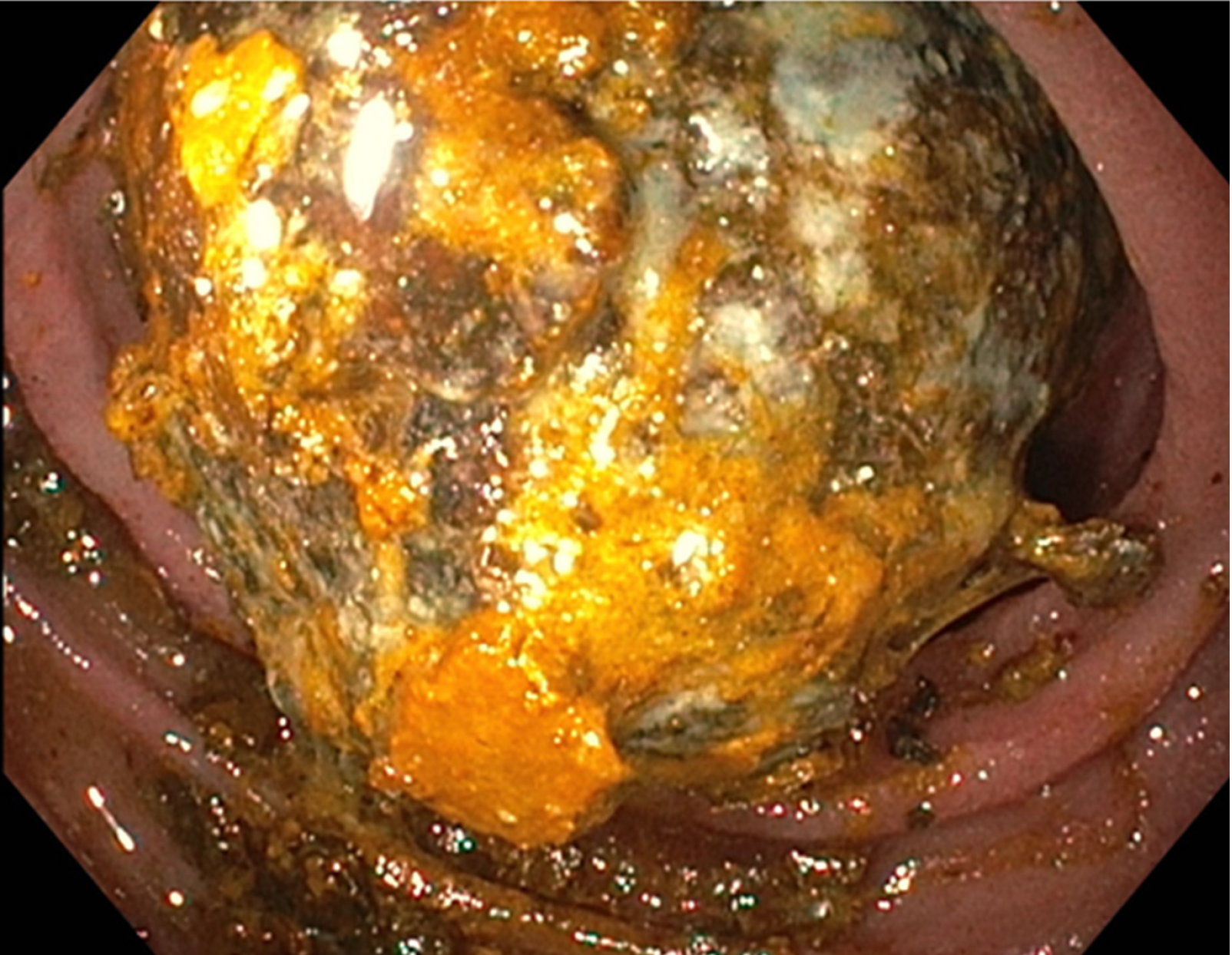
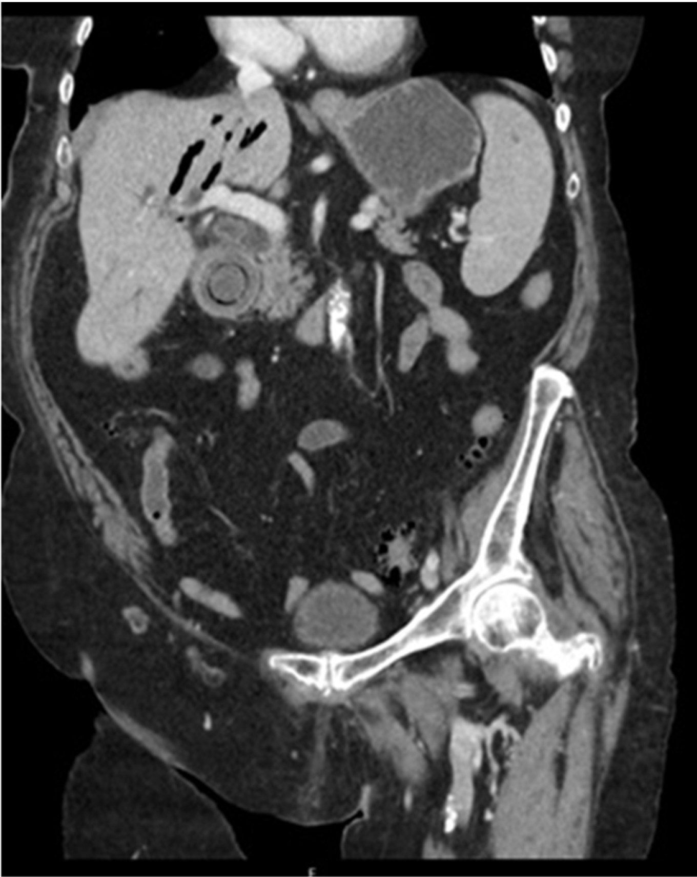
Abdominal ultrasound showed a gallstone in the gallbladder and widened intrahepatic bile ducts, and the distal part of the common bile duct could not be visualized. Gastroscopy (Figure 1) revealed fluid retention caused by a massive obstructive stone in the bulbus. Abdominal CT scanning demonstrated a fistula between the gallbladder and the bulbus and a large impacted stone in the bulbus (Figure 2).
The patient was diagnosed with Bouveret’s syndrome, a gastric outlet obstruction after passage of a gallstone through a fistula into the duodenum.
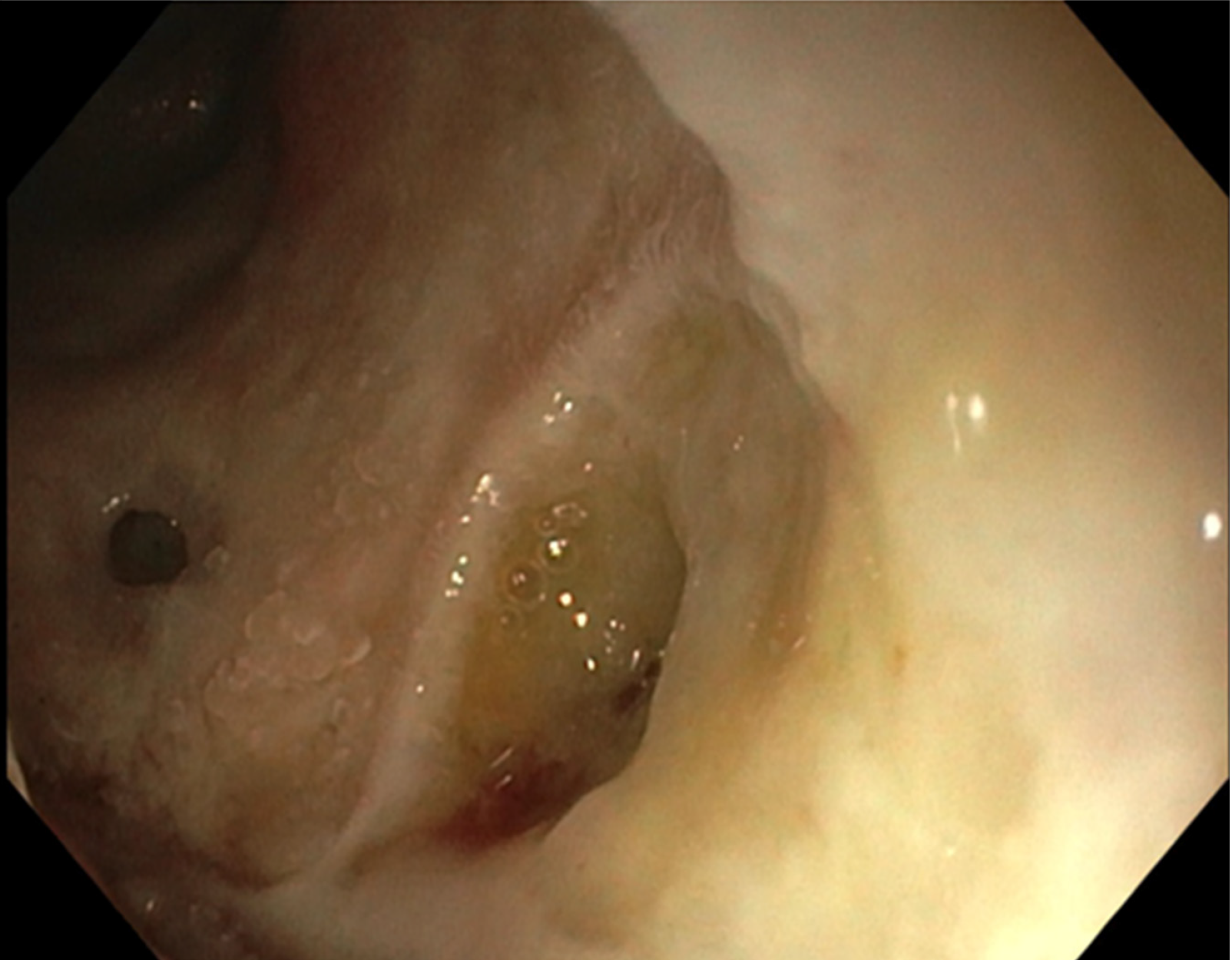
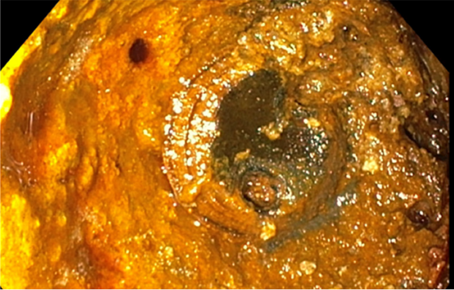
After multidisciplinary consultation between the surgeon and gastroenterologist, endoscopic laser lithotripsy of the impacted stone was planned under general anesthesia. A Dornier Medilas H Solvo lithotripsy 350 µm laser fiber (10 Hz, 2 Joules) was used to disintegrate the stone into smaller pieces. Larger parts were then placed into the stomach with large forceps and eventually removed with a Roth Net. Laser lithotripsy caused some mucosal damage in the duodenum; however, no signs of a perforation were visible. This was confirmed using contrast-enhanced fluoroscopy (Figures 3 and 4). The total endoscopy time was approximately 150 min.
Postoperatively, the patient recovered well. She was treated with antibiotics due to a urinary tract infection. After 1 wk, she was discharged in good clinical condition. Unfortunately, she developed a mood disorder several weeks after admission, which could be attributed to the general anesthesia given during the procedure. Months later, she recovered completely and is now in relatively good health. Due to her older age, clinical condition and the patient’s wishes, cholecystectomy was not performed.
Gastric outlet obstruction caused by large gallstones was first described by Bonnet in 1841 in 2 patients at autopsy. The first preoperative diagnosis was made by Leon August Bouveret in 1896 and since then bears his name[2]. A mechanical obstruction due to a gallstone that has entered the gastrointestinal tract is a complication that appears in 0.3%-0.5% of patients with cholelithiasis. Adhesions between the gallbladder and the digestive tract can occur due to acute cholecystitis. The combination of adhesions and pressure of larger calculi can lead to necrosis and the formation of a cholecystoenteric fistula. Most of these stones pass without producing any obstruction. Stones larger than 2 cm can be impacted in the digestive tract, which occurs mostly in the terminal ileum[1].
In approximately 1%-3% of cases, the stones cause obstruction in the duodenum. This phenomenon is called Bouveret’s syndrome. Patients are mostly female of advanced age and present with signs of nausea, vomiting and abdominal pain in approximately 70% of patients[3]. Additional examinations, such as CT or MRI, can show Rigler’s triad: A dilated stomach, pneumobilia, and a radio-opaque shadow suggesting an enteric gallstone[3]. As shown in Figure 2, Rigler’s triad was present in our patient. Multiple imaging modalities can be used in patients with Bouveret’s syndrome as symptoms can be nonspecific. The preferred additional examinations are computed tomography and esophagogastroduodenoscopy due to their high sensitivity and specificity for diagnosing Bouveret’s syndrome[4]. As this condition is mostly seen in elderly individuals with multiple comorbidities, treatment by an open surgical approach is unsuitable. In recent decades, several case reports of successful endoscopic removal have been published[5]. The first successful case was reported by Bedogni in 1985, who used an endoscopic basket[6]. Using endoscopic baskets or nets is relatively simple and quick but ineffective at removing larger stones.
If a stone cannot be removed with an endoscopic basket or net, mechanical lithotripsy, electrohydraulic or laser lithotripsy can be useful techniques to disintegrate the stone. The problem with electrohydraulic lithotripsy, however, is that incidental focusing of the shock waves on the intestine wall may cause bleeding and perforation[7]. The benefit of using laser lithotripsy is precise targeting of energy onto the stone with minimal tissue injury. The direct visual control of the laser application makes the treatment technically easy and safe. As in our case, no complications were described in previous cases[8-10].
Despite the successful reports regarding endoscopic treatment, visualization of the gallstone only occurred in approximately two-thirds of cases. Unfortunately, even fewer than two-thirds can be removed successfully[3]. In addition, it is important to remove all stone fragments to avoid postoperative gallstone ileus[8].
Full resolution of the symptoms of Bouveret's syndrome was achieved by endoscopic treatment with laser lithotripsy in an elderly patient with a high surgical risk. Laser lithotripsy can be considered an effective non-invasive therapeutic alternative to surgical treatment in Bouveret's syndrome.
| 1. | Qasaimeh GR, Bakkar S, Jadallah K. Bouveret's Syndrome: An Overlooked Diagnosis. A Case Report and Review of Literature. Int Surg. 2014;99:819-823. [RCA] [PubMed] [DOI] [Full Text] [Cited by in Crossref: 53] [Cited by in RCA: 48] [Article Influence: 4.0] [Reference Citation Analysis (0)] |
| 2. | Lawther RE, Diamond T. Bouveret's syndrome: gallstone ileus causing gastric outlet obstruction. Ulster Med J. 2000;69:69-70. [PubMed] |
| 3. | Cappell MS, Davis M. Characterization of Bouveret's syndrome: a comprehensive review of 128 cases. Am J Gastroenterol. 2006;101:2139-2146. [RCA] [PubMed] [DOI] [Full Text] [Cited by in Crossref: 161] [Cited by in RCA: 150] [Article Influence: 7.5] [Reference Citation Analysis (0)] |
| 4. | Caldwell KM, Lee SJ, Leggett PL, Bajwa KS, Mehta SS, Shah SK. Bouveret syndrome: current management strategies. Clin Exp Gastroenterol. 2018;11:69-75. [RCA] [PubMed] [DOI] [Full Text] [Full Text (PDF)] [Cited by in Crossref: 56] [Cited by in RCA: 90] [Article Influence: 11.3] [Reference Citation Analysis (0)] |
| 5. | Dumonceau JM, Devière J. Novel treatment options for Bouveret's syndrome: a comprehensive review of 61 cases of successful endoscopic treatment. Expert Rev Gastroenterol Hepatol. 2016;10:1245-1255. [RCA] [PubMed] [DOI] [Full Text] [Cited by in Crossref: 13] [Cited by in RCA: 29] [Article Influence: 2.9] [Reference Citation Analysis (0)] |
| 6. | Bedogni G, Contini S, Meinero M, Pedrazzoli C, Piccinini GC. Pyloroduodenal obstruction due to a biliary stone (Bouveret's syndrome) managed by endoscopic extraction. Gastrointest Endosc. 1985;31:36-38. [RCA] [PubMed] [DOI] [Full Text] [Cited by in Crossref: 38] [Cited by in RCA: 38] [Article Influence: 0.9] [Reference Citation Analysis (2)] |
| 7. | Binmoeller KF, Brückner M, Thonke F, Soehendra N. Treatment of difficult bile duct stones using mechanical, electrohydraulic and extracorporeal shock wave lithotripsy. Endoscopy. 1993;25:201-206. [RCA] [PubMed] [DOI] [Full Text] [Cited by in Crossref: 202] [Cited by in RCA: 171] [Article Influence: 5.2] [Reference Citation Analysis (0)] |
| 8. | Alsolaiman MM, Reitz C, Nawras AT, Rodgers JB, Maliakkal BJ. Bouveret's syndrome complicated by distal gallstone ileus after laser lithotropsy using Holmium: YAG laser. BMC Gastroenterol. 2002;2:15. [RCA] [PubMed] [DOI] [Full Text] [Full Text (PDF)] [Cited by in Crossref: 37] [Cited by in RCA: 44] [Article Influence: 1.8] [Reference Citation Analysis (0)] |
| 9. | Neuhaus H, Zillinger C, Born P, Ott R, Allescher H, Rösch T, Classen M. Randomized study of intracorporeal laser lithotripsy versus extracorporeal shock-wave lithotripsy for difficult bile duct stones. Gastrointest Endosc. 1998;47:327-334. [RCA] [PubMed] [DOI] [Full Text] [Cited by in Crossref: 118] [Cited by in RCA: 97] [Article Influence: 3.5] [Reference Citation Analysis (0)] |
| 10. | Rogart JN, Perkal M, Nagar A. Successful Multimodality Endoscopic Treatment of Gastric Outlet Obstruction Caused by an Impacted Gallstone (Bouveret's Syndrome). Diagn Ther Endosc. 2008;2008:471512. [RCA] [PubMed] [DOI] [Full Text] [Full Text (PDF)] [Cited by in Crossref: 7] [Cited by in RCA: 11] [Article Influence: 0.7] [Reference Citation Analysis (0)] |
Open-Access: This article is an open-access article that was selected by an in-house editor and fully peer-reviewed by external reviewers. It is distributed in accordance with the Creative Commons Attribution NonCommercial (CC BY-NC 4.0) license, which permits others to distribute, remix, adapt, build upon this work non-commercially, and license their derivative works on different terms, provided the original work is properly cited and the use is non-commercial. See:
Manuscript source: Unsolicited manuscript
Specialty type: Gastroenterology and hepatology
Country/Territory of origin: Netherlands
Peer-review report’s scientific quality classification
Grade A (Excellent): A, A
Grade B (Very good): B
Grade C (Good): 0
Grade D (Fair): 0
Grade E (Poor): 0
P-Reviewer: Demetrashvili Z, Lima M, Sun WB S-Editor: Wang J L-Editor: Webster J E-Editor: Ma YJ


