Published online Jan 7, 2020. doi: 10.3748/wjg.v26.i1.70
Peer-review started: September 20, 2019
First decision: November 22, 2019
Revised: December 11, 2019
Accepted: December 21, 2019
Article in press: December 22, 2019
Published online: January 7, 2020
Processing time: 108 Days and 10.6 Hours
Faecal immunochemical test (FIT) has been recommended to assess symptomatic patients for colorectal cancer (CRC) detection. Nevertheless, some conditions could theoretically favour blood originating in proximal areas of the gastrointestinal tract passing through the colon unmetabolized. A positive FIT result could be related to other gastrointestinal cancers (GIC).
To assess the risk of GIC detection and related death in FIT-positive symptomatic patients (threshold 10 μg Hb/g faeces) without CRC.
Post hoc cohort analysis performed within two prospective diagnostic test studies evaluating the diagnostic accuracy of different FIT analytical systems for CRC and significant colonic lesion detection. Ambulatory patients with gastrointestinal symptoms referred consecutively for colonoscopy from primary and secondary healthcare, underwent a quantitative FIT before undergoing a complete colonoscopy. Patients without CRC were divided into two groups (positive and negative FIT) using the threshold of 10 μg Hb/g of faeces and data from follow-up were retrieved from electronic medical records of the public hospitals involved in the research. We determined the cumulative risk of GIC, CRC and upper GIC. Hazard rate (HR) was calculated adjusted by age, sex and presence of significant colonic lesion.
We included 2709 patients without CRC and a complete baseline colonoscopy, 730 (26.9%) with FIT ≥ 10 µgr Hb/gr. During a mean time of 45.5 ± 20.0 mo, a GIC was detected in 57 (2.1%) patients: An upper GIC in 35 (1.3%) and a CRC in 14 (0.5%). Thirty-six patients (1.3%) died due to GIC: 22 (0.8%) due to an upper GIC and 9 (0.3%) due to CRC. FIT-positive subjects showed a higher CRC risk (HR 3.8, 95%CI: 1.2-11.9) with no differences in GIC (HR 1.5, 95%CI: 0.8-2.7) or upper GIC risk (HR 1.0, 95%CI: 0.5-2.2). Patients with a positive FIT had only an increased risk of CRC-related death (HR 10.8, 95%CI: 2.1-57.1) and GIC-related death (HR 2.2, 95%CI: 1.1-4.3), with no differences in upper GIC-related death (HR 1.4, 95%CI: 0.6-3.3). An upper GIC was detected in 22 (0.8%) patients during the first year. Two variables were independently associated: anaemia (OR 5.6, 95%CI: 2.2-13.9) and age ≥ 70 years (OR 2.7, 95%CI: 1.1-7.0).
Symptomatic patients without CRC have a moderate risk increase in upper GIC, regardless of the FIT result. Patients with a positive FIT have an increased risk of post-colonoscopy CRC.
Core tip: Our study, evaluates for the first time whether symptomatic patients with a positive faecal immunochemical test (FIT) result, no colorectal cancer (CRC) and a complete exploration of the colon have increased risk of related gastrointestinal cancer (GIC) detection or death. We found that this cohort of patients only have an increased risk of related CRC and death when compared with the cohort with a negative FIT result. Although the risk of upper GIC is higher than expected, the probability of detecting an upper GIC is unrelated to the FIT result and only associated with anaemia and advanced age.
- Citation: Pin-Vieito N, Iglesias MJ, Remedios D, Rodríguez-Alonso L, Rodriguez-Moranta F, Álvarez-Sánchez V, Fernández-Bañares F, Boadas J, Martínez-Bauer E, Campo R, Bujanda L, Ferrandez Á, Piñol V, Rodríguez-Alcalde D, Guardiola J, Cubiella J, on behalf of the COLONPREDICT study investigators. Risk of gastrointestinal cancer in a symptomatic cohort after a complete colonoscopy: Role of faecal immunochemical test. World J Gastroenterol 2020; 26(1): 70-85
- URL: https://www.wjgnet.com/1007-9327/full/v26/i1/70.htm
- DOI: https://dx.doi.org/10.3748/wjg.v26.i1.70
The use of quantitative faecal immunochemical test (FIT) is increasing outside the screening setting. FIT has proved its ability to identify which symptomatic patients are more likely to have an underlying colorectal cancer (CRC) or even other significant colonic lesions (SCL). Therefore, it is useful to improve the suitability of referrals for investigation of abdominal symptoms[1].
In this sense, the National Institute for Health and Care Excellence (NICE) has recently recommended adoption of FIT in primary care to guide referral for suspected CRC in people without rectal bleeding who have unexplained symptoms but do not meet the criteria for a suspected cancer pathway referral, and results should be reported using a threshold of 10 μg Hb/g faeces[2,3].
This has been possible due to the progressive replacement of the guaiac-based faecal occult blood test by the immunochemical-based test. FIT reacts with human globin, a protein digested by enzymes in the upper gastrointestinal tract (GIT), so it should have greater specificity to detect lower GIT lesions than guaiac-based tests and is not modified by diet[4,5]. Nevertheless, some conditions (e.g., altered bowel habit, prior gastrectomy) could theoretically favour blood originating in proximal areas of the GIT passing through the colon unmetabolized. A previous systematic review led to the conclusion that there is insufficient evidence to recommend for or against routine esophagogastroduodenoscopy (EGD) in patients with a positive faecal occult blood test followed by negative colonoscopy[6].
However, all the studies included were mainly based on faecal occult blood test that used the guaiac method or had been performed in a screening setting. Thus, conclusions drawn from these data cannot be extrapolated to the application of FIT in symptomatic patients. These patients may require additional diagnostic workup as long as complaints could be related to bleeding lesions located in the GIT proximal to the colon[7]. Thus, we aim to assess the risk of gastrointestinal cancers (GIC) detection and related death in symptomatic patients with a positive determination of FIT (≥ 10 μg Hb/g faeces) without CRC at baseline quality colonoscopy and to evaluate whether it might be worthwhile to perform additional evaluations to detect an upper GIC.
This is a post hoc cohort analysis performed within two prospective diagnostic test studies evaluating the diagnostic accuracy of different FIT analytical systems for CRC and SCL detection[8,9].
We followed the Strengthening the Reporting of Observational studies in Epidemiology statement to conduct and report our study[10]. The main characteristics of the different cohorts have been detailed elsewhere[8,9].
The study population consisted of ambulatory patients with gastrointestinal symptoms referred consecutively for colonoscopy from primary and secondary healthcare in ten out of the thirteen hospitals that took part in the primary studies. Patients included in the analysis underwent a quantitative FIT before undergoing a complete colonoscopy. Patients were excluded from this analysis if a CRC was detected on baseline exploration or the colonoscopy was incomplete. A colonoscopy was considered complete if more than 90% of the mucosa could be evaluated according to the Aronchick scale and caecal intubation was achieved[11]. In addition, patients were excluded from this analysis if follow-up after colonoscopy was insufficient (< 2 years) or a GIC was diagnosed before basal colonoscopy.
Patients were divided into two groups (positive and negative FIT) using the threshold of 10 μg Hb/g of faeces. All individuals collected a stool sample from one bowel movement without specific diet or medication restrictions before colonoscopy. Characteristics of the different FIT system used are shown in Table 1. Estimates of faecal haemoglobin (f-Hb) were quantitated as µg Hb/g of faeces so that results could be compared across analytical systems[12].
| Ref. | Country | Analytical system for estimation of faecal haemoglobin concentration |
| Cubiella et al[9], 2016 (DC) | Spain | OC-Sensor: 100% |
| Rodríguez-Alonso et al[8], 2015 | Spain | OC-Sensor: 100% |
| Cubiella et al[9], 2016 (VC) | Spain | OC-Sensor: 49.7%; OC-Auto 3 Latex 13.8%; FOB Gold 2.4%; Linear i-FOB 34.1% |
| Overall | Spain | OC-Sensor: 81.8%; OC-Auto 3 Latex 5.0%; FOB Gold 0.9%; Linear i-FOB 12.3% |
The colonoscopist was blinded to the FIT results. The bowel was cleansed and sedated as previously reported and all colonoscopies were performed by experienced endoscopists who reported any colorectal lesion and obtained biopsies if appropriate[13]. All polyps were removed either upon baseline exploration or afterwards.
SCL was defined as advanced adenoma (any adenoma ≥ 10 mm, with high-grade dysplasia or villous histology), histologically confirmed colitis (any aetiology), polyps ≥ 10 mm, polyposis (> 10 polyps of any histology), complicated diverticular disease (bleeding, diverticulitis), bleeding angiodysplasia and colonic ulcer. Any other colonic lesion was considered non-significant.
The main outcomes of this analysis are GIC detection and GIC-related death. Data from follow-up were retrieved from electronic medical records of the public hospitals involved in the research. For all patients, cancer diagnoses of any aetiology were recorded. We classified all cancers that could justify the presence of blood in the GIT as a GIC: Oral, throat, oesophageal, gastric, intestinal and CRC. We defined an upper GIC as a cancer that can be detected in an EGD exploration: Oesophageal, gastric, duodenal or ampullary cancer. The cause and date of death were recorded. We pooled the different causes of death into five categories: related to (1) GIC, (2) Upper GIC, (3) CRC, (4) Global cancer or (5) Global death.
We first performed a descriptive analysis of the cohorts included in the analysis. We determined whether there were differences using the Chi-square and student t test in the qualitative and quantitative variables, respectively. We calculated cumulative risk and number of cases per 1000 patient-years and its 95% confidence interval (CI). Differences in cumulative risk were analysed with the Chi-square test and Cochran–Mantel–Haenszel statistics and expressed as the risk ratio (RR) and its 95%CI. In order to control confounding variables, age, sex and SCL, we performed a Cox regression analysis to determine the hazard ratio (HR) of detecting a new cancer and cancer-related death respectively.
In order to determine whether there was an association between the baseline faecal haemoglobin concentration and length of time to GIC detection, we performed a descriptive analysis and a correlation analysis. We determined the Spearman correlation coefficient (r).
Finally, we evaluated which variables were associated with detection of any upper GIC during the first year after baseline colonoscopy. In this respect, we determined which variables had a statistically significant association with detection of an upper GIC using the Chi-square and the Cochran–Mantel–Haenszel statistics and expressed the differences as RR and its 95%CI. We included variables with a statistically significant association (P < 0.05) in a multivariate logistic regression analysis and expressed the association as the odds ratio (OR) and 95%CI. Statistical analysis was performed using SPSS statistical software, version 15.0 (SPSS Inc., Chicago, IL, USA)
The statistical methods of this study were reviewed by Noel Pin Vieito from Complexo Hospitalario Universitario de Ourense.
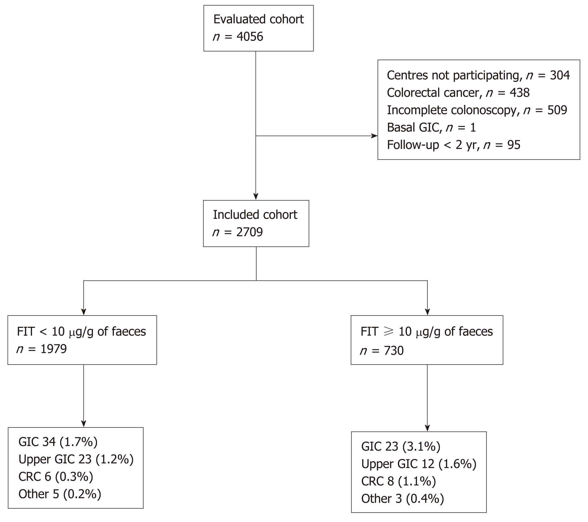
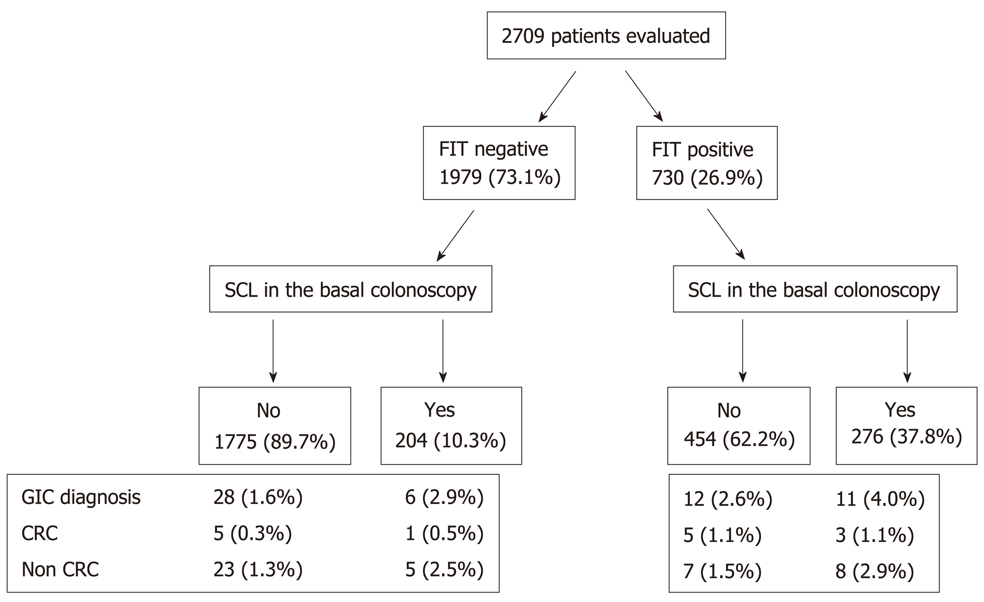
We excluded 1347 patients out of the 4056 symptomatic patients initially included in both studies, yielding a final sample of 2709 (Figure 1).
Of these participants, 1979 (73.1%) and 730 (26.9%) had a negative and positive FIT, respectively. The cohorts included were different in terms of age, sex, healthcare referring to colonoscopy, colonoscopy indication, findings in baseline exploration and length of follow-up as shown in Table 2.
| Characteristics | Overall (n = 2709) | FIT < 10 µg/g (n = 1979) | FIT ≥ 10 µg/g (n = 730) | P value8 |
| Demographic | ||||
| Age (yr) | 62.9 ± 13.5 | 62.0 ± 13.5 | 65.5 ± 13.0 | < 0.001 |
| Female sex | 1432 (52.9) | 1084 (54.8) | 348 (47.7) | 0.001 |
| Primary healthcare referral1 | 617 (24.2) | 397 (21.2) | 220 (32.2) | < 0.001 |
| Previous colonoscopy2 | 444 (25.9) | 287 (25.8) | 157 (26.0) | 0.9 |
| Daily using ASA2 | 330 (19.2) | 193 (17.3) | 137 (22.7) | 0.01 |
| Indications | ||||
| Rectal bleeding1 | 1234 (48.3) | 843 (45.1) | 391 (57.2) | < 0.001 |
| Change of bowel habit1 | 1271 (49.8) | 913 (48.8) | 358 (52.4) | 0.1 |
| Anaemia3,4 | 368 (16.2) | 236 (13.9) | 132 (23.1) | < 0.001 |
| Abdominal pain5 | 766 (41.3) | 587 (41.8) | 179 (40.0) | 0.4 |
| Weight loss5 | 391 (21.1) | 301 (21.4) | 90 (20.1) | 0.6 |
| Basal colonoscopy findings | ||||
| Benign anorectal lesion2 | 756 (44.0) | 495 (44.4) | 261 (43.3) | 0.6 |
| Significant colonic lesions6 | 480 (17.7) | 204 (10.3) | 276 (37.8) | < 0.001 |
| Advanced adenoma1,7 | 337 (13.2) | 139 (7.4) | 198 (29.0) | < 0.001 |
| Follow-up (mo) | 45.5 ± 20.0 | 47.9 ± 21.2 | 39.2 ± 14.1 | < 0.001 |
During a mean time of 45.5 ± 20.0 mo, a GIC was detected in 57 (2.1%) patients: An upper GIC (six oesophageal carcinomas, 25 gastric carcinomas, one duodenal adenocarcinoma, two ampullary carcinomas and one duodenal GIST) in 35 (1.3%), a CRC in 14 (0.5%) and other GIC (three cholangiocarcinomas, two small bowel adenocarcinomas, one small bowel lymphoma, one lingual carcinoma and one piriform sinus carcinoma) in 8 (0.3%). The distribution of the GIC according to the FIT result is shown in Figure 1. Thirty-six patients (1.3%) died due to GIC: 22 (0.8%) due to an upper GIC and 9 (0.3%) due to CRC. Finally, 205 (7.6%) patients developed a cancer and 197 (7.3%) died, 98 (3.5%) due to cancer. Cumulative risk and number of cancers and death per 1000 patient-years is shown in Table 3.
| Event | Risk | Overall (n = 2709) | FIT < 10 µg/g (n = 1979) | FIT ≥10 µg/g (n = 730) | RR1 (95%CI) | HR2 (95%CI) |
| GIC | Cumulative3 | 2.1% (1.6-2.6) | 1.7% (1.1-2.3) | 3.2% (1.9-4.4) | 1.9 (1.1-3.2) | 1.5 (0.8-2.7) |
| GIC | Density4 | 5.6 (4.1-7.0) | 4.3 (2.9-5.8) | 9.7 (5.8-13.7) | ||
| GIC | Cumulative death3 | 1.3% (0.9-1.8) | 1.0% (0.5-1.4) | 2.3% (1.2-3.4) | 2.5 (1.3-4.7) | 2.2 (1.1-4.3) |
| GIC | Death density4 | 3.5 (2.4-4.6) | 2.4 (1.3-3.5) | 7.1 (3.7-10.5) | ||
| Up GIC5 | Cumulative3 | 1.3% (0.9-1.7) | 1.2% (0.7-1.6) | 1.6% (0.7-2.6) | 1.4 (0.7-2.8) | 1.0 (0.5-2.2) |
| Up GIC5 | Density4 | 3.4 (2.3-4.6) | 2.9 (1.7-4.1) | 5.1 (2.2-8.0) | ||
| Up GIC5 | Cumulative death3 | 0.8% (0.5-1.2) | 0.7% (0.3-1.0) | 1.2% (0.4-2.0) | 1.6 (0.7-3.7) | 1.4 (0.6-3.3) |
| Up GIC5 | Death density4 | 2.1 (1.2-3.0) | 1.6 (0.8-2.5) | 3.8 (1.3-6.2) | ||
| CRC | Cumulative3 | 0.5% (0.2-0.8) | 0.3% (0.1-0.5) | 1.1% (0.3-1.9) | 3.6 (1.3-10.5) | 3.8 (1.2-11.9) |
| CRC | Density4 | 1.4 (0.7-2.1) | 0.8 (0.2-1.4) | 3.4 (1.0-5.7) | ||
| CRC | Cumulative death3 | 0.3% (0.1-0.5) | 0.1% (0.0-0.2) | 1.0% (0.3-1.7) | 9.5 (2.0-46.2) | 10.8 (2.1-57.1) |
| CRC | Death density4 | 0.9 (0.3-1.4) | 0.3 (-0.1-0.6) | 2.9 (0.8-5.1) | ||
| Cancer | Cumulative3 | 7.6% (6.6-8.6) | 7.3% (6.2-8.5) | 8.2% (6.2-10.2) | 1.1 (0.8-1.5) | 1.1 (0.8-1.5) |
| Cancer | Density4 | 20.5 (17.7-23.3) | 18.8 (15.8-21.9) | 25.9 (19.4-32.5) | ||
| Cancer | Cumulative death3 | 3.6% (2.9-4.3) | 3.2% (2.4-4.0) | 4.8% (3.2-6.3) | 1.5 (1.0-2.3) | 1.4 (0.9-2.2) |
| Cancer | Death density4 | 9.5 (7.6-11.4) | 8.0 (6.0-9.9) | 14.7 (9.8-19.6) | ||
| Death | Cumulative3 | 7.3% (6.3-8.2) | 7.0% (5.9-8.1) | 7.9% (6.0-9.9) | 1.1 (0.8-1.5) | 1.1 (0.8-1.5) |
| Death | Density4 | 19.2 (16.5-21.8) | 17.6 (14.7-20.5) | 24.3 (18.1-30.6) |
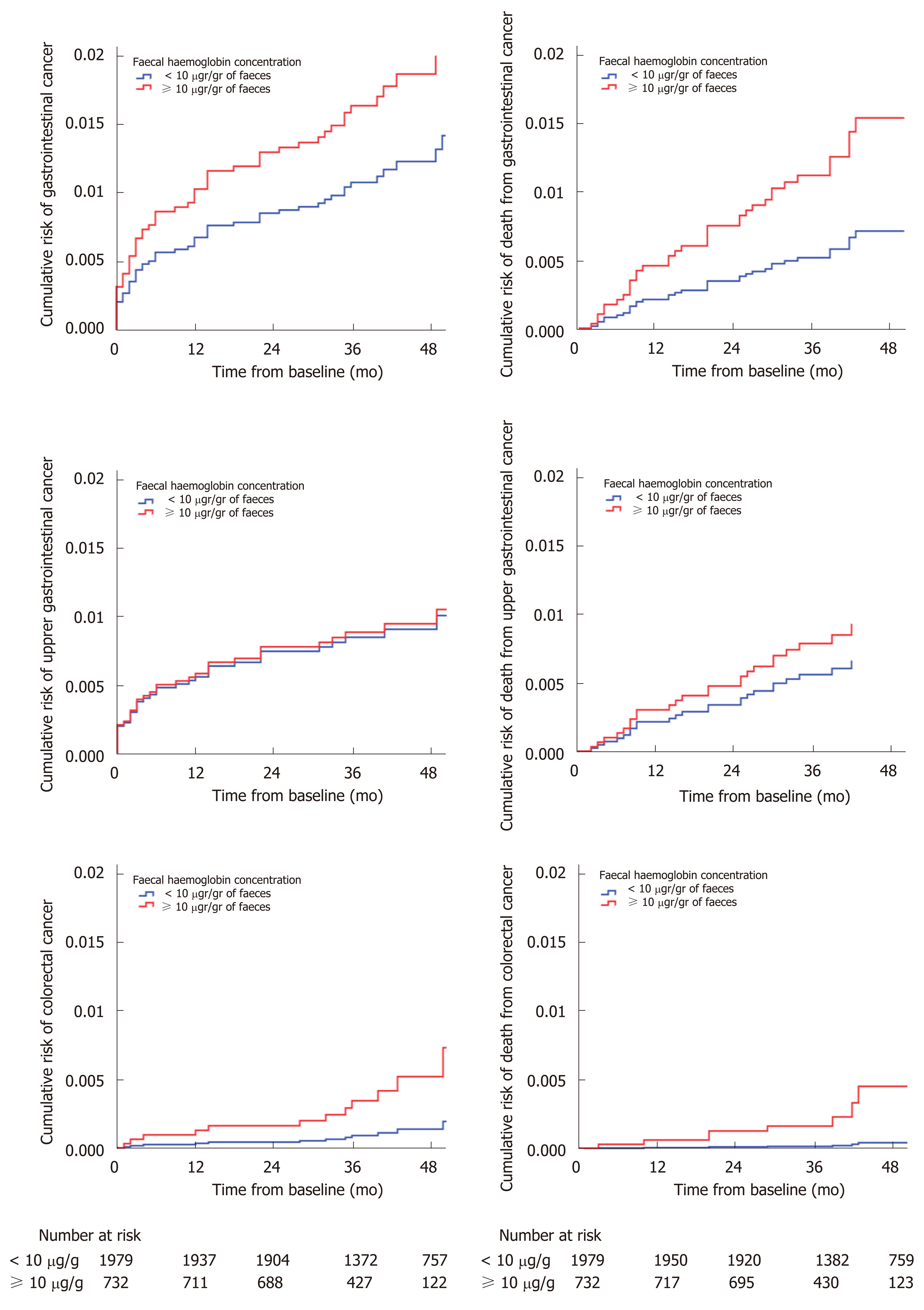
Patients with positive FIT showed greater GIC risk (≥ 10 µg/g of faeces = 3.2%, < 10 µg/g of faeces = 1.7%; RR 1.9, 95%CI: 1.1-3.2) and GIC-related mortality (≥ 10 µg/g of faeces = 2.3%, < 10 µg/g of faeces = 1.0%; OR 2.5, 95%CI: 1.3-4.6). In the subgroup analysis, patients in the positive FIT cohort had an increased risk of CRC (≥ 10 µg/g of faeces = 1.1%, < 10 µg/g of faeces = 0.3%; RR 3.6, 95%CI: 1.3-10.5) and CRC-related mortality (≥ 10 µg/g of faeces = 1.0%, < 10 µg/g of faeces = 0.1%; RR 9.5, 95%CI: 2.0-46.2) but no differences in upper GIC or upper GIC-related mortality as shown in Table 3. However, in the Cox’s proportional multivariate regression analysis, patients with a positive FIT had only an increased risk of CRC (HR 3.8, 95% CI 1.2-11.9), CRC-related death (HR 10.8, 95%CI: 2.1-57.1) and GIC-related death (HR 2.2, 95%CI: 1.1-4.3), after adjusting for confounding variables. The cumulative risk of cancer and related death calculated in the Cox’s multivariate regression analysis is shown in Figure 2.
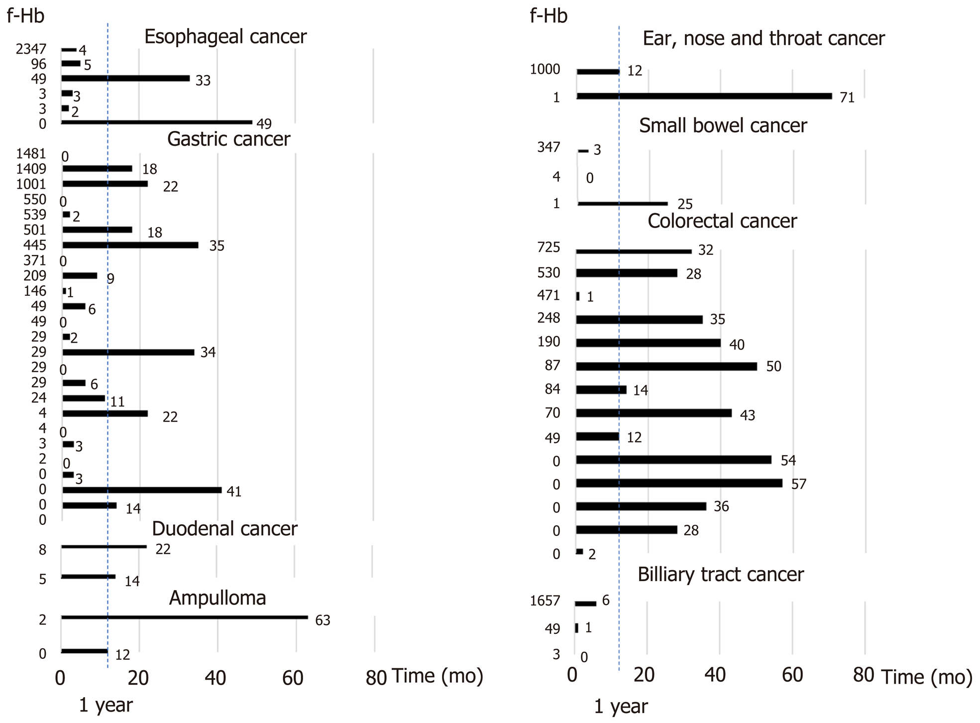
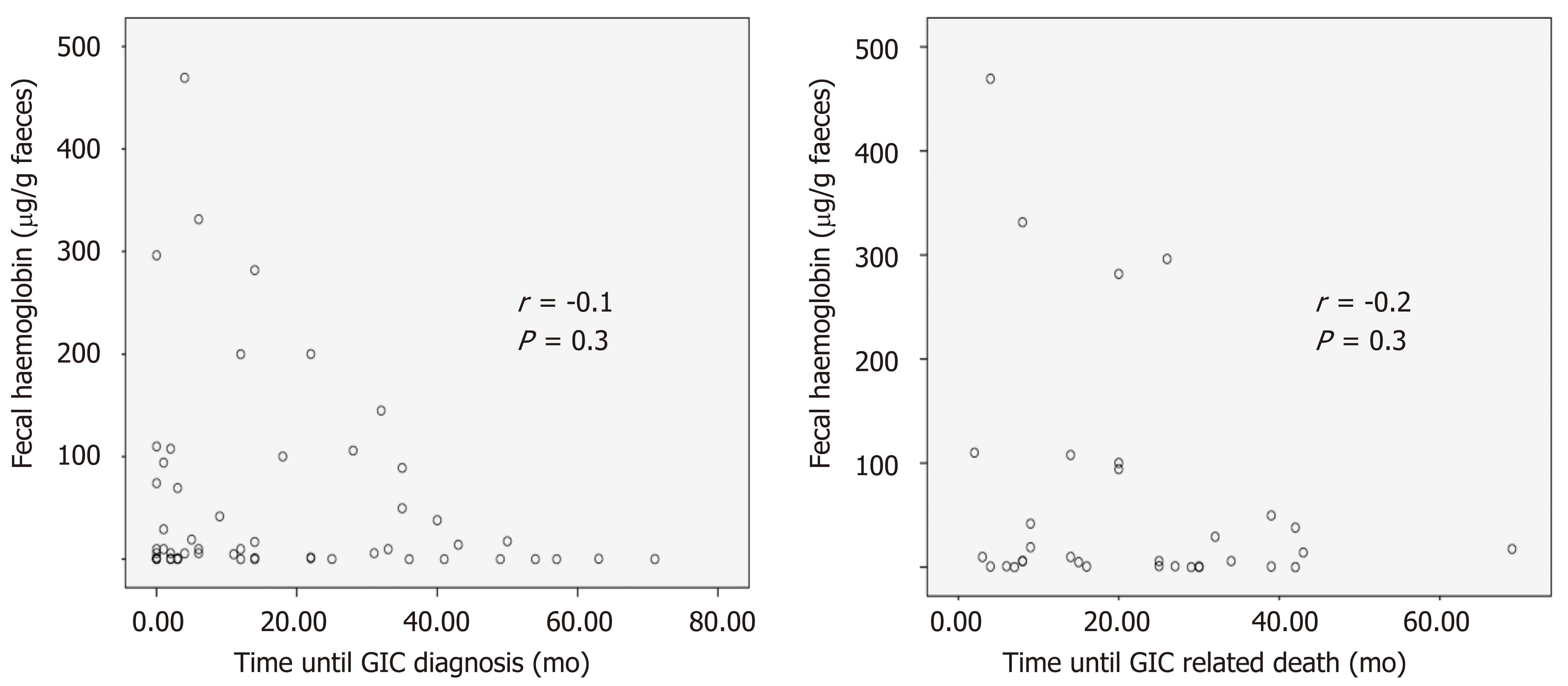
Figure 3 links time elapsed until diagnosis of each GIC throughout follow-up with the FIT result. We did not detect a correlation between time to GIC diagnosis (r = -0.1; P = 0.4) or related death (r = -0.2; P = 0.3) and FIT result as shown in Figure 4.
During the first year after baseline colonoscopy, 22 (0.8%) upper GIC were detected: 17 cases of gastric carcinomas, 4 oesophageal carcinomas and one ampullary carcinoma. Only two variables were independently associated with detection of an upper GIC during the first year: anaemia (OR 5.6, 95%CI: 2.2-13.9), defined as < 11 g/100 mL in men and < 10 g/100 mL in non-menstruating women, and age ≥ 70 years (OR 2.7, 95%CI: 1.1-7.0), as shown in Table 4.
| Upper gastrointestinal cancer | Odds ratio (95 %CI)1 | Odds ratio (95 %CI)2 | |
| Sex | |||
| Female (n = 1432) | 10 (0.7) | 1 | |
| Male (n = 1277) | 12 (0.9) | 1.3 (0.6-3.1) | |
| Age | |||
| < 70 yr (n = 1757) | 8 (0.5) | 1 | 1 |
| ≥ 70 yr (n = 952) | 14 (1.5) | 3.3 (1.4-7.8) | 2.7 (1.1-7.0) |
| Primary healthcare referral | |||
| No (n = 1936) | 19 (1.0) | 1 | |
| Yes (n = 617) | 3 (0.5) | 0.5 (0.1-1.7) | |
| Rectal bleeding | |||
| No (n = 1319) | 16 (1.2) | 1 | |
| Yes (n = 1234) | 6 (0.5) | 0.4 (0.1-1.0) | |
| Change of bowel habit | |||
| No (n = 1282) | 12 (0.9) | 1 | |
| Adequate (n = 1271) | 10 (0.8) | 0.8 (0.4-1.9) | |
| Anaemia3 | |||
| No (n = 2077) | 13 (0.6) | 1 | 1 |
| Yes (n = 191) | 8 (4.2) | 6.9 (2.8-17.0) | 5.6 (2.2-13.9) |
| Abdominal pain | |||
| No (n = 1319) | 12 (1.1) | 1 | |
| Yes (n = 1234) | 5 (0.7) | 0.6 (0.2-1.7) | |
| Weight loss | |||
| No (n = 1462) | 12 (0.8) | 1 | |
| Yes (n = 391) | 5 (1.3) | 1.5 (0.5-4.4) | |
| Faecal immunochemical test | |||
| < 10 µg/g (n = 1979) | 14 (0.7) | 1 | |
| ≥ 10 µg/g (n = 730) | 8 (1.1%) | 1.5 (0.6-3.7) | |
| Benign anorectal lesion | |||
| No (n = 961) | 7 (0.7) | 1 | |
| Yes (n = 756) | 6 (0.8) | 1.1 (0.4-3.2) | |
| Significant colonic lesion4 | |||
| No (n = 2216) | 16 (0.7) | 1 | |
| Yes (n = 480) | 6 (1.3) | (0.7-4.5) | |
| Advanced adenoma5 | |||
| No (n = 2968) | 16 (0.7) | 1 | |
| Yes (n = 337) | 6 (1.8) | 2.5 (1.0-6.4) |
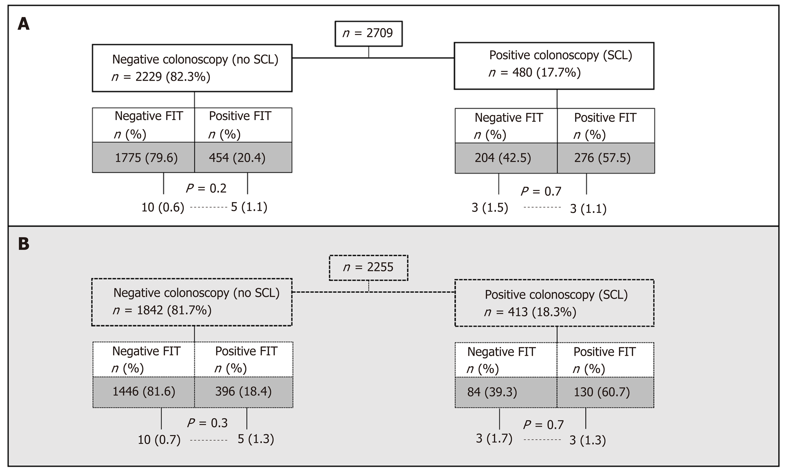
The distribution of GIC according to FIT result and presence of SCL at baseline colonoscopy is shown in Figure 5. For each subgroup, the minimum diagnostic yield of an upper endoscopy performed at the time of FIT determination, has been calculated assuming a theoretical 100% sensitivity for any esophageal or gastric bleeding tumor developed over the first year since performing baseline colonoscopy.
There were no significant differences in gastroesophageal cancer (GEC) diagnoses irrespective of FIT result, both in the subgroup of patients with SCL as well as in the subgroup with normal baseline colonoscopy.
Those results were similar when the analysis was limited to people aged 50 and older (Figure 6).
Our study, for the first time, evaluates whether symptomatic patients with a positive FIT result, no CRC and a complete exploration of the colon have increased risk of related GIC detection or death. We found that this cohort of patients only have an increased risk of related CRC and death when compared with the cohort with a negative FIT result. There are no differences in the risk of upper GIC between both cohorts. In addition, we have identified two variables independently associated with detection of an upper GIC during the first year: Anaemia and advanced age.
Our analysis has several strengths. The main one is that we have included a wide number of symptomatic patients who underwent FIT and colonoscopy in several public hospitals in Spain. In this sense, we have limited our analysis to subjects with complete baseline colonoscopy and resection of pre-neoplastic lesions. On the other hand, we performed follow-up analysis by means of search in the electronic medical records of our centres linked to the National Health System′s Hospital Discharge Records Database (CMBD in Spanish), which receives notifications from around 98% of Spanish public hospitals that have seen to more than 99% of the Spanish population[14]. Since 2005, the CMBD also has partial coverage from private hospitals[15].
However, the main weakness of our analysis arises from differences between the cohorts in terms of demographics, basal symptoms, endoscopic findings or follow-up. Moreover, the risk of GIC during follow-up, as expected, is low. To solve this limitation, we performed a Cox multivariate regression analysis controlling by confounding variables and final results are consistent.
Our study detected a higher than expected risk of GIC in the patients evaluated, mainly related to upper GIC and CRC. Estimated 30-year risk of developing an upper GIC in the United States is 0.98%[16], which is lower than the risk detected in our symptomatic cohort. Moreover, the incidence of GEC is also notable even in patients with a positive FIT result who were diagnosed with a SCL in the baseline colonoscopy, which could theoretically justify the presence of haemoglobin in faeces. This is related to the lack of specificity of symptoms related to diagnosis of cancer. In this sense, we believe that most GIC detected are prevalent. As an example, anaemia, although mainly related to CRC, is related to any GIC with positive predictive values ranging between 1% and 5% of the population seen in primary healthcare[2].
FIT has been recommended for adoption in primary care to guide referral for suspected colorectal cancer in people without rectal bleeding who have unexplained symptoms but do not meet the criteria for a suspected cancer pathway referral. Furthermore, NICE has recommended 10 µg Hb/g of faeces as the threshold for further evaluation referral[3]. This recommendation is based on the high accuracy of the test for CRC detection in symptomatic patients[1,17]. However, one practical doubt when using FIT in symptomatic patients is what to do with “false positive” results. Most evidence available comes from asymptomatic patients and suggests that a positive FIT is not predictive of prevalent GIC[7,18,19]. A recently published study revealed that only 0.14% of all persons with a positive FIT result were diagnosed with gastric or oesophageal cancer within 3 years and the risk was similar to the group with negative FIT[20]. Our study evaluates, for the first time, the risk of GIC after a false positive FIT result. In this sense, the probability of detecting an upper GIC is not modified by the FIT result.
It is noteworthy that our study did not exclude patients with high risk symptoms as rectal bleeding which are outside of NICE recommendation. However, most of the studies included in the meta-analysis that supports NICE recommendation[1], were not only concerned with patients with low risk symptoms (i.e., rectal bleeding is described in several patients in those studies). That clinical concern was highlighted by Fraser[21] and led to the development of an additional review and meta-analysis to obtain more information about the accuracy of FIT through the broad spectrum of symptomatic patients[17]. In our cohort, the risk of GIC cancer tends to be lower in patients with rectal bleeding. Probably, this is due to this symptom’s being less subjective than others like abdominal pain and more specific to the colon. Thus, unlike other indications, patients with overt bleeding who underwent a quality colonoscopy that ruled out CRC were less likely to be diagnosed with an upper GIC.
Although the risk is low, CRC risk is increased in symptomatic subjects with positive FIT even after a high-quality colonoscopy when compared to patients with a negative test. This finding is worthy of several comments. CRC detected fall into the definition of a post-colonoscopy colorectal cancer (PCCRC)[22]. In fact, the rate of PCCRC detected, approximately 3%, is located in the expected segment between 2.5% and 7.7%. However, we must highlight that the risk of PCCRC is higher after a positive FIT, probably due to the higher prevalence in this group of patients. This finding should be taken into account by physicians if symptoms persist after a normal colonoscopy. Finally, the risk of PCCRC calculated per 1000 colonoscopies is higher than the risk previously documented ranging between 0.8 and 2.4[23]. Our population consists of symptomatic patients with a CRC prevalence in the original studies ranging between 3.0% and 13.7%. We therefore suggest that the risk of PCCRC should be evaluated on the basis of the colonoscopy indication. However, the sample size of our analysis and the low number of PCCRC detected did not enable us to analyse additional factors that could predict the risk of PCCRC, such as age, comorbidity and diverticular disease, or the relationship with baseline symptoms[24].
A recent study conducted in patients taking part in CRC screening has associated the presence of detectable f-Hb with increased risk of death from a wide range of causes unrelated to CRC or even GIC[25]. In that study, Libby et al[26] consider the possibility of detectable f-Hb originating from subclinical colonic inflammation due to a generalised inflammatory state. We did not find such an association. However, the threshold used in our study (10 μg Hb/g faeces) is much lower than the concentration of approximately 80 μg Hb/g faeces required to attain a positive result by means of the qualitative method used by Libby et al[26].
Early diagnosis of GIC is challenging as long as abdominal symptoms are common, mostly related to benign diseases and non-specific to a particular cancer. In fact, abdominal symptoms are very common among patients with cancer (23%), mainly related to GIC and CRC in particular[27]. In contrast with breast cancer or melanoma, GIC have a broad symptom signature with varying predictive value[28]. In order to reduce delays in patients with lower abdominal symptoms with a low positive predictive value for CRC, FITs are recommended for adoption in primary care to guide referral for suspected CRC[3]. Our analysis aims to resolve a frequent issue that will take place when patients with lower abdominal symptoms are evaluated with a FIT. Hypothetically, 179-229 out of 1000 symptomatic patients will have a positive FIT and colonoscopy without CRC[2]. As our results show, this patient cohort has a similar risk of GIC as the cohort with a negative FIT. In this situation, an EGD should be recommended in patients with anaemia especially if they are elderly. However, special caution should be taken with the risk of PCCRC after positive FIT and normal colonoscopy if abdominal symptoms persist or reappear.
Our results are the basis to design a large prospective follow-up study including patients treated in primary healthcare with abdominal symptoms. In these patients, diagnostic evaluation should not be restricted to GIC. Other abdominal cancers in addition to benign gastrointestinal diseases should be evaluated to determine the positive predictive value and the best diagnostic strategy for each group of symptoms.
Additionally, a recent study concluded that endoscopic gastric cancer screening could be cost-effective if combined with a screening colonoscopy in countries with a gastric cancer risk ≥ 10 per 100000[29]. Given the gastroesophageal cancer incidences shown during the first year since FIT determination in our cohort irrespective of SCL finding in the basal colonoscopy, the cost-utility of combining upper and lower endoscopies should be investigated also in this setting.
To summarise, the risk of GIC is higher than expected in patients with low gastrointestinal symptoms and no CRC detected in a complete colonoscopy. The probability of detecting an upper GIC is unrelated to the FIT result and only associated with the presence of anaemia and advanced age. Finally, the risk of PCCRC in our study is within the ranges expected and clearly associated with the FIT result.
Faecal immunochemical test for haemoglobin (FIT) is more specific and appears to be equal to or more sensitive than guaiac-based tests when used for colorectal cancer (CRC) screening. FIT reacts with human globin, so it should have greater specificity to detect lower gastrointestinal tract (GIT) lesions than guaiac-based tests. However, a previous systematic review led to the conclusion that there is insufficient evidence to recommend for or against routine esophagogastroduodenoscopy in asymptomatic patients with a positive faecal occult blood test followed by negative colonoscopy.
Out of a screening setting, several approaches have been developed to improve the suitability of referrals for investigation of symptoms suggestive of CRC and reduce delays in diagnosis and some include using FIT. Therefore, it will be increasingly common for clinicians to face the uncertainty of a patient with non-specific digestive symptoms, a positive FIT result and normal colonoscopy.
We aim to assess the risk of gastrointestinal cancer (GIC) detection and related death in symptomatic patients with a positive determination of FIT (threshold 10 μg Hb/g faeces) without CRC at baseline quality colonoscopy.
We performed a post hoc cohort analysis within two prospective diagnostic test studies evaluating the diagnostic accuracy of FIT for CRC detection. Outpatients with gastrointestinal symptoms referred consecutively for colonoscopy from primary and secondary healthcare were divided into two groups (positive and negative FIT) using the threshold of 10 μg Hb/g of faeces and data from follow-up were retrieved from their electronic medical records. We determined the cumulative risk of GIC, CRC and upper GIC. Hazard rate was calculated adjusted by age, sex and presence of significant colonic lesion on basal colonoscopy.
This study revealed high neoplasia and death rates in our cohort (n = 2709) of people consulting with a physician for non-acute symptoms suggestive of lower gastrointestinal tract disorders. FIT-positive patients have higher incidence of GIC during follow-up. However, this did not result in a statistically significant increase in the risk of upper GIC development after multivariate adjustment. Moreover, we found that this cohort of patients only has an increased risk of related CRC and death when compared to the cohort with a negative FIT result.
This study suggests that FIT positivity using the threshold of 10 μg Hb/g of faeces is not enough to differentiate which patients would benefit from continuing workup to rule out a GIC out of screening setting. Nevertheless, small amounts of f-Hb may originate in the upper GI tract or the small bowel and this possibility must be considered along with other false-positive risk factors when interpreting FIT requested to rule out CRC or another significant colonic lesion.
We hypothesize that benign lesions (i.e. due to non-steroid anti-inflammatory drugs) are much more prevalent than GIC in the upper tract regardless of symptoms. Thus, it is much more likely that a small amount of detectable (unmetabolized) haemoglobin, originally from any kind of lesion located in the upper tract or the small bowel will be unrelated to a GIC. However, the study design is not suitable to prove this hypothesis. A large prospective follow-up study which takes competitive FIT positive causes and other risk factors into consideration would provide a predictive model to guide decision-making.
Part of our data was previously presented as an oral presentation at a Spanish scientific meeting (Asociación Española de Gastroenterología; March 2017) and was accepted for poster presentation at the United European Gastroenterology Week in 2017 (Barcelona).
The contents of this manuscript have not been published previously in any journal nor deposited on a pre-print server. Furthermore, this paper has not been presented at any scientific meeting in abstract form.
Hospital Universitario de Canarias: Natalia González-López, Enrique Quintero.
Donostia Hospital: Jesús Bañales, Luis Bujanda, María J Perugorria.
Registre del Càncer de Catalunya Pla Director d'Oncologia de Catalunya, Hospital Duran i Reynals, L’Hospitalet de Llobregat: Ramón Cleries, Josepa Ribes, Xavier Sanz.
Hospital Universitario de Móstoles: Jorge López-Vicente , Daniel Rodriguez-Alcalde.
Hospital Dr. Josep Trueta: Virginia Piñol, Leyanira Torrealba.
Complexo Hospitalario Universitario de Ourense: Irene Blanco , Joaquín Cubiella, Marta Díaz-Ondina, María Salve, Javier Fernández-Seara, María José Iglesias, Pedro Macía, David Remedios , Eloy Sánchez, Pablo Vega.
Corporació Sanitària i Universitària Parc Taulí: Rafel Campo, Eva Martínez-Bauer, Marta Pujol.
Complejo Hospitalario de Pontevedra: Victoria Álvarez Sánchez, José Mera, Juan Turnes.
Hospital de Sagunto: Joan Clofent, Ana Garayoa.
Hospital Universitari Mútua de Terrassa: Fernando Fernández-Bañares, Victoria Gonzalo, Mar Pujals.
Consorci Sanitari de Terrassa: Jaume Boadas, Sara Galter, Eva Garcia-Lanuza, Rebeca Gimeno.
Departamento de Bioquímica, CATLAB, Viladecavalls, Barcelona: Antonio Alsius.
Hospital Clínico Universitario de Zaragoza: Ángel Ferrández, Marina Solano Sánchez.
Manuscript source: Unsolicited manuscript
Specialty type: Gastroenterology and hepatology
Country of origin: Spain
Peer-review report classification
Grade A (Excellent):
Grade B (Very good):
Grade C (Good): C
Grade D (Fair):
Grade E (Poor):
P-Reviewer: Fedeli U S-Editor: Gong ZM L-Editor: A E-Editor: Zhang YL
| 1. | Westwood M, Lang S, Armstrong N, van Turenhout S, Cubiella J, Stirk L, Ramos IC, Luyendijk M, Zaim R, Kleijnen J, Fraser CG. Faecal immunochemical tests (FIT) can help to rule out colorectal cancer in patients presenting in primary care with lower abdominal symptoms: a systematic review conducted to inform new NICE DG30 diagnostic guidance. BMC Med. 2017;15:189. [RCA] [PubMed] [DOI] [Full Text] [Full Text (PDF)] [Cited by in Crossref: 75] [Cited by in RCA: 89] [Article Influence: 9.9] [Reference Citation Analysis (0)] |
| 2. | National Institute for Health and Care Excellence. NICE Guideline 12. Suspected cancer: recognition and referral 2015. Accessed July 8, 2018 Available from: https://www.nice.org.uk/guidance/ng12. |
| 3. | NICE. Diagnostics guidance DG30. Quantitative faecal immunochemical tests to guide referral for colorectal cancer in primary care. 2017, accessed July 8, 2018 Available from: https://www.nice.org.uk/guidance/dg30. |
| 4. | Harewood GC, McConnell JP, Harrington JJ, Mahoney DW, Ahlquist DA. Detection of occult upper gastrointestinal tract bleeding: performance differences in fecal occult blood tests. Mayo Clin Proc. 2002;77:23-28. [RCA] [PubMed] [DOI] [Full Text] [Cited by in Crossref: 15] [Cited by in RCA: 19] [Article Influence: 0.8] [Reference Citation Analysis (0)] |
| 5. | Levi Z, Hazazi R, Rozen P, Vilkin A, Waked A, Niv Y. A quantitative immunochemical faecal occult blood test is more efficient for detecting significant colorectal neoplasia than a sensitive guaiac test. Aliment Pharmacol Ther. 2006;23:1359-1364. [RCA] [PubMed] [DOI] [Full Text] [Cited by in Crossref: 37] [Cited by in RCA: 35] [Article Influence: 1.8] [Reference Citation Analysis (0)] |
| 6. | Allard J, Cosby R, Del Giudice ME, Irvine EJ, Morgan D, Tinmouth J. Gastroscopy following a positive fecal occult blood test and negative colonoscopy: systematic review and guideline. Can J Gastroenterol. 2010;24:113-120. [RCA] [PubMed] [DOI] [Full Text] [Full Text (PDF)] [Cited by in Crossref: 25] [Cited by in RCA: 29] [Article Influence: 1.8] [Reference Citation Analysis (0)] |
| 7. | McLoughlin MT, Telford JJ. Positive occult blood and negative colonoscopy--should we perform gastroscopy? Can J Gastroenterol. 2007;21:633-636. [RCA] [PubMed] [DOI] [Full Text] [Cited by in Crossref: 3] [Cited by in RCA: 7] [Article Influence: 0.4] [Reference Citation Analysis (0)] |
| 8. | Rodríguez-Alonso L, Rodríguez-Moranta F, Ruiz-Cerulla A, Lobatón T, Arajol C, Binefa G, Moreno V, Guardiola J. An urgent referral strategy for symptomatic patients with suspected colorectal cancer based on a quantitative immunochemical faecal occult blood test. Dig Liver Dis. 2015;47:797-804. [RCA] [PubMed] [DOI] [Full Text] [Cited by in Crossref: 45] [Cited by in RCA: 46] [Article Influence: 4.2] [Reference Citation Analysis (0)] |
| 9. | Cubiella J, Vega P, Salve M, Díaz-Ondina M, Alves MT, Quintero E, Álvarez-Sánchez V, Fernández-Bañares F, Boadas J, Campo R, Bujanda L, Clofent J, Ferrandez Á, Torrealba L, Piñol V, Rodríguez-Alcalde D, Hernández V, Fernández-Seara J; COLONPREDICT study investigators. Development and external validation of a faecal immunochemical test-based prediction model for colorectal cancer detection in symptomatic patients. BMC Med. 2016;14:128. [RCA] [PubMed] [DOI] [Full Text] [Full Text (PDF)] [Cited by in Crossref: 60] [Cited by in RCA: 55] [Article Influence: 5.5] [Reference Citation Analysis (0)] |
| 10. | Vandenbroucke JP, von Elm E, Altman DG, Gøtzsche PC, Mulrow CD, Pocock SJ, Poole C, Schlesselman JJ, Egger M; STROBE Initiative. Strengthening the Reporting of Observational Studies in Epidemiology (STROBE): explanation and elaboration. Epidemiology. 2007;18:805-835. [RCA] [PubMed] [DOI] [Full Text] [Cited by in Crossref: 1209] [Cited by in RCA: 2021] [Article Influence: 106.4] [Reference Citation Analysis (0)] |
| 11. | Aronchick CA, Lipshutz WH, Wright SH, Dufrayne F, Bergman G. A novel tableted purgative for colonoscopic preparation: efficacy and safety comparisons with Colyte and Fleet Phospho-Soda. Gastrointest Endosc. 2000;52:346-352. [RCA] [PubMed] [DOI] [Full Text] [Cited by in Crossref: 283] [Cited by in RCA: 328] [Article Influence: 12.6] [Reference Citation Analysis (0)] |
| 12. | Fraser CG, Allison JE, Halloran SP, Young GP; Expert Working Group on Fecal Immunochemical Tests for Hemoglobin, Colorectal Cancer Screening Committee, World Endoscopy Organization. A proposal to standardize reporting units for fecal immunochemical tests for hemoglobin. J Natl Cancer Inst. 2012;104:810-814. [RCA] [PubMed] [DOI] [Full Text] [Cited by in Crossref: 116] [Cited by in RCA: 127] [Article Influence: 9.1] [Reference Citation Analysis (0)] |
| 13. | Jover R, Herráiz M, Alarcón O, Brullet E, Bujanda L, Bustamante M, Campo R, Carreño R, Castells A, Cubiella J, García-Iglesias P, Hervás AJ, Menchén P, Ono A, Panadés A, Parra-Blanco A, Pellisé M, Ponce M, Quintero E, Reñé JM, Sánchez del Río A, Seoane A, Serradesanferm A, Soriano Izquierdo A, Vázquez Sequeiros E; Spanish Society of Gastroenterology; Spanish Society of Gastrointestinal Endoscopy Working Group. Clinical practice guidelines: quality of colonoscopy in colorectal cancer screening. Endoscopy. 2012;44:444-451. [RCA] [PubMed] [DOI] [Full Text] [Cited by in Crossref: 107] [Cited by in RCA: 104] [Article Influence: 7.4] [Reference Citation Analysis (4)] |
| 14. | Ministerio de Sanidad, Consumo y Bienestar social. Registro de Altas de los Hospitales del Sistema Nacional de Salud. CMBD. Accessed May 10, 2019 Available from: http://www.mscbs.gob.es/estadEstudios/estadisticas/cmbdhome.htm. |
| 15. | Ministerio de Sanidad, Consumoy Bienestar social. Explotación estadística del Conjunto Mínimo Básico de Datos Hospitalarios. Norma estatal 2013. Methodological Note. 2015, accessed May 10, 2019 Available from: https://www.mscbs.gob.es/estadEstudios/estadisticas/docs/NORMAGRD2013/Nota_metNormaEstatal2013.pdf. |
| 16. | Gupta N, Bansal A, Wani SB, Gaddam S, Rastogi A, Sharma P. Endoscopy for upper GI cancer screening in the general population: a cost-utility analysis. Gastrointest Endosc. 2011;74:610-624.e2. [RCA] [PubMed] [DOI] [Full Text] [Cited by in Crossref: 62] [Cited by in RCA: 84] [Article Influence: 5.6] [Reference Citation Analysis (0)] |
| 17. | Pin Vieito N, Zarraquiños S, Cubiella J. High-risk symptoms and quantitative faecal immunochemical test accuracy: Systematic review and meta-analysis. World J Gastroenterol. 2019;25:2383-2401. [RCA] [PubMed] [DOI] [Full Text] [Full Text (PDF)] [Cited by in CrossRef: 38] [Cited by in RCA: 41] [Article Influence: 5.9] [Reference Citation Analysis (1)] |
| 18. | Levi Z, Vilkin A, Niv Y. Esophago-gastro-duodenoscopy is not indicated in patients with positive immunochemical test and nonexplanatory colonoscopy. Eur J Gastroenterol Hepatol. 2010;22:1431-1434. [RCA] [PubMed] [DOI] [Full Text] [Cited by in Crossref: 4] [Cited by in RCA: 7] [Article Influence: 0.4] [Reference Citation Analysis (0)] |
| 19. | Rivero-Sánchez L, Grau J, Augé JM, Moreno L, Pozo A, Serradesanferm A, Díaz M, Carballal S, Sánchez A, Moreira L, Balaguer F, Pellisé M, Castells A; PROCOLON group. Colorectal cancer after negative colonoscopy in fecal immunochemical test-positive participants from a colorectal cancer screening program. Endosc Int Open. 2018;6:E1140-E1148. [RCA] [PubMed] [DOI] [Full Text] [Full Text (PDF)] [Cited by in Crossref: 11] [Cited by in RCA: 20] [Article Influence: 2.5] [Reference Citation Analysis (0)] |
| 20. | van der Vlugt M, Grobbee EJ, Bossuyt PM, Bos ACRK, Kuipers EJ, Lansdorp-Vogelaar I, Spaander MCW, Dekker E. Risk of Oral and Upper Gastrointestinal Cancers in Persons With Positive Results From a Fecal Immunochemical Test in a Colorectal Cancer Screening Program. Clin Gastroenterol Hepatol. 2018;16:1237-1243.e2. [RCA] [PubMed] [DOI] [Full Text] [Cited by in Crossref: 14] [Cited by in RCA: 25] [Article Influence: 3.1] [Reference Citation Analysis (0)] |
| 21. | Fraser CG. Faecal immunochemical tests (FIT) in the assessment of patients presenting with lower bowel symptoms: Concepts and challenges. Surgeon. 2018;16:302-308. [RCA] [PubMed] [DOI] [Full Text] [Cited by in Crossref: 24] [Cited by in RCA: 28] [Article Influence: 3.5] [Reference Citation Analysis (0)] |
| 22. | Morris EJ, Rutter MD, Finan PJ, Thomas JD, Valori R. Post-colonoscopy colorectal cancer (PCCRC) rates vary considerably depending on the method used to calculate them: a retrospective observational population-based study of PCCRC in the English National Health Service. Gut. 2015;64:1248-1256. [RCA] [PubMed] [DOI] [Full Text] [Full Text (PDF)] [Cited by in Crossref: 81] [Cited by in RCA: 113] [Article Influence: 10.3] [Reference Citation Analysis (0)] |
| 23. | le Clercq CM, Bouwens MW, Rondagh EJ, Bakker CM, Keulen ET, de Ridder RJ, Winkens B, Masclee AA, Sanduleanu S. Postcolonoscopy colorectal cancers are preventable: a population-based study. Gut. 2014;63:957-963. [RCA] [PubMed] [DOI] [Full Text] [Cited by in Crossref: 236] [Cited by in RCA: 279] [Article Influence: 23.3] [Reference Citation Analysis (0)] |
| 24. | Singh S, Singh PP, Murad MH, Singh H, Samadder NJ. Prevalence, risk factors, and outcomes of interval colorectal cancers: a systematic review and meta-analysis. Am J Gastroenterol. 2014;109:1375-1389. [RCA] [PubMed] [DOI] [Full Text] [Cited by in Crossref: 228] [Cited by in RCA: 221] [Article Influence: 18.4] [Reference Citation Analysis (0)] |
| 25. | Libby G, Fraser CG, Carey FA, Brewster DH, Steele RJC. Occult blood in faeces is associated with all-cause and non-colorectal cancer mortality. Gut. 2018;67:2116-2123. [RCA] [PubMed] [DOI] [Full Text] [Full Text (PDF)] [Cited by in Crossref: 33] [Cited by in RCA: 40] [Article Influence: 5.0] [Reference Citation Analysis (0)] |
| 26. | McDonald PJ, Strachan JA, Digby J, Steele RJ, Fraser CG. Faecal haemoglobin concentrations by gender and age: implications for population-based screening for colorectal cancer. Clin Chem Lab Med. 2011;50:935-940. [RCA] [PubMed] [DOI] [Full Text] [Cited by in Crossref: 39] [Cited by in RCA: 51] [Article Influence: 3.4] [Reference Citation Analysis (0)] |
| 27. | Koo MM, von Wagner C, Abel GA, McPhail S, Hamilton W, Rubin GP, Lyratzopoulos G. The nature and frequency of abdominal symptoms in cancer patients and their associations with time to help-seeking: evidence from a national audit of cancer diagnosis. J Public Health (Oxf). 2018;40:e388-e395. [RCA] [PubMed] [DOI] [Full Text] [Full Text (PDF)] [Cited by in Crossref: 25] [Cited by in RCA: 26] [Article Influence: 3.3] [Reference Citation Analysis (0)] |
| 28. | Koo MM, Hamilton W, Walter FM, Rubin GP, Lyratzopoulos G. Symptom Signatures and Diagnostic Timeliness in Cancer Patients: A Review of Current Evidence. Neoplasia. 2018;20:165-174. [RCA] [PubMed] [DOI] [Full Text] [Full Text (PDF)] [Cited by in Crossref: 98] [Cited by in RCA: 133] [Article Influence: 16.6] [Reference Citation Analysis (0)] |
| 29. | Areia M, Spaander MC, Kuipers EJ, Dinis-Ribeiro M. Endoscopic screening for gastric cancer: A cost-utility analysis for countries with an intermediate gastric cancer risk. United European Gastroenterol J. 2018;6:192-202. [RCA] [PubMed] [DOI] [Full Text] [Cited by in Crossref: 44] [Cited by in RCA: 92] [Article Influence: 10.2] [Reference Citation Analysis (0)] |


