Minireviews Open Access
Copyright ©The Author(s) 2018. Published by Baishideng Publishing Group Inc. All rights reserved.
World J Gastroenterol. Jul 21, 2018; 24(27): 2984-2994
Published online Jul 21, 2018. doi: 10.3748/wjg.v24.i27.2984
Role of aldehyde dehydrogenase 2 in ischemia reperfusion injury: An update
Arnau Panisello-Roselló, Emma Folch-Puy, Joan Roselló-Catafau, Department of Experimental Pathology, Institute of Biomedical Research of Barcelona (IIBB)-CSIC, Barcelona 08036, Spain
Alexandre Lopez, René Adam, Centre Hepatobiliare, AP-HP Hôpital Paul Brousse, Villejuif 75008, France
Teresa Carbonell, Department of Physiology, Faculty of Biology, Universitat de Barcelona, Barcelona 08036, Spain
Anabela Rolo, Carlos Palmeira, Center for Neuroscience and Cell Biology, University of Coimbra, Coimbra 3004-517, Portugal
Marc Net, Institute Georges Lopez, Lissieu 69380, France
ORCID number: Arnau Panisello Roselló (0000-0003-2062-6134); Alexandre Lopez (0000-0001-8978-4486); Emma Folch-Puy (0000-0002-6277-9027); Teresa Carbonell (0000-0002-7131-3667); Anabela Rolo (0000-0003-3535-9630); Carlos M Palmeira (0000-0002-2639-7697); Marc Net Abraham (0000-0003-0906-6404); Joan Rosello-Catafau (0000-0001-7127-4883).
Author contributions: All authors contributed equally to the writing of the manuscript and approved the final version.
Conflict-of-interest statement: The authors have no conflict of interests to declare.
Open-Access: This article is an open-access article which was selected by an in-house editor and fully peer-reviewed by external reviewers. It is distributed in accordance with the Creative Commons Attribution Non Commercial (CC BY-NC 4.0) license, which permits others to distribute, remix, adapt, build upon this work non-commercially, and license their derivative works on different terms, provided the original work is properly cited and the use is non-commercial. See: http://creativecommons.org/licenses/by-nc/4.0/
Correspondence to: Joan Roselló-Catafau PhD, Adjunct Professor, Doctor, Department of Experimental Pathology, Institute of Biomedical Research of Barcelona (IIBB)-CSIC, C/Roselló 161, Barcelona 08036, Spain. jrcbam@iibb.csic.es
Telephone: +34-93-3638333 Fax: +34-93-3638301
Received: April 12, 2018
Peer-review started: April 12, 2018
First decision: May 21, 2018
Revised: June 28, 2018
Accepted: June 30, 2018
Article in press: June 30, 2018
Published online: July 21, 2018
Processing time: 98 Days and 21.8 Hours

Abstract

Aldehyde dehydrogenase 2 (ALDH2) is best known for its critical detoxifying role in liver alcohol metabolism. However, ALDH2 dysfunction is also involved in a wide range of human pathophysiological situations and is associated with complications such as cardiovascular diseases, diabetes mellitus, neurodegenerative diseases and aging. A growing body of research has shown that ALDH2 provides important protection against oxidative stress and the subsequent loading of toxic aldehydes such as 4-hydroxy-2-nonenal and adducts that occur in human diseases, including ischemia reperfusion injury (IRI). There is increasing evidence of its role in IRI pathophysiology in organs such as heart, brain, small intestine and kidney; however, surprisingly few studies have been carried out in the liver, where ALDH2 is found in abundance. This study reviews the role of ALDH2 in modulating the pathways involved in the pathophysiology of IRI associated with oxidative stress, autophagy and apoptosis. Special emphasis is placed on the role of ALDH2 in different organs, on therapeutic “preconditioning” strategies, and on the use of ALDH2 agonists such as Alda-1, which may become a useful therapeutic tool for preventing the deleterious effects of IRI in organ transplantation.

Key Words: Aldehyde dehydrogenase 2; 4-hydroxy-2-nonenal autophagy; Apoptosis; Ischemia reperfusion injury; Preconditioning

Core tip: Aldehyde dehydrogenase 2 (ALDH2) plays a crucial role not only in liver ethanol metabolism but also in diverse pathophysiological dysfunctions including cardiovascular diseases, stroke, diabetes, neuro-degenerative dysfunctions, and aging. Its involvement has recently been identified in ischemia reperfusion injury (IRI). The present study provides an updated review of the literature on the role of ALDH2 in ischemia-reperfusion injury and its activation in different organs (heart, brain, kidney, intestine, etc) focusing especially on its possible use as a potential therapeutic target for preventing IRI associated with organ transplantation.


INTRODUCTION

Aldehyde dehydrogenase 2 (ALDH2), a tetrameric allosteric mitochondrial enzyme, is one of the 19 different ALDH enzymes ubiquitously expressed in all human tissues. It is most abundant in the liver, where it plays an essential role in the ethanol detoxifying pathway[1]. This pathway comprises two enzymatic steps. In the first, ethanol is metabolized by alcohol dehydrogenase (ADH) to form acetaldehyde, which is highly diffusible and crosses biological membranes, then circulates in the blood and is metabolized by ALDH2 to acetic acid[1,2]. ALDH2 is also responsible for the detoxification of other toxic short-term aldehydes, including some aromatic and polycyclic types[1,3]. In addition, ALDH2 oxidizes endogenous aldehydic products from lipid peroxidation of mitochondrial and plasma membranes under oxidative stress conditions, such as 4-hydroxy-2-nonenal (4-HNE) and lipoperoxides (malondialdehyde, MDA)[4,5]. The clearance of these harmful 4-HNE and aldehyde adducts, performed by ALDH2, is crucial for cell survival[1,4-6], since it is well known that 4-HNE affects mitochondrial and membrane integrity and other functions like apoptosis[7,8]. Although ALDH2 was initially known for its role in ethanol metabolism in liver[1], it has also been implicated in several pathologies, such as cardiovascular diseases[1,9], diabetes[1,10], neurologic dysfunctions[1,11] and more recently, in ischemia reperfusion injury (IRI) in organs such as heart[11-14], brain and eyes[15-19], intestine[20,21], kidney[22,23] and spinal cord[24].

IRI, a process inherent in organ tumor resection and transplantation, affects organ quality and transplant outcomes. During the process, the organ deteriorates due to the lack of blood flow and oxygen deprivation (ischemic injury) and subsequently after the restoration of blood flow and oxygen supply (reperfusion injury). While some damage occurs during the ischemia phase, reperfusion by itself triggers a new set of detrimental cellular processes that provoke the energy metabolism breakdown exacerbating the injury and cell death. With the entry of oxygen to the organ, reactive oxygen species (ROS), lipoperoxides and toxic aldehydes are generated, inflammatory mediators are released and cell signaling pathways are activated, extending cell damage[25,26], exacerbating cell death processes, and eventually leading to organ failure.

In this review, we first present some general considerations on the role of ALDH2 in the complex pathophysiology of IRI/oxidative mechanisms, focusing on 4-HNE and its consequences for autophagy/apoptosis processes. Then we discuss the use of therapeutic strategies, such as “surgical preconditioning” (remote ischemic preconditioning and post-conditioning) and “pharmacological preconditioning” (isoflurane, ethanol; nitrite/nitrate strategies and ALDH2 agonists) for the prevention of IRI. In the final section, we offer an update on specific ALDH2 investigations carried out in different organs subjected to I/R such as heart, brain, eyes, intestine and kidney, and assess the possibility of using ALDH2 agonists to prevent cold IRI in the future, with special emphasis on liver transplantation.

ALDH2 AND 4-HNE

The mitochondria are the main source of the ROS generated during reperfusion. ROS interacts with polyunsaturated fatty acids in biological membranes, producing high toxic and reactive molecules such as 4-HNE[4,5], which can trigger the opening of mitochondrial permeability transition pores and inhibit the electron transport chain, thus contributing to the extension of the damage.

The specific electrophilic features of 4-HNE confer on this molecule a high reactivity and capacity to modify enzymes like kinases, belonging to sensitive pathways of cell homeostasis such as mitochondrial bioenergetics, redox balance, ROS formation, autophagy, apoptosis, and so on.

ALDH2 overexpression delays the formation of 4-HNE and toxic aldehyde adducts, and thus preserves the mitochondrial function during IRI[27,28]. Nevertheless, despite the ability of ALDH2 to remove these aldehyde adducts, when 4-HNE is accumulated at high concentrations, it may act as an ALDH2 inhibitor in vitro, countering its beneficial effects and worsening the impact of I/R insult[12,29]. 4-HNE has been shown to inflict organelle damage in a wide variety of cell components, exposing the cell to a high level of stress and finally leading to its death. In this regard, it is clear that 4-HNE may modify essential cell-signaling molecules of survival mechanisms, as it does in autophagy (AMPK) and apoptosis (Akt)[27,28,30].

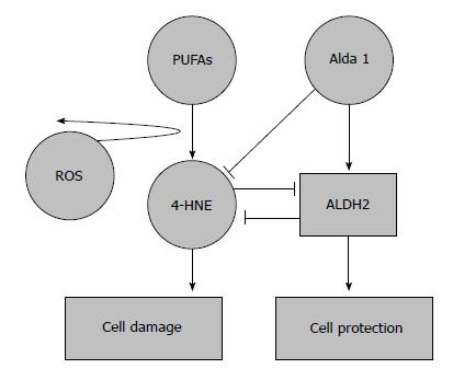
Ultimately, the clearance ratio of 4-HNE-adducts by ALDH2 and the ALDH2-4-HNE balance is crucial in order to prevent the modulation of survival mechanisms by 4-HNE and thus minimize some of the deleterious effects of IRI caused by the formation of these toxic aldehydes (Figure 1).

Figure 1
Figure 1 Protective effects of aldehyde dehydrogenase 2 on 4-hydroxy-2-nonenal accumulation in ischemia reperfusion injury. 4-hydroxy-2-nonenal (4-HNE) is a pivotal marker for cell damage associated with oxidative stress; its accumulation is prevented by aldehyde dehydrogenase 2 (ALDH2) activation and the action of its agonists. An overwhelming 4-HNE accumulation may also inhibit ALDH2 action. 4-HNE: 4-hydroxy-2-nonenal.
ALDH2 AND AUTOPHAGY

Autophagy is believed to play a key role in cell survival, with distinct functions depending on the event the cell is facing. In fact, autophagy is a cellular housekeeping mechanism, which in physiological conditions removes long-lived, aggregated and misfolded proteins, clears damaged organelles and plays an important role in the “survival and death” strategies of cellular stress responses, as occurs in the I/R condition[31,32-34].

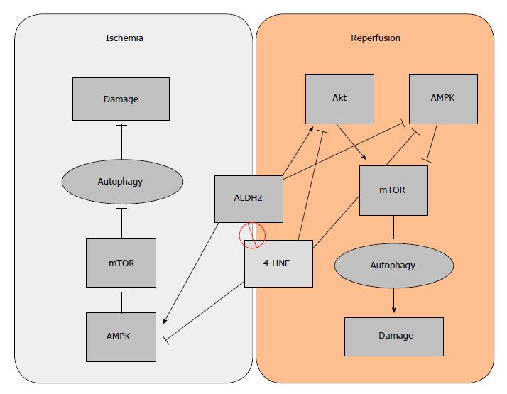
Autophagy has two paradoxically opposite behaviors, depending on the stage of the I/R condition: during ischemia, certain levels of autophagy are cytoprotective[35], but during reperfusion it is detrimental[32,33]. One of the key elements that regulate autophagy is the mammalian target of rapamycin (mTOR), an autophagy inhibitor. The inhibitory effect of mTOR over autophagy can be enhanced by Akt during reperfusion, or inhibited by AMPK (in both ischemia and reperfusion)[31]. Taking into account the dual behavior of autophagy and seeing some of its regulators, the role that ALDH2 plays in I/R is also dual: During ischemia, ALDH2 benefits AMPK activation, thus contributing to the inhibition of mTOR and subsequently increasing cytoprotective autophagy. On the other hand, during the reperfusion phase, cytoprotective autophagy is no longer active. Under reperfusion conditions, ALDH2 inhibits AMPK but also activates Akt, whose phosphorylation is kicked on to activate mTOR, thus resulting in a detrimental Akt-dependent inhibition of autophagy[28]. The ALDH2 dual regulatory effect on autophagy mechanisms through the Akt/AMPK/mTOR pathway could be a useful tool for preventing IRI. Considering that both AMPK and Akt are targets for 4-HNE, the beneficial effects that ALDH2 exerts on autophagy may be due both to its interaction with AMPK and to its capacity to remove 4-HNE (Figure 2).

Figure 2
Figure 2 Dual regulatory effect of aldehyde dehydrogenase 2 (ALDH2) on autophagy. During ischemia, AMPK is activated to promote “cytoprotective” autophagy due to mTOR inhibition. On the other hand, the activation of Akt during the phase of reperfusion inhibits “deleterious” autophagy, which is associated with apoptosis, thus reducing organ damage.

We have seen that autophagy is a mechanism which, during periods of scarce resources, enables the cell to recycle its own products in order to survive. Most of this survival potential is performed through the preservation of the mitochondria, which are among the organelles most in need of protection. Therefore, mitophagy, the selective process to auto-phagocytize mitochondria, should be carefully regulated[36]. Some IRI-related studies report that after ALDH2 activation, the mitophagy regulators tensin homolog-induced putative kinase 1 (PINK1)/Parkin expression is suppressed, resulting in reduced injury. Even though the specific mechanism of action remains unclear (either via direct inhibition or via 4-HNE cleansing), ALDH2 clearly exerts a beneficial effect on mitophagy[37].

We can conclude that ALDH2 plays a positive role in the modulation of cyto-protective autophagy. It is an especially promising agent for the regulation of the deleterious effects of autophagy in cold IRI processes associated with organ transplantation.

ALDH2: NECROPTOSIS AND APOPTOSIS

Programmed cell death results in either a lytic or a non-lytic morphology, which includes different cell signaling pathways. It can be caused by well-known mechanisms such as autophagy, apoptosis, necrosis and necroptosis[19,38,39], which are determinant for organ preservation/cell survival associated with ischemia reperfusion injury.

Briefly, “necrosis” is one of the most frequent consequences of metabolic injury due to the organ oxygen deprivation during ischemia, leading to ATP depletion. It is characterized by cell swelling, membrane rupture and release of cell contents with the subsequent inflammatory response[40]. By contrast, “apoptosis” is a non-lytic form of silent programmed cell death in which the individual dying cells separate from their neighbors and shrink rather than swell[40]. Cell death by necrosis may also be programmed, as also occurs in apoptosis, and is then called “necroptosis”. In any case, distinct signaling events drive the lytic cell and non-lytic cell death processes (necroptosis and apoptosis, respectively). Thus, necroptosis is similar in nature to necrosis but it is induced by the activation of RIPK1 (receptor-interacting protein kinase 1) and RIPK3 (receptor- interacting protein kinase 1), a caspase-independent form of programmed cell death. Studies by Cheng Shen et al[41] showed that ALDH2 deficiency promotes necroptosis through the activation of the RIPK1/RIPK3/MLKL pathway.

In contrast to necroptosis, there is no inflammation during apoptosis; the process is dependent on caspases 3 and 9. Apoptosis is a process controlled at multiple checkpoints by cell signaling expression, sometimes with opposite actions; examples are Bax (pro-apoptotic) and Bcl-2 (anti-apoptotic). The ratio between the two is decisive for the apoptotic response, which is regulated by an upstream mediator, Akt, which ultimately determines the fate of the cell[42,43].

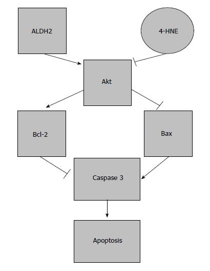
Due to its cytotoxic characteristics, 4-HNE is an important mediator of oxidative stress-induced apoptosis[42]. As previously mentioned, 4-HNE inhibits a group of a several kinases, one of which is Akt. Under high concentrations of 4-HNE, a dramatic apoptotic response is induced. This effect has been suggested to occur through the inhibition of Akt, which increases the Bax/Bcl-2 ratio and activates a caspase-3 apoptotic cascade[43,44]. This Akt inhibition in the presence of 4-HNE can be totally reversed by using an ALDH2 agonist, such as Alda-1[45]. Further investigations have evidenced that increased ALDH2 expression through Alda-1 treatment protects I/R-induced brain cell necrosis and apoptosis[16,24]. Besides these benefits of ALDH2 observed in the heart and brain, benefits have also been shown for limiting neuronal apoptosis in spinal cord IRI[24]. Recently, Zhong et al[45] reported that low ALDH2 promotes liver apoptosis through the MAPK pathway when ALDH2 agonists are used, in which ALDH2 action is not only based on the 4-HNE clearance ratio, but also on its subsidiary involvement in controlling indirect 4-HNE pathways.

Again, the ratio of 4-HNE formation and its cleansing by ALDH2 is essential. High 4-HNE levels will limit ALDH2 activity, avoiding subsequent 4-HNE clearance and producing a positive 4-HNE feedback that ultimately causes apoptosis and cell death[1,42] (Figure 3).

Figure 3
Figure 3 Effects of aldehyde dehydrogenase 2 on apoptosis in ischemia reperfusion injury. Protection induced by aldehyde dehydrogenase 2 (ALDH2) on apoptosis. The balance between ALDH2/4-HNE is responsible for the modulation of apoptosis through Akt and caspase 3 expression.
ALDH2 ACTIVATION AND IRI PREVENTION IN DIFFERENT ORGANS: SOME CONSIDERATIONS

In general, ALDH2 activation plays a protective role in the complex physiopathology of IRI, whose beneficial effects have mostly been studied in the heart, inducing cardio-protection as well as a protective role against the I/R insult[12,46]. This protection has been evidenced in other organs such as brain, intestine, kidney and spinal cord[16,20,22,24]. Surprisingly, the role of ALDH2 in hepatic I/R has not been studied in depth, even though the liver is one of the organs where ALDH2 is most abundant[1]. This is probably due to the fact that most studies of ALDH2 in liver have focused on ethanol metabolism disorders.

In the following lines, we describe the most important ongoing research into the role of ALDH2 in IRI in heart, brain, eyes, intestine, and kidney.

ALDH2 and heart

The most important feature of ALDH2 myocardial cardio-protection is the clearance of toxic aldehydes (such as 4-HNE) and its adducts[12,46]; Alda-1, an activator of ALDH2, raises this reactive aldehyde clearance even further, increasing the protection against myocardial IRI disorders[14,15]. Moreover, ALDH2 activation is associated with an improved mitochondrial function and a remodeling of the ventricular function[47,48]. ALDH2-induced cardio-protection (with the subsequent detoxification of toxic aldehydes) against IRI also occurs through a differential regulation of autophagy, involving both AMPK and Akt-mTOR signaling mechanisms, produced during ischemia and reperfusion respectively[28,49]. Guo et al[50] recently described a novel protective ALDH2 mechanism in type I diabetes, which defends against the induced myocardial dysfunction via an AMPK-dependent regulation of autophagy. These benefits are confirmed by the fact that ALDH2 inhibition by O-linked-N-acetylglucosamine (O-GlcNAC) acylation contributes to hyperglycemic exacerbation of myocardial IRI, which is prevented by Alda-1[51]. Even more recently, it has been reported that ALDH2 induces cardio-protection through the regulation of mitophagy by suppressing tensin homolog-induced putative kinase 1 (PINK1)/Parkin expression, thus preventing the accumulation of 4-HNE and other toxic species[37].

ALDH2 in brain, intestine, kidney and eyes

The role of ALDH2 in other organs, such as brain, eyes, intestine, and kidney, has been poorly investigated. In brain and intestine, ALDH2 activation contributes to the clearance of the 4-HNE accumulation and MDA generation against IRI. Alda-1, an ALDH2 agonist, increases the 4-HNE adduct clearance, subsequently promoting protection against brain IRI and preventing the deleterious effects of apoptosis[15,16]. In kidney, after bilateral ischemia, ALDH2 activation is involved in the protection induced by ethanol at physiological levels[23]. Interestingly, an increased ALDH2 expression also has a protective effect, reducing renal cell apoptosis by inhibiting the MAPK pathway, after using hypothermic machine perfusion[22].

Recent investigations have shown the existence of ALDH2 expression in adult rat retinal tissues, which may be involved in the retina redox balance. However, its role in the regulatory lipoperoxidation mechanisms associated with retinal ischemia reperfusion injury needs to be investigated in depth[52,53].

ALDH2 ACTIVATION: ISCHEMIC PRECONDITIONING

Several therapeutic surgical and pharmacological strategies have been used to protect the organ against I/R insult. As an example, “ischemic preconditioning” (IPC) is based on the application of a previous transient ischemia and/or reperfusion that will prepare the organ prior to a sustained I/R. IPC was initially evidenced in the heart by Murry et al[54] but has also been found to be protective for other organs such as kidney, intestine and liver[55-57]. Another surgical strategy is “remote ischemic preconditioning” (RIPC)[58], which consists in the application of brief ischemia in one organ to confer protection on distant organs as well[59]. The involvement of ALDH2 as an important mediator in the benefits conferred by RIPC has also been discussed by Contractor et al[59].

ALDH2 ACTIVATION: PHARMACOLOGICAL PRECONDITIONING

The use of pharmacological agents to promote organ protection (pharmacological preconditioning) is limited when it is mandatory to avoid their potential undesirable secondary effects. We have assessed the protective role of ALDH2 when using different protective strategies against IRI based on the administration of volatile anesthetics (isoflurane), nitrites/nitrates and ALDH2 agonists (Alda-1).

ALDH2 and isoflurane preconditioning

Recent investigations in the heart and other organs have demonstrated that the use of isoflurane, a volatile anesthetic agent, is an effective preconditioning agent that mimics the protective effects of IPC but avoids some of its disadvantages caused by the reduction of the blood flow, and presents greater ethical acceptability and clinical safety[60-62]. ALDH2 is involved in the cardio-protection induced by isoflurane preconditioning, and this ALDH2/isoflurane-induced cardio-protection is substantially blocked by a PKCε inhibitor, suggesting that mitochondrial PKCε plays an important role in isoflurane-induced protection mechanisms. It has been discussed whether the phosphorylation of ALDH2 increases PKCε mitochondrial translocation, and the inhibition of mitochondrial translocation by other protein kinases such as PKC delta, contribute even more to isoflurane protection against I/R insult[63].

ALDH2 and ethanol preconditioning

Ethanol, at low doses, also acts as a preconditioning agent[51]. Its acute cardio-protective effects are critically modulated by the dose used and by whether it remains present during the ischemic period. In these conditions of ethanol preconditioning, ALDH2 is activated to induce protection against IRI through the mitochondrial translocation of PKCε, which is responsible for the increased metabolism from HNE to HNA, thus limiting the accumulation of HNE protein adducts and improving cardiac function[64,65].

In this context, it has been suggested that the use of ALDH2 activators mimics ethanol cardio-protection. In addition, ALDH2 protection against ethanol toxicity is regulated by Akt and AMPK, and subsequently, autophagy and apoptosis through their downstream substrate mTOR[28].

ALDH2 and NO/nitrite preconditioning

Nitric oxide (NO) is a free-radical gas, considered as a relevant therapeutic tar- get during IRI. Its effects on I/R injury are probably related to the dose or to the conditions during ischemia and reperfusion, usually at nanomolar or low micromolar concentrations[66]. Apart from its role in tissue protection during ischemia, and due to its volatile nature, NO is a fast-response cell signaling molecule and a well-known vasodilator during hypoxic events, also regulating mitochondrial oxygen consumption[66]. There is growing evidence that eNOS-derived NO is a critical component in IPC and APC signal transduction[67,68]. Last but not least, due to its reductive capacity, NO is a potent ROS scavenger, and thus, performs one of the most important functions during reperfusion[69].

As a volatile molecule, the half-life of NO is not very long; this is why there is a need for a pool of nitrogen in a more stable form, i.e., nitrites (NO2-) and nitrates (NO3-). Nitrites are the reduced form of nitrates, and under physiological conditions they can be recycled in blood and tissues, where NO generation from nitrites is linearly dependent on oxygen and pH levels[66,69].

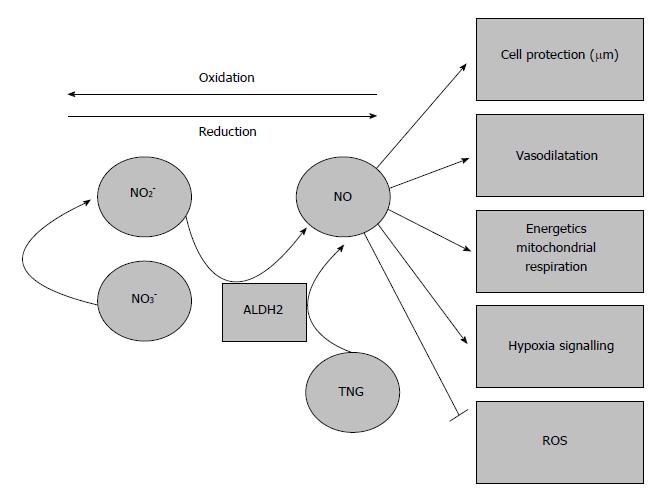
It has been widely shown that ALDH2 has a reductase-dependent activity that produces the bioconversion of nitroglycerin to 1, 2-glyceryl denigrates, resulting in the release of NO[1]. Further experiments have shown that in vitro, ALDH2 is partially responsible for nitrite bioactivation, promoting vasodilation during hypoxic episodes (Figure 4). However, Oelze et al[70] showed that the peroxynitrite generated from NO impairs the enzymatic activity of ALDH2, and therefore the scavenging activity of NO may result in a suicide inhibition of ALDH2 in later stages.

Figure 4
Figure 4 Aldehyde dehydrogenase 2 and vasodilation due to nitrates/nitrites/nitric oxide generation. The generation of nitric oxide from nitrates/nitrites and its vasodilatory properties may contribute to prevent the deleterious effects of ischemia-reperfusion injury. ALDH2: Aldehyde dehydrogenase 2.

The role of ALDH2 cannot be conclusively confirmed, due to the impossibility of ruling out other non- enzymatic or nitrite reductase species (e.g., eNOS)[71-73]. However, it can be concluded that ALDH2 is closely related to NO and nitrogen metabolism and exerts positive effects[72,73].

ALDH2 agonist preconditioning: Alda-1

With this in mind, it has been proposed that new therapeutic ALDH2 approaches might be based on promoting ALDH2 over-expression by using ALDH2 activators and determining the role of ALDH2 in a variety of human pathologies such as IRI. Here we focus specifically on the use of Alda-1, a selective ALDH2 agonist, in IRI in different organs.

Small molecules called ALDAs have been used as activators of ALDH2. One of the most frequently used is Alda-1. Defined as N-(1,3-benzodioxol-5-ylmethyl)-2,6 di-chloro-benzamide, Alda-1 increases ALDH2 activity in humans twofold with its wild-type and 11-fold with its defective variant ALDH2*2[1].

Experiments have shown that the administration of Alda-1 just before the ischemic insult reduces infarct size by 60%, most likely through ALDH2 activation[12]. Alda-1 also has beneficial effects on the heart and brain by inhibiting 4-HNE and related adducts[15]. A potential therapeutic value of Alda-1 during the progression of post-myocardial infarction cardiomyopathy has also been suggested[29]. Alda-1 may be useful for treating heart failure patients, since ALDH2 activation in heart failure restores mitochondrial function and improves ventricular function and remodeling[48]. It was recently reported that ALDH2 activation regulates mitophagy by preventing 4-HNE, ROS and superoxide dismutase (SOD) accumulation[37]. Along these lines, the recent investigations by Zhu et al[20] have shown that Alda-1 pre-treatment reduces the intestinal injury induced by I/R in mice. Benefits were associated with the prevention of 4-HNE and MDA accumulation, suggesting a potential clinical application in the near future[21].

FUTURE PERSPECTIVES OF ALDH2 IN COLD ISCHEMIA REPERFUSION INJURY/ORGAN TRANSPLANTATION

ROS play a major role in the progression of IRI, which is inherent to organ transplantation. ROS are responsible for the generation of MDA, as well as other toxic aldehydes like 4-HNE and its adducts. During graft reperfusion, ROS are mainly produced in the mitochondria, becoming the main source of lipoperoxides and toxic aldehydes. Therefore, the prevention and clearance of those toxic elements generated during an episode of ischemia-reperfusion is decisive in organ transplantation, and the role of ALDH2 is crucial.

So far, the vast majority of investigations on the role of ALDH2 activation in organ preservation are limited to the heart and kidney[22,74]. Gong et al[74] demonstrated that addition of Alda-1 (ALDH2 activator) to histidine-ketoglutarate-tryptophan (HTK; a solution regularly used in heart surgery) improved cardio-protection through the subsequent ALDH2 activation and toxic aldehyde removal. The fact that Alda-1 ameliorates the quality of cardioplegic solutions such as HTK suggests that ALDH2 activators could be used to improve preservation solutions meant for other organs besides the heart, such as UW, HTK, Celsior and IGL-1, which are used for clinical transplantation in liver, where ALDH2 has an important presence[75].

These findings have a bearing on the consideration of the potential use of ALDH2 activators as additives in organ preservation solutions in order to improve their protective quality. They are also relevant to the assessment of the contribution of various commercial solutions to the preservation of mitochondrial ALDH2 activity during graft cold storage, especially when expanded criteria donors are used.

Recent studies have reported higher mitochondrial ALDH2 activity in liver grafts preserved in IGL-1 (24 h at 4 °C) than in livers preserved in UW or HTK solutions[76]. These results are consistent with the increases reported in renal ALDH2 activity by Zhong et al[22], who subjected kidney grafts to machine perfusion preservation before transplantation.

These results confirm that ALDH2 plays a role in both static and dynamic graft preservation. It exerts a protective effect, during the ischemic phase by tackling ATP breakdown and during the reperfusion phase by reducing oxidative damage, which are inherent conditions of IRI. In this regard, further studies of ALDH2 using machine perfusion strategies in subnormothermic conditions should be carried out in order to improve the subsequent graft outcome.

Finally, although ALDH2 plays a role in distinct organs with well-defined physiological functions (heart, brain, eyes, liver, kidney and intestine), each organ contains multiple different cell types which, in turn, may give specific responses under ALDH2 activation/administration. Few studies have sought to clarify the relationship between ALDH2 and specific cell types or its influence on the substantial cross-talk between the different cells in each organ under ALDH2 administration[77]. Therefore, further investigation of this issue is now warranted.

CONCLUSION

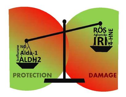
In recent years, the ability of ALDH2 to modify the activity of some key enzymes and essential survival pathways of the cell has been demonstrated (autophagy, apoptosis, necroptosis, etc). Some of its effects are exerted directly through its catalytic center, in a one-to-one interaction with those key enzymes that modifies the whole metabolic pathway. However, it may also regulate the metabolism indirectly, removing and cleansing toxic sub-products resulting from pathologies like IRI, which compromise cell viability, as we have seen in the case of 4-HNE (Figure 5).

Figure 5
Figure 5 Aldehyde dehydrogenase 2 and 4-hydroxy-2-nonenal balance in ischemia reperfusion injury. Red areas represent damage, whereas green ones depict protection of the cell through the various mechanisms. ALDH2: Aldehyde dehydrogenase 2; 4-HNE: 4-hydroxy-2-nonenal.

As a result, ALDH2 will affect any metabolic pathway in which 4-HNE is involved, and so its importance lies not only in its ability to activate other enzymes, but also in the collateral effects of those metabolic processes regulated by 4-HNE. In these processes, the regulation levels will depend on the balance of several factors such as the aldehyde/ALDH2 production ratio (where ALDH2 agonists take on a major role), the metabolization ratio of 4-HNE by ALDH2 and whether 4-HNE reaches the level of no-return where it inhibits all the ALDH2 present and the balance is definitely broken (Figure 5).

ALDH2 affects a wide range spectrum of mechanisms, among which ethanol metabolism has been the most studied. Yet, its role in important pathophysiological processes, such as IRI, remains unclear. With regard to IRI, the behavior of ALDH2 differs according to the phase (ischemia or reperfusion). Given that a variety of harmful elements such as 4-HNE are produced during an I/R episode, ALDH2 may play a critical role in the liver. This role should be further elucidated in future studies.

However, just as most studies have focused on the role of ALDH2 in ethanol metabolism rather than IRI, studies of ALDH2 in IRI are mainly focused on heart, brain or kidney and have paid hardly any attention to the liver. This is especially surprising in view of the fact that the liver is the organ with the highest ALDH2 concentration recorded so far due its detoxifying role. Just before this review was completed, a study evidencing the protective role of ALDH2 activation by Alda-1 in a model of warm ischemia reperfusion in liver was published[78].

ACKNOWLEDGMENTS

This work was supported by Instituto de Salud Carlos III through FIS project PI 15/00110 co-funded by FEDER from Regional Development European Funds (European Union) and the FOIE GRAS project, which has received funding from the European Union’s Horizon 2020 Research and Innovation programme under the Marie Sklodowska-Curie Grant (Agreement No. 722619).

Footnotes

Manuscript source: Invited manuscript

Specialty type: Gastroenterology and hepatology

Country of origin: Spain

Peer-review report classification

Grade A (Excellent): 0

Grade B (Very good): 0

Grade C (Good): C, C, C

Grade D (Fair): 0

Grade E (Poor): 0

P- Reviewer: Abdel-Hamid SMM, Jin J, Katada K S- Editor: Wang XJ L- Editor: Filipodia E- Editor: Huang Y

References
1.  Chen CH, Ferreira JC, Gross ER, Mochly-Rosen D. Targeting aldehyde dehydrogenase 2: new therapeutic opportunities. Physiol Rev. 2014;94:1-34.  [PubMed]  [DOI]  [Cited in This Article: ]  [Cited by in Crossref: 350]  [Cited by in F6Publishing: 444]  [Article Influence: 40.4]  [Reference Citation Analysis (0)]
2.  Klyosov AA, Rashkovetsky LG, Tahir MK, Keung WM. Possible role of liver cytosolic and mitochondrial aldehyde dehydrogenases in acetaldehyde metabolism. Biochemistry. 1996;35:4445-4456.  [PubMed]  [DOI]  [Cited in This Article: ]  [Cited by in Crossref: 146]  [Cited by in F6Publishing: 146]  [Article Influence: 5.0]  [Reference Citation Analysis (0)]
3.  Klyosov AA. Kinetics and specificity of human liver aldehyde dehydrogenases toward aliphatic, aromatic, and fused polycyclic aldehydes. Biochemistry. 1996;35:4457-4467.  [PubMed]  [DOI]  [Cited in This Article: ]  [Cited by in Crossref: 116]  [Cited by in F6Publishing: 120]  [Article Influence: 4.1]  [Reference Citation Analysis (0)]
4.  Mali VR, Palaniyandi SS. Regulation and therapeutic strategies of 4-hydroxy-2-nonenal metabolism in heart disease. Free Radic Res. 2014;48:251-263.  [PubMed]  [DOI]  [Cited in This Article: ]  [Cited by in Crossref: 66]  [Cited by in F6Publishing: 84]  [Article Influence: 7.0]  [Reference Citation Analysis (0)]
5.  Schneider C, Porter NA, Brash AR. Routes to 4-hydroxynonenal: fundamental issues in the mechanisms of lipid peroxidation. J Biol Chem. 2008;283:15539-15543.  [PubMed]  [DOI]  [Cited in This Article: ]  [Cited by in Crossref: 220]  [Cited by in F6Publishing: 186]  [Article Influence: 10.9]  [Reference Citation Analysis (0)]
6.  Milkovic L, Cipak Gasparovic A, Zarkovic N. Overview on major lipid peroxidation bioactive factor 4-hydroxynonenal as pluripotent growth-regulating factor. Free Radic Res. 2015;49:850-860.  [PubMed]  [DOI]  [Cited in This Article: ]  [Cited by in Crossref: 54]  [Cited by in F6Publishing: 55]  [Article Influence: 5.5]  [Reference Citation Analysis (0)]
7.  Kristal BS, Park BK, Yu BP. 4-Hydroxyhexenal is a potent inducer of the mitochondrial permeability transition. J Biol Chem. 1996;271:6033-6038.  [PubMed]  [DOI]  [Cited in This Article: ]
8.  Kruman I, Bruce-Keller AJ, Bredesen D, Waeg G, Mattson MP. Evidence that 4-hydroxynonenal mediates oxidative stress-induced neuronal apoptosis. J Neurosci. 1997;17:5089-5100.  [PubMed]  [DOI]  [Cited in This Article: ]
9.  Zhang Y, Ren J. ALDH2 in alcoholic heart diseases: molecular mechanism and clinical implications. Pharmacol Ther. 2011;132:86-95.  [PubMed]  [DOI]  [Cited in This Article: ]  [Cited by in Crossref: 110]  [Cited by in F6Publishing: 131]  [Article Influence: 9.4]  [Reference Citation Analysis (0)]
10.  Zhang Y, Babcock SA, Hu N, Maris JR, Wang H, Ren J. Mitochondrial aldehyde dehydrogenase (ALDH2) protects against streptozotocin-induced diabetic cardiomyopathy: role of GSK3β and mitochondrial function. BMC Med. 2012;10:40.  [PubMed]  [DOI]  [Cited in This Article: ]  [Cited by in Crossref: 106]  [Cited by in F6Publishing: 119]  [Article Influence: 9.2]  [Reference Citation Analysis (0)]
11.  Hao PP, Chen YG, Wang JL, Wang XL, Zhang Y. Meta-analysis of aldehyde dehydrogenase 2 gene polymorphism and Alzheimer’s disease in East Asians. Can J Neurol Sci. 2011;38:500-506.  [PubMed]  [DOI]  [Cited in This Article: ]
12.  Chen CH, Budas GR, Churchill EN, Disatnik MH, Hurley TD, Mochly-Rosen D. Activation of aldehyde dehydrogenase-2 reduces ischemic damage to the heart. Science. 2008;321:1493-1495.  [PubMed]  [DOI]  [Cited in This Article: ]  [Cited by in Crossref: 604]  [Cited by in F6Publishing: 625]  [Article Influence: 36.8]  [Reference Citation Analysis (0)]
13.  Pang JJ, Barton LA, Chen YG, Ren J. Mitochondrial aldehyde dehydrogenase in myocardial ischemia-reperfusion injury: from bench to bedside. Sheng Li Xue Bao. 2015;67:535-544.  [PubMed]  [DOI]  [Cited in This Article: ]
14.  Budas GR, Disatnik MH, Mochly-Rosen D. Aldehyde dehydrogenase 2 in cardiac protection: a new therapeutic target? Trends Cardiovasc Med. 2009;19:158-164.  [PubMed]  [DOI]  [Cited in This Article: ]  [Cited by in Crossref: 84]  [Cited by in F6Publishing: 93]  [Article Influence: 6.2]  [Reference Citation Analysis (0)]
15.  Luo XJ, Liu B, Ma QL, Peng J. Mitochondrial aldehyde dehydrogenase, a potential drug target for protection of heart and brain from ischemia/reperfusion injury. Curr Drug Targets. 2014;15:948-955.  [PubMed]  [DOI]  [Cited in This Article: ]  [Cited by in Crossref: 23]  [Cited by in F6Publishing: 29]  [Article Influence: 2.9]  [Reference Citation Analysis (0)]
16.  Fu SH, Zhang HF, Yang ZB, Li TB, Liu B, Lou Z, Ma QL, Luo XJ, Peng J. Alda-1 reduces cerebral ischemia/reperfusion injury in rat through clearance of reactive aldehydes. Naunyn Schmiedebergs Arch Pharmacol. 2014;387:87-94.  [PubMed]  [DOI]  [Cited in This Article: ]  [Cited by in Crossref: 53]  [Cited by in F6Publishing: 61]  [Article Influence: 5.1]  [Reference Citation Analysis (0)]
17.  Qi Y, Zhao M, Bai Y, Huang L, Yu W, Bian Z, Zhao M, Li X. Retinal ischemia/reperfusion injury is mediated by Toll-like receptor 4 activation of NLRP3 inflammasomes. Invest Ophthalmol Vis Sci. 2014;55:5466-5475.  [PubMed]  [DOI]  [Cited in This Article: ]  [Cited by in Crossref: 57]  [Cited by in F6Publishing: 61]  [Article Influence: 5.5]  [Reference Citation Analysis (0)]
18.  Baker ML, Hand PJ, Wang JJ, Wong TY. Retinal signs and stroke: revisiting the link between the eye and brain. Stroke. 2008;39:1371-1379.  [PubMed]  [DOI]  [Cited in This Article: ]  [Cited by in Crossref: 134]  [Cited by in F6Publishing: 143]  [Article Influence: 8.4]  [Reference Citation Analysis (0)]
19.  Gao S, Andreeva K, Cooper NG. Ischemia-reperfusion injury of the retina is linked to necroptosis via the ERK1/2-RIP3 pathway. Mol Vis. 2014;20:1374-1387.  [PubMed]  [DOI]  [Cited in This Article: ]
20.  Zhu Q, He G, Wang J, Wang Y, Chen W. Pretreatment with the ALDH2 agonist Alda-1 reduces intestinal injury induced by ischaemia and reperfusion in mice. Clin Sci (Lond). 2017;131:1123-1136.  [PubMed]  [DOI]  [Cited in This Article: ]  [Cited by in Crossref: 32]  [Cited by in F6Publishing: 40]  [Article Influence: 5.0]  [Reference Citation Analysis (0)]
21.  Dollé L. Pharmacological enrollment of aldehyde dehydrogenase modulators to assist treating ischemia reperfusion-induced intestinal injury: is there a gap to be bridged? Clin Sci (Lond). 2017;131:1137-1140.  [PubMed]  [DOI]  [Cited in This Article: ]  [Cited by in Crossref: 1]  [Cited by in F6Publishing: 1]  [Article Influence: 0.1]  [Reference Citation Analysis (0)]
22.  Zhong Z, Hu Q, Fu Z, Wang R, Xiong Y, Zhang Y, Liu Z, Wang Y, Ye Q. Increased Expression of Aldehyde Dehydrogenase 2 Reduces Renal Cell Apoptosis During Ischemia/Reperfusion Injury After Hypothermic Machine Perfusion. Artif Organs. 2016;40:596-603.  [PubMed]  [DOI]  [Cited in This Article: ]  [Cited by in Crossref: 22]  [Cited by in F6Publishing: 23]  [Article Influence: 2.3]  [Reference Citation Analysis (0)]
23.  Yuan Q, Hong S, Han S, Zeng L, Liu F, Ding G, Kang Y, Mao J, Cai M, Zhu Y. Preconditioning with physiological levels of ethanol protect kidney against ischemia/reperfusion injury by modulating oxidative stress. PLoS One. 2011;6:e25811.  [PubMed]  [DOI]  [Cited in This Article: ]  [Cited by in Crossref: 42]  [Cited by in F6Publishing: 46]  [Article Influence: 3.3]  [Reference Citation Analysis (0)]
24.  Liu XZ, Sun X, Shen KP, Jin WJ, Fu ZY, Tao HR, Xu ZX. Aldehyde dehydrogenase 2 overexpression inhibits neuronal apoptosis after spinal cord ischemia/reperfusion injury. Neural Regen Res. 2017;12:1166-1171.  [PubMed]  [DOI]  [Cited in This Article: ]  [Cited by in Crossref: 15]  [Cited by in F6Publishing: 15]  [Article Influence: 1.9]  [Reference Citation Analysis (0)]
25.  Casillas-Ramírez A, Mosbah IB, Ramalho F, Roselló-Catafau J, Peralta C. Past and future approaches to ischemia-reperfusion lesion associated with liver transplantation. Life Sci. 2006;79:1881-1894.  [PubMed]  [DOI]  [Cited in This Article: ]  [Cited by in Crossref: 139]  [Cited by in F6Publishing: 154]  [Article Influence: 8.1]  [Reference Citation Analysis (0)]
26.  Zaouali MA, Ben Abdennebi H, Padrissa-Altés S, Mahfoudh-Boussaid A, Roselló-Catafau J. Pharmacological strategies against cold ischemia reperfusion injury. Expert Opin Pharmacother. 2010;11:537-555.  [PubMed]  [DOI]  [Cited in This Article: ]  [Cited by in Crossref: 48]  [Cited by in F6Publishing: 52]  [Article Influence: 3.5]  [Reference Citation Analysis (0)]
27.  Mali VR, Deshpande M, Pan G, Thandavarayan RA, Palaniyandi SS. Impaired ALDH2 activity decreases the mitochondrial respiration in H9C2 cardiomyocytes. Cell Signal. 2016;28:1-6.  [PubMed]  [DOI]  [Cited in This Article: ]  [Cited by in Crossref: 19]  [Cited by in F6Publishing: 22]  [Article Influence: 2.2]  [Reference Citation Analysis (0)]
28.  Ma H, Guo R, Yu L, Zhang Y, Ren J. Aldehyde dehydrogenase 2 (ALDH2) rescues myocardial ischaemia/reperfusion injury: role of autophagy paradox and toxic aldehyde. Eur Heart J. 2011;32:1025-1038.  [PubMed]  [DOI]  [Cited in This Article: ]  [Cited by in Crossref: 231]  [Cited by in F6Publishing: 271]  [Article Influence: 18.1]  [Reference Citation Analysis (0)]
29.  Gomes KM, Bechara LR, Lima VM, Ribeiro MA, Campos JC, Dourado PM, Kowaltowski AJ, Mochly-Rosen D, Ferreira JC. Aldehydic load and aldehyde dehydrogenase 2 profile during the progression of post-myocardial infarction cardiomyopathy: benefits of Alda-1. Int J Cardiol. 2015;179:129-138.  [PubMed]  [DOI]  [Cited in This Article: ]  [Cited by in Crossref: 41]  [Cited by in F6Publishing: 44]  [Article Influence: 4.0]  [Reference Citation Analysis (0)]
30.  Liu W, Akhand AA, Takeda K, Kawamoto Y, Itoigawa M, Kato M, Suzuki H, Ishikawa N, Nakashima I. Protein phosphatase 2A-linked and -unlinked caspase-dependent pathways for downregulation of Akt kinase triggered by 4-hydroxynonenal. Cell Death Differ. 2003;10:772-781.  [PubMed]  [DOI]  [Cited in This Article: ]  [Cited by in Crossref: 76]  [Cited by in F6Publishing: 76]  [Article Influence: 3.5]  [Reference Citation Analysis (0)]
31.  Van Erp AC, Hoeksma D, Rebolledo RA, Ottens PJ, Jochmans I, Monbaliu D, Pirenne J, Leuvenink HGD, Decuypere JP. The Crosstalk between ROS and Autophagy in the Field of Transplantation Medicine. Oxid Med Cell Longev. 2017;2017:7120962.  [PubMed]  [DOI]  [Cited in This Article: ]  [Cited by in Crossref: 30]  [Cited by in F6Publishing: 30]  [Article Influence: 3.8]  [Reference Citation Analysis (0)]
32.  Decuypere JP, Ceulemans LJ, Agostinis P, Monbaliu D, Naesens M, Pirenne J, Jochmans I. Autophagy and the Kidney: Implications for Ischemia-Reperfusion Injury and Therapy. Am J Kidney Dis. 2015;66:699-709.  [PubMed]  [DOI]  [Cited in This Article: ]  [Cited by in Crossref: 79]  [Cited by in F6Publishing: 108]  [Article Influence: 10.8]  [Reference Citation Analysis (0)]
33.  Cursio R, Colosetti P, Gugenheim J. Autophagy and liver ischemia-reperfusion injury. Biomed Res Int. 2015;2015:417590.  [PubMed]  [DOI]  [Cited in This Article: ]  [Cited by in Crossref: 71]  [Cited by in F6Publishing: 96]  [Article Influence: 9.6]  [Reference Citation Analysis (0)]
34.  Go KL, Lee S, Zendejas I, Behrns KE, Kim JS. Mitochondrial Dysfunction and Autophagy in Hepatic Ischemia/Reperfusion Injury. Biomed Res Int. 2015;2015:183469.  [PubMed]  [DOI]  [Cited in This Article: ]  [Cited by in Crossref: 70]  [Cited by in F6Publishing: 91]  [Article Influence: 9.1]  [Reference Citation Analysis (0)]
35.  Panisello-Roselló A, Verde E, Lopez A, Flores M, Folch-Puy E, Rolo A, Palmeira C, Hotter G, Carbonell T, Adam R. Cytoprotective Mechanisms in Fatty Liver Preservation against Cold Ischemia Injury: A Comparison between IGL-1 and HTK. Int J Mol Sci. 2018;19.  [PubMed]  [DOI]  [Cited in This Article: ]  [Cited by in Crossref: 13]  [Cited by in F6Publishing: 13]  [Article Influence: 1.9]  [Reference Citation Analysis (0)]
36.  Poole AC, Thomas RE, Andrews LA, McBride HM, Whitworth AJ, Pallanck LJ. The PINK1/Parkin pathway regulates mitochondrial morphology. Proc Natl Acad Sci USA. 2008;105:1638-1643.  [PubMed]  [DOI]  [Cited in This Article: ]  [Cited by in F6Publishing: 1]  [Reference Citation Analysis (0)]
37.  Ji W, Wei S, Hao P, Xing J, Yuan Q, Wang J, Xu F, Chen Y. Aldehyde Dehydrogenase 2 Has Cardioprotective Effects on Myocardial Ischaemia/Reperfusion Injury via Suppressing Mitophagy. Front Pharmacol. 2016;7:101.  [PubMed]  [DOI]  [Cited in This Article: ]  [Cited by in Crossref: 51]  [Cited by in F6Publishing: 72]  [Article Influence: 8.0]  [Reference Citation Analysis (0)]
38.  Wang S, Pacher P, De Lisle RC, Huang H, Ding WX. A Mechanistic Review of Cell Death in Alcohol-Induced Liver Injury. Alcohol Clin Exp Res. 2016;40:1215-1223.  [PubMed]  [DOI]  [Cited in This Article: ]  [Cited by in Crossref: 69]  [Cited by in F6Publishing: 96]  [Article Influence: 10.7]  [Reference Citation Analysis (0)]
39.  Rosenbaum DM, Degterev A, David J, Rosenbaum PS, Roth S, Grotta JC, Cuny GD, Yuan J, Savitz SI. Necroptosis, a novel form of caspase-independent cell death, contributes to neuronal damage in a retinal ischemia-reperfusion injury model. J Neurosci Res. 2010;88:1569-1576.  [PubMed]  [DOI]  [Cited in This Article: ]  [Cited by in Crossref: 28]  [Cited by in F6Publishing: 126]  [Article Influence: 8.4]  [Reference Citation Analysis (0)]
40.  Malhi H, Gores GJ, Lemasters JJ. Apoptosis and necrosis in the liver: a tale of two deaths? Hepatology. 2006;43:S31-S44.  [PubMed]  [DOI]  [Cited in This Article: ]
41.  Shen C, Wang C, Han S, Wang Z, Dong Z, Zhao X, Wang P, Zhu H, Sun X, Ma X. Aldehyde dehydrogenase 2 deficiency negates chronic low-to-moderate alcohol consumption-induced cardioprotecion possibly via ROS-dependent apoptosis and RIP1/RIP3/MLKL-mediated necroptosis. Biochim Biophys Acta. 2017;1863:1912-1918.  [PubMed]  [DOI]  [Cited in This Article: ]  [Cited by in Crossref: 31]  [Cited by in F6Publishing: 24]  [Article Influence: 3.0]  [Reference Citation Analysis (0)]
42.  Awasthi YC, Sharma R, Cheng JZ, Yang Y, Sharma A, Singhal SS, Awasthi S. Role of 4-hydroxynonenal in stress-mediated apoptosis signaling. Mol Aspects Med. 2003;24:219-230.  [PubMed]  [DOI]  [Cited in This Article: ]  [Cited by in Crossref: 258]  [Cited by in F6Publishing: 431]  [Article Influence: 19.6]  [Reference Citation Analysis (0)]
43.  Teijido O, Dejean L. Upregulation of Bcl2 inhibits apoptosis-driven BAX insertion but favors BAX relocalization in mitochondria. FEBS Lett. 2010;584:3305-3310.  [PubMed]  [DOI]  [Cited in This Article: ]  [Cited by in Crossref: 70]  [Cited by in F6Publishing: 86]  [Article Influence: 5.7]  [Reference Citation Analysis (0)]
44.  Ji GR, Yu NC, Xue X, Li ZG. 4-Hydroxy-2-nonenal induces apoptosis by inhibiting AKT signaling in human osteosarcoma cells. ScientificWorldJournal. 2014;2014:873525.  [PubMed]  [DOI]  [Cited in This Article: ]  [Cited by in Crossref: 19]  [Cited by in F6Publishing: 22]  [Article Influence: 2.0]  [Reference Citation Analysis (0)]
45.  Zhong Z, Ye S, Xiong Y, Wu L, Zhang M, Fan X, Li L, Fu Z, Wang H, Chen M. Decreased expression of mitochondrial aldehyde dehydrogenase-2 induces liver injury via activation of the mitogen-activated protein kinase pathway. Transpl Int. 2016;29:98-107.  [PubMed]  [DOI]  [Cited in This Article: ]  [Cited by in Crossref: 12]  [Cited by in F6Publishing: 13]  [Article Influence: 1.3]  [Reference Citation Analysis (0)]
46.  Liu X, Sun A. Aldehyde Dehydrogenase-2 Roles in Ischemic Cardiovascular Disease. Curr Drug Targets. 2017;18:1817-1823.  [PubMed]  [DOI]  [Cited in This Article: ]  [Cited by in Crossref: 10]  [Cited by in F6Publishing: 10]  [Article Influence: 1.4]  [Reference Citation Analysis (0)]
47.  Mali VR, Pan G, Deshpande M, Thandavarayan RA, Xu J, Yang XP, Palaniyandi SS. Cardiac Mitochondrial Respiratory Dysfunction and Tissue Damage in Chronic Hyperglycemia Correlate with Reduced Aldehyde Dehydrogenase-2 Activity. PLoS One. 2016;11:e0163158.  [PubMed]  [DOI]  [Cited in This Article: ]  [Cited by in Crossref: 24]  [Cited by in F6Publishing: 25]  [Article Influence: 2.8]  [Reference Citation Analysis (0)]
48.  Gomes KM, Campos JC, Bechara LR, Queliconi B, Lima VM, Disatnik MH, Magno P, Chen CH, Brum PC, Kowaltowski AJ. Aldehyde dehydrogenase 2 activation in heart failure restores mitochondrial function and improves ventricular function and remodelling. Cardiovasc Res. 2014;103:498-508.  [PubMed]  [DOI]  [Cited in This Article: ]  [Cited by in Crossref: 93]  [Cited by in F6Publishing: 108]  [Article Influence: 9.8]  [Reference Citation Analysis (0)]
49.  Zhang Y, Ren J. Autophagy in ALDH2-elicited cardioprotection against ischemic heart disease: slayer or savior? Autophagy. 2010;6:1212-1213.  [PubMed]  [DOI]  [Cited in This Article: ]
50.  Guo Y, Yu W, Sun D, Wang J, Li C, Zhang R, Babcock SA, Li Y, Liu M, Ma M. A novel protective mechanism for mitochondrial aldehyde dehydrogenase (ALDH2) in type i diabetes-induced cardiac dysfunction: role of AMPK-regulated autophagy. Biochim Biophys Acta. 2015;1852:319-331.  [PubMed]  [DOI]  [Cited in This Article: ]  [Cited by in Crossref: 71]  [Cited by in F6Publishing: 75]  [Article Influence: 6.8]  [Reference Citation Analysis (0)]
51.  Liu B, Wang J, Li M, Yuan Q, Xue M, Xu F, Chen Y. Inhibition of ALDH2 by O-GlcNAcylation contributes to the hyperglycemic exacerbation of myocardial ischemia/reperfusion injury. Oncotarget. 2017;8:19413-19426.  [PubMed]  [DOI]  [Cited in This Article: ]  [Cited by in Crossref: 17]  [Cited by in F6Publishing: 18]  [Article Influence: 2.3]  [Reference Citation Analysis (0)]
52.  Pinazo-Durán MD, Verdejo C, Azorín I, Renau-Piqueras J, Iborra FJ. Colocalization of aldehyde dehydrogenases and Fe/NADPH-induced lipid peroxidation in tissue sections of rat retina. Ophthalmic Res. 2000;32:61-68.  [PubMed]  [DOI]  [Cited in This Article: ]
53.  Galbis-Estrada C, Pons-Vázquez S, Gallego-Pinazo R, Lleó-Perez A, Garcia-Medina JJ, Bou VV, Sanz-Solana P, Pinazo-Durán MD. Glutathione-dependent formaldehyde dehydrogenase (ADH3) and low km mitochondrial aldehyde dehydrogenase (ALDH2). New evidence for differential expression in the rat retina in response to oxidative stress. Free Radic Res. 2012;46:77-84.  [PubMed]  [DOI]  [Cited in This Article: ]  [Cited by in Crossref: 9]  [Cited by in F6Publishing: 11]  [Article Influence: 0.8]  [Reference Citation Analysis (0)]
54.  Murry CE, Jennings RB, Reimer KA. Preconditioning with ischemia: a delay of lethal cell injury in ischemic myocardium. Circulation. 1986;74:1124-1136.  [PubMed]  [DOI]  [Cited in This Article: ]  [Cited by in Crossref: 5406]  [Cited by in F6Publishing: 5506]  [Article Influence: 141.2]  [Reference Citation Analysis (0)]
55.  Riera M, Herrero I, Torras J, Cruzado JM, Fatjo M, Lloberas N, Alsina J, Grinyo JM. Ischemic preconditioning improves postischemic acute renal failure. Transplant Proc. 1999;31:2346-2347.  [PubMed]  [DOI]  [Cited in This Article: ]  [Cited by in Crossref: 19]  [Cited by in F6Publishing: 21]  [Article Influence: 0.8]  [Reference Citation Analysis (0)]
56.  Sola A, De Oca J, González R, Prats N, Roselló-Catafau J, Gelpí E, Jaurrieta E, Hotter G. Protective effect of ischemic preconditioning on cold preservation and reperfusion injury associated with rat intestinal transplantation. Ann Surg. 2001;234:98-106.  [PubMed]  [DOI]  [Cited in This Article: ]
57.  Peralta C, Bartrons R, Riera L, Manzano A, Xaus C, Gelpí E, Roselló-Catafau J. Hepatic preconditioning preserves energy metabolism during sustained ischemia. Am J Physiol Gastrointest Liver Physiol. 2000;279:G163-G171.  [PubMed]  [DOI]  [Cited in This Article: ]  [Cited by in Crossref: 76]  [Cited by in F6Publishing: 83]  [Article Influence: 3.3]  [Reference Citation Analysis (0)]
58.  Tapuria N, Kumar Y, Habib MM, Abu Amara M, Seifalian AM, Davidson BR. Remote ischemic preconditioning: a novel protective method from ischemia reperfusion injury--a review. J Surg Res. 2008;150:304-330.  [PubMed]  [DOI]  [Cited in This Article: ]  [Cited by in Crossref: 253]  [Cited by in F6Publishing: 264]  [Article Influence: 15.5]  [Reference Citation Analysis (0)]
59.  Contractor H, Støttrup NB, Cunnington C, Manlhiot C, Diesch J, Ormerod JO, Jensen R, Bøtker HE, Redington A, Schmidt MR. Aldehyde dehydrogenase-2 inhibition blocks remote preconditioning in experimental and human models. Basic Res Cardiol. 2013;108:343.  [PubMed]  [DOI]  [Cited in This Article: ]  [Cited by in Crossref: 34]  [Cited by in F6Publishing: 34]  [Article Influence: 2.8]  [Reference Citation Analysis (0)]
60.  Kersten JR, Schmeling TJ, Pagel PS, Gross GJ, Warltier DC. Isoflurane mimics ischemic preconditioning via activation of K(ATP) channels: reduction of myocardial infarct size with an acute memory phase. Anesthesiology. 1997;87:361-370.  [PubMed]  [DOI]  [Cited in This Article: ]
61.  Tsutsumi YM, Patel HH, Lai NC, Takahashi T, Head BP, Roth DM. Isoflurane produces sustained cardiac protection after ischemia-reperfusion injury in mice. Anesthesiology. 2006;104:495-502.  [PubMed]  [DOI]  [Cited in This Article: ]
62.  Li H, Lang XE. Protein kinase C signaling pathway involvement in cardioprotection during isoflurane pretreatment. Mol Med Rep. 2015;11:2683-2688.  [PubMed]  [DOI]  [Cited in This Article: ]  [Cited by in Crossref: 10]  [Cited by in F6Publishing: 10]  [Article Influence: 0.9]  [Reference Citation Analysis (0)]
63.  Lang XE, Wang X, Zhang KR, Lv JY, Jin JH, Li QS. Isoflurane preconditioning confers cardioprotection by activation of ALDH2. PLoS One. 2013;8:e52469.  [PubMed]  [DOI]  [Cited in This Article: ]  [Cited by in Crossref: 35]  [Cited by in F6Publishing: 35]  [Article Influence: 2.9]  [Reference Citation Analysis (0)]
64.  Churchill EN, Disatnik MH, Mochly-Rosen D. Time-dependent and ethanol- induced cardiac protection from ischemia mediated by mitochondrial translocation of epsilon PKC and activation of aldehyde dehydrogenase 2. J Mol Cell Cardiol. 2009;46:278-284.  [PubMed]  [DOI]  [Cited in This Article: ]
65.  Budas GR, Disatnik MH, Chen CH, Mochly-Rosen D. Activation of aldehyde dehydrogenase 2 (ALDH2) confers cardioprotection in protein kinase C epsilon knockout mice. J Mol Cell Cardiol. 2010;48:757-764.  [PubMed]  [DOI]  [Cited in This Article: ]
66.  Duranski MR, Greer JJ, Dejam A, Jaganmohan S, Hogg N, Langston W, Patel RP, Yet SF, Wang X, Kevil CG. Cytoprotective effects of nitrite during in vivo ischemia-reperfusion of the heart and liver. J Clin Invest. 2005;115:1232-1240.  [PubMed]  [DOI]  [Cited in This Article: ]  [Cited by in Crossref: 484]  [Cited by in F6Publishing: 532]  [Article Influence: 26.6]  [Reference Citation Analysis (0)]
67.  Chiari PC, Bienengraeber MW, Weihrauch D, Krolikowski JG, Kersten JR, Warltier DC, Pagel PS. Role of endothelial nitric oxide synthase as a trigger and mediator of isoflurane-induced delayed preconditioning in rabbit myocardium. Anesthesiology. 2005;103:74-83.  [PubMed]  [DOI]  [Cited in This Article: ]
68.  Amour J, Brzezinska AK, Weihrauch D, Billstrom AR, Zielonka J, Krolikowski JG, Bienengraeber MW, Warltier DC, Pratt PF Jr, Kersten JR. Role of heat shock protein 90 and endothelial nitric oxide synthase during early anesthetic and ischemic preconditioning. Anesthesiology. 2009;110:317-325.  [PubMed]  [DOI]  [Cited in This Article: ]  [Cited by in Crossref: 42]  [Cited by in F6Publishing: 49]  [Article Influence: 3.1]  [Reference Citation Analysis (0)]
69.  Lundberg JO, Weitzberg E, Gladwin MT. The nitrate-nitrite-nitric oxide pathway in physiology and therapeutics. Nat Rev Drug Discov. 2008;7:156-167.  [PubMed]  [DOI]  [Cited in This Article: ]  [Cited by in Crossref: 1721]  [Cited by in F6Publishing: 1853]  [Article Influence: 109.0]  [Reference Citation Analysis (0)]
70.  Oelze M, Knorr M, Schell R, Kamuf J, Pautz A, Art J, Wenzel P, Münzel T, Kleinert H, Daiber A. Regulation of human mitochondrial aldehyde dehydrogenase (ALDH-2) activity by electrophiles in vitro. J Biol Chem. 2011;286:8893-8900.  [PubMed]  [DOI]  [Cited in This Article: ]  [Cited by in Crossref: 36]  [Cited by in F6Publishing: 35]  [Article Influence: 2.5]  [Reference Citation Analysis (0)]
71.  Arif S, Borgognone A, Lin EL, O’Sullivan AG, Sharma V, Drury NE, Menon A, Nightingale P, Mascaro J, Bonser RS. Role of aldehyde dehydrogenase in hypoxic vasodilator effects of nitrite in rats and humans. Br J Pharmacol. 2015;172:3341-3352.  [PubMed]  [DOI]  [Cited in This Article: ]  [Cited by in Crossref: 11]  [Cited by in F6Publishing: 11]  [Article Influence: 1.1]  [Reference Citation Analysis (0)]
72.  Rassaf T, Totzeck M, Hendgen-Cotta UB, Shiva S, Heusch G, Kelm M. Circulating nitrite contributes to cardioprotection by remote ischemic preconditioning. Circ Res. 2014;114:1601-1610.  [PubMed]  [DOI]  [Cited in This Article: ]  [Cited by in Crossref: 238]  [Cited by in F6Publishing: 271]  [Article Influence: 24.6]  [Reference Citation Analysis (0)]
73.  Ormerod JO, Evans JD, Contractor H, Beretta M, Arif S, Fernandez BO, Feelisch M, Mayer B, Kharbanda RK, Frenneaux MP. Human Second Window Pre-Conditioning and Post-Conditioning by Nitrite Is Influenced by a Common Polymorphism in Mitochondrial Aldehyde Dehydrogenase. JACC Basic Transl Sci. 2017;2:13-21.  [PubMed]  [DOI]  [Cited in This Article: ]  [Cited by in Crossref: 5]  [Cited by in F6Publishing: 6]  [Article Influence: 0.8]  [Reference Citation Analysis (0)]
74.  Gong D, Zhang Y, Zhang H, Gu H, Jiang Q, Hu S. Aldehyde dehydrogenase-2 activation during cardioplegic arrest enhances the cardioprotection against myocardial ischemia-reperfusion injury. Cardiovasc Toxicol. 2012;12:350-358.  [PubMed]  [DOI]  [Cited in This Article: ]  [Cited by in Crossref: 20]  [Cited by in F6Publishing: 16]  [Article Influence: 1.3]  [Reference Citation Analysis (0)]
75.  Adam R, Delvart V, Karam V, Ducerf C, Navarro F, Letoublon C, Belghiti J, Pezet D, Castaing D, Le Treut YP, Gugenheim J, Bachellier P, Pirenne J, Muiesan P; ELTR contributing centres, the European Liver, Intestine Transplant Association (ELITA). Compared efficacy of preservation solutions in liver transplantation: a long-term graft outcome study from the European Liver Transplant Registry. Am J Transplant. 2015;15:395-406.  [PubMed]  [DOI]  [Cited in This Article: ]  [Cited by in Crossref: 106]  [Cited by in F6Publishing: 101]  [Article Influence: 10.1]  [Reference Citation Analysis (0)]
76.  Arnau Panisello-Rosello, Alexandre Lopez, Carlos Castro, Sravan Balloji, Emma Folch-Puy, Anabela Rolo, Carlos Palmeira, René Adam and Joan Roselló-Catafau Involvement of aldehyde dehydrogenase 2 (ALDH2) in fatty liver graft protection mechanisms against cold ischemic injury. Poster session presented at: American Transplant Congress 2018; June 2-6; Seattle, Washington, USA.  [PubMed]  [DOI]  [Cited in This Article: ]
77.  Marino A, Levi R. Salvaging the ischemic heart: Gi-coupled receptors in mast cells activate a PKC/ALDH2 pathway providing anti-RAS cardioprotection. Curr Med Chem. 2018;.  [PubMed]  [DOI]  [Cited in This Article: ]  [Cited by in Crossref: 12]  [Cited by in F6Publishing: 13]  [Article Influence: 1.9]  [Reference Citation Analysis (0)]
78.  Zhang T, Zhao Q, Ye F, Huang CY, Chen WM, Huang WQ. Alda-1, an ALDH2 activator, protects against hepatic ischemia/reperfusion injury in rats via inhibition of oxidative stress. Free Radic Res. 2018;52:629-638.  [PubMed]  [DOI]  [Cited in This Article: ]  [Cited by in Crossref: 29]  [Cited by in F6Publishing: 35]  [Article Influence: 5.0]  [Reference Citation Analysis (0)]