Published online Jul 28, 2011. doi: 10.3748/wjg.v17.i28.3342
Revised: February 26, 2011
Accepted: March 5, 2011
Published online: July 28, 2011
AIM: To clarify the usefulness of the self-expanding metallic stents (SEMS) in the management of acute proximal colon obstruction due to colon carcinoma before curative surgery.
METHODS: Eighty-one colon (proximal to spleen flex) carcinoma patients (47 males and 34 females, aged 18-94 years, mean = 66.2 years) treated between September 2004 and June 2010 for acute colon obstruction were enrolled to this study, and their clinical and radiological features were reviewed. After a cleaning enema was administered, urgent colonoscopy was performed. Subsequently, endoscopic decompression using SEMS placement was attempted.
RESULTS: Endoscopic decompression using SEMS placement was technically successful in 78 (96.3%) of 81 patients. Three patients’ symptoms could not be relieved after SEMS placement and emergent operation was performed 1 d later. The site of obstruction was transverse colon in 18 patients, the hepatic flex in 42, and the ascending colon in 21. Following adequate cleansing of the colon, patients’ abdominal girth was decreased from 88 ± 3 cm before drainage to 72 ± 6 cm 7 d later, and one-stage surgery after 8 ± 1 d (range, 7-10 d) was performed. No anastomotic leakage or postoperative stenosis occurred after operation.
CONCLUSION: SEMS placement is effective and safe in the management of acute proximal colon obstruction due to colon carcinoma, and is considered as a bridged method before curative surgery.
- Citation: Yao LQ, Zhong YS, Xu MD, Xu JM, Zhou PH, Cai XL. Self-expanding metallic stents drainage for acute proximal colon obstruction. World J Gastroenterol 2011; 17(28): 3342-3346
- URL: https://www.wjgnet.com/1007-9327/full/v17/i28/3342.htm
- DOI: https://dx.doi.org/10.3748/wjg.v17.i28.3342
At the time of diagnosis, 7%-29% colorectal cancer patients present with an emergent bowel obstruction, which necessitates emergency colectomy with an unprepared bowel (proximal colon cancer) or colostomy (left colon and rectum cancer)[1]. For acute obstruction of colon and rectum cancer, various methods have been reported for a single-stage operation to reduce the cost and to improve patient care, including intraoperative colonic lavage[2-4], decompression with a metallic stent[5-7], and decompression with a transanal drainage tube[8-10]. But less than 4% of all reported cases shared their experiences in self-expanding metallic stents (SEMS) placement in the colon proximal to the splenic flexure. This is mainly because of the concern about the safety of SEMS in the proximal colon and because acute obstruction of the right colon has traditionally been managed by resection and primary anastomosis[11]. However, recent studies suggest that emergency right-sided colonic resections resulted in a significantly higher morbidity and mortality when compared with elective resections[12].
Endoscopic Center of Zhongshan Hospital is one of the largest centers in the world, which performed more than 15 000 colonoscopies each year. Since 2004, we have used metallic stent placement for 300 cases of acute colorectal obstruction. We retrospectively reviewed the outcomes of the patients with acute proximal colon obstruction treated with SEMS placement as a bridge to curative surgery.
Eighty-one colon (proximal to spleen flex) carcinoma patients (47 males and 34 females, aged 18-94 years, mean, 66.2 years) treated between September 2004 and June 2010 for acute colon obstruction were enrolled to this study. The symptoms in these patients were abdominal pain, abdominal fullness, vomiting and constipation. Physical examination showed a distended and tympanic abdomen. Plain abdominal X-ray revealed a distended large bowel and an air-fluid level displaying an acute lower bowel obstruction. After the cleaning enema was administered, urgent colonoscopy was performed for the diagnosis and SEMS placement. Informed consent was obtained from each patient.
SEMS used in the present study was 20 mm in diameter and 60 mm, 80 mm and 100 mm in length, depending on the length and caliber of the stricture. These stents have a unique one-step-through-the-scope delivery system (7.3 mm in outer diameter and 190 cm in length) that enables the stent to be passed through the 3.7 mm working channel of the colonoscope before deployment (Micro-Tech Co., Nanjing, China).
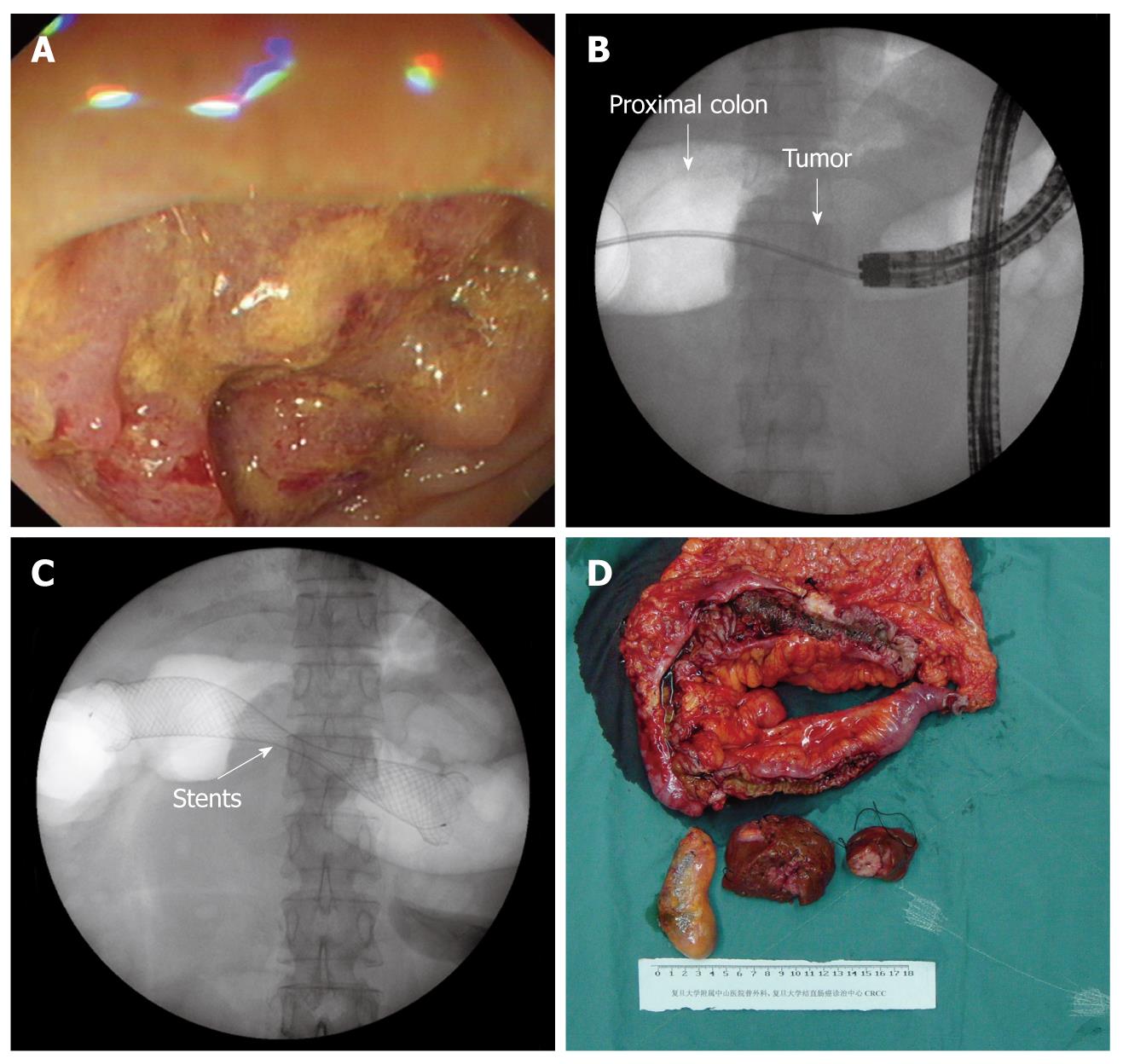
A colonoscope (CF260I; Olympus, Tokyo, Japan) was inserted and advanced to the site of the tumor. Combined with fluoroscopy, the site and etiology of acute bowel obstruction can be revealed. Under fluoroscopic and endoscopic guidance, a hydrophilic biliary guidewire (Jagwire, Boston Scientific, Natick, MA, USA) preloaded through a standard biliary catheter was then introduced through the tumor beyond the point of obstruction. After recognizing fluoroscopically the anatomically correct position of the guidewire passing into an air-filled, dilated proximal bowel, water-soluble contrast was injected proximally to the stricture to evaluate the length of the stricture, the degree and the anatomy of the obstruction, and whether a synchronous lesion existed. After the guidewire was positioned, suitable stents were inserted and placed under fluoroscopy. The immediate escape of air and liquid feces through the stents indicated successful decompression.
The patients were asked to take 150 mL paraffine orally to help colonic cleaning. A series of examinations, including chest X-ray, abdominal ultrasound or abdominal computed tomography scan, and blood tests for carcinoembryonic antigen, were performed. After the colon obstruction was relieved 7-10 d later, mechanical bowel preparation using polyethylene glycol or sodium phosphate and one-stage surgery was performed.
Endoscopic decompression by means of SEMS placement was technically successful in 78 (96.3%) of our 81 patients. Three patients’ symptoms could not be relieved after SEMS placement and emergent operation was performed 1 d later.
Emergency colonoscopy for initial diagnosis was very useful in differentiating acute colorectal obstruction from obstruction of the small intestine as well as for evaluating the etiology of the obstruction. The obstruction occurred in the transverse colon of 18 patients, in the hepatic flex of 42, and the ascending colon of 21 patients.
All 78 successful patients showed marked improvement in abdominal symptoms shortly after the SEMS placement, and repeated abdominal X-ray showed a reduction of the colonic distention. Following adequate cleansing of the colon, patients’ abdominal girth was decreased from 88 ± 3 cm before drainage to 72 ± 6 cm 7 d later (Table 1).
| 0 d1 | 1 d | 2 d | 3 d | 4 d | 5 d | 6 d | 7 d | |
| Abdominal girth (cm) | 88 ± 3 | 86 ± 4 | 80 ± 4 | 78 ± 3 | 77 ± 5 | 73 ± 6 | 73 ± 5 | 72 ± 6 |
Following appropriate staging and adequate cleansing of the colon, 72 patients received one-stage surgery after 8 ± 1 d (range, 7-10 d), including 5 patients receiving synchronous liver metastasis resection (Figure 1) and 3 receiving synchronous partial duodenal resection. Six patients, who had lung and liver metastasis, avoided major surgeries and accepted SEMS placement as palliative treatment.
The morbidity was 3.8% (3/78), including one case of wound dehiscence and two cases of cardiac complications. No anastomotic leakage and stricture were found in these patients.
Acute large-bowel obstruction in primary colorectal carcinoma is an emergent onset and has a poor prognosis, which necessitates immediate surgical treatment[13,14]. The mortality rate of emergent surgery for patients with acute obstruction caused by colorectal carcinoma has been reported to be 17% compared with 7.7% for elective surgery[14]. The use of metallic stent and transanal decompression drainage tube has been reported to avoid emergent surgery, but the majority of reported cases of colonic stenting have involved the distal colon. This is mainly due to the concern about technical safety and different surgical approaches for the right-sided colon obstruction.
Due to the site of proximal colon obstruction, if the patients had a tortuous colon, it will be difficult to deploy the stent to the appropriate site. The literature, largely on distal colon stents, has shown that the technical success rate with colonic SEMS is typically higher than 90%[15-17]. Repici et al[11] reported a series of 21 proximal colon obstruction cases treated with SEMS placement. Twenty (95%) cases were technically successful and 85% cases had their symptoms relieved. No early complications (perforation, hemorrhage or deaths) occurred. Dronamraju et al[18] reported 16 cases of proximal colon obstruction, including 8 cases with lesions in the ascending colon and 8 with lesions in the transverse colon. The placement of SEMS was technically successful in 15 (94%) patients, which relieved the bowel obstruction (passing stool and flatus) in 14 (87.5%) patients. One patient had post-stent bleeding that was managed conservatively, and there were no perforations or procedure-related deaths, stent dislodgements, or reocclusions. Recently, a multicenter randomized control trial[19] comparing SEMS drainage and emergent surgery for colonic obstruction, ended earlier due to the high incidence of adverse events in SEMS group (6/11 cases, 54.5%). The limited experience of doctors (63 collaborators only finished 11 stents placement in 1 year) may be the reason why the result was different from our data.
Our data suggest that similar outcomes can be seen with proximally placed colon stents. SEMS was deployed successfully in all the patients, regardless of the site of obstruction: transverse colon, hepatic flex or the ascending colon. Three patients still had the obstruction symptoms and had emergent surgery 1 d after SEMS placement. As seen in the operation, the bowl function of the three patients were destroyed (without any bowl movement) due to the obstruction. So for the patients after SEMS placement, if the symptoms cannot be relieved in 1 d, presence of bowl paralysis should be considered and an emergent surgery should be performed immediately.
In the cases with a tortuous colon, the additional twists and turns in the colonoscope itself sometimes need increased force on the stent delivery system before it actually begins to deploy despite the use of the large-channel colonoscope. In case the colonoscope was looped, manual reduction was sometimes required before the undeployed stent catheter could be fully advanced out of the colonoscope and across the stricture. A relatively straight endoscope was also associated with less resistance during stent deployment.
Surgically, right colonic obstruction is managed differently from the left colonic obstruction. Right sided lesions can be managed with a one-stage operation and ileocolonic anastomosis without the need for formal bowel preparation. However, some patients with right colonic obstruction are elderly persons and have some comorbidities which can increase the postoperative complications. Recent studies suggest that emergency right-sided colonic resections had a significantly higher morbidity and mortality compared with elective resections[12].
On the other hand, right colon cancer with acute obstruction can not be resected in the emergent surgery. For those patients, emergent operation does not benefit patients’ survival, while SEMS placement can transfer the emergency situation to a selective state, permitting patients to receive a new adjuvant therapy before surgery. In our study, two patients underwent synchronous liver metastasis resection, two synchronous partial duodenal resection and one patient with lung and liver metastasis received chemotherapy and avoided a major surgery. These patients had a similar postoperative morbidity rate compared with those undergoing elective surgeries.
In conclusion, management of acute proximal colon obstruction due to colon cancer using SEMS placement is safe and effective, which can provide an opportunity for preoperative staging and/or new adjuvant therapy. It is a useful bridge to curative surgery, and should be applicated widely.
Acute large-bowel obstruction in primary colorectal carcinoma is an emergent onset and has a poor prognosis, which necessitates immediate surgical treatment. The mortality rate of emergent surgery for patients with acute obstruction caused by colorectal carcinoma has been reported to be 17% compared with 7.7% for elective surgery.
Endoscopic self-expanding metallic stents (SEMS) drainage for proximal colon obstruction has been scarcely reported. The authors retrospectively reviewed the outcomes of the patients with acute proximal colon obstruction treated with SEMS placement as a bridge to curative surgery.
Endoscopic decompression using SEMS placement for proximal colon obstruction had a high successful rate (96.3%). Resectable liver metastasis could be treated after SEMS drainage.
SEMS placement is effective and safe in the management of acute proximal colon obstruction due to colon carcinoma, and is considered as a bridged method before curative surgery.
SEMS drainage: Self-expanded metal stents used to treat acute colorectal obstruction.
The authors have done a good job in describing their experiences about SEMS for proximal colon obstruction so that the readers can easily put the findings into the context of their own practice.
Peer reviewer: Ralf G Jakobs, Professer, Department of Medicine, Klinikum Ludwigshafen, Academic Hospital of the University of Mainz, Bremserstrasse 79, Ludwigshafen, 67063, Germany
S- Editor Sun H L- Editor Ma JY E- Editor Zheng XM
| 1. | Poultsides GA, Servais EL, Saltz LB, Patil S, Kemeny NE, Guillem JG, Weiser M, Temple LK, Wong WD, Paty PB. Outcome of primary tumor in patients with synchronous stage IV colorectal cancer receiving combination chemotherapy without surgery as initial treatment. J Clin Oncol. 2009;27:3379-3384. |
| 2. | Murray JJ, Schoetz DJ, Coller JA, Roberts PL, Veidenheimer MC. Intraoperative colonic lavage and primary anastomosis in nonelective colon resection. Dis Colon Rectum. 1991;34:527-531. |
| 3. | Edino ST, Mohammed AZ, Anumah M. Intraoperative colonic lavage in emergency surgical treatment of left-sided large bowel lesions. Trop Doct. 2005;35:37-38. |
| 4. | Lim JF, Tang CL, Seow-Choen F, Heah SM. Prospective, randomized trial comparing intraoperative colonic irrigation with manual decompression only for obstructed left-sided colorectal cancer. Dis Colon Rectum. 2005;48:205-209. |
| 5. | Baik SH, Kim NK, Cho HW, Lee KY, Sohn SK, Cho CH, Kim TI, Kim WH. Clinical outcomes of metallic stent insertion for obstructive colorectal cancer. Hepatogastroenterology. 2006;53:183-187. |
| 6. | Meisner S, Hensler M, Knop FK, West F, Wille-Jørgensen P. Self-expanding metal stents for colonic obstruction: experiences from 104 procedures in a single center. Dis Colon Rectum. 2004;47:444-450. |
| 8. | Xu JM, Zhong YS, Xu MD, Zhou PH, Liu FL, Wei Y, Yao LQ, Qin XY. [Clinical use of endoscopic ileus tube drainage in preoperative therapy for acute low malignant colorectal obstruction]. Zhonghua Weichangwaike Zazhi. 2006;9:308-310. |
| 9. | Horiuchi A, Nakayama Y, Tanaka N, Kajiyama M, Fujii H, Yokoyama T, Hayashi K. Acute colorectal obstruction treated by means of transanal drainage tube: effectiveness before surgery and stenting. Am J Gastroenterol. 2005;100:2765-2770. |
| 10. | Geller A, Petersen BT, Gostout CJ. Endoscopic decompression for acute colonic pseudo-obstruction. Gastrointest Endosc. 1996;44:144-150. |
| 11. | Repici A, Adler DG, Gibbs CM, Malesci A, Preatoni P, Baron TH. Stenting of the proximal colon in patients with malignant large bowel obstruction: techniques and outcomes. Gastrointest Endosc. 2007;66:940-944. |
| 12. | Hsu TC. Comparison of one-stage resection and anastomosis of acute complete obstruction of left and right colon. Am J Surg. 2005;189:384-387. |
| 13. | Baccari P, Bisagni P, Crippa S, Sampietro R, Staudacher C. Operative and long-term results after one-stage surgery for obstructing colonic cancer. Hepatogastroenterology. 2006;53:698-701. |
| 14. | Pavlidis TE, Marakis G, Ballas K, Rafailidis S, Psarras K, Pissas D, Papanicolaou K, Sakantamis A. Safety of bowel resection for colorectal surgical emergency in the elderly. Colorectal Dis. 2006;8:657-662. |
| 15. | García-Cano J, González-Huix F, Juzgado D, Igea F, Pérez-Miranda M, López-Rosés L, Rodríguez A, González-Carro P, Yuguero L, Espinós J. Use of self-expanding metal stents to treat malignant colorectal obstruction in general endoscopic practice (with videos). Gastrointest Endosc. 2006;64:914-920. |
| 16. | van Hooft JE, Bemelman WA, Breumelhof R, Siersema PD, Kruyt PM, van der Linde K, Veenendaal RA, Verhulst ML, Marinelli AW, Gerritsen JJ. Colonic stenting as bridge to surgery versus emergency surgery for management of acute left-sided malignant colonic obstruction: a multicenter randomized trial (Stent-in 2 study). BMC Surg. 2007;7:12. |
| 17. | Kim JS, Hur H, Min BS, Sohn SK, Cho CH, Kim NK. Oncologic outcomes of self-expanding metallic stent insertion as a bridge to surgery in the management of left-sided colon cancer obstruction: comparison with nonobstructing elective surgery. World J Surg. 2009;33:1281-1286. |
| 18. | Dronamraju SS, Ramamurthy S, Kelly SB, Hayat M. Role of self-expanding metallic stents in the management of malignant obstruction of the proximal colon. Dis Colon Rectum. 2009;52:1657-1661. |


