Published online Nov 28, 2008. doi: 10.3748/wjg.14.6873
Revised: September 13, 2008
Accepted: September 20, 2008
Published online: November 28, 2008
Lymphangioma of the pancreas is an extremely rare benign tumour of lymphatic origin, with fewer than 60 published cases. Histologically, it is polycystic, with the cysts separated by thin septa and lined with endothelial cells. Though congenital, it can affect all age groups, and occurs more frequently in females. Patients usually present with epigastric pain and an associated palpable mass. Complete excision is curative, even though, depending on the tumour location, surgery may be simple or involve extensive pancreatic resection and anastomoses. The authors present a 49-year-old woman in whom a polycystic septated mass, 35 mm x 35 mm in size, was discovered by ultrasonography (US) in the body of the pancreas during investigations for epigastric pain and nausea. At surgery, a well circumscribed polycystic tumor was completely excised, with preservation of the pancreatic duct. The postoperative recovery was uneventful. Histology confirmed a microcystic lymphangioma of the pancreas. Immunohistochemistry showed cystic endothelial cells reactivity to factor VIII-RA (++), CD31 (+++) and CD34 (-). Postoperatively, abdominal pain disappeared and the patient remained symptomfree for 12 mo until now. Although extremely rare, lymphangioma of the pancreas should be taken into consideration as a differential diagnosis of a pancreatic cystic lesion, especially in women.
- Citation: Colovic RB, Grubor NM, Micev MT, Atkinson HDE, Rankovic VI, Jagodic MM. Cystic lymphangioma of the pancreas. World J Gastroenterol 2008; 14(44): 6873-6875
- URL: https://www.wjgnet.com/1007-9327/full/v14/i44/6873.htm
- DOI: https://dx.doi.org/10.3748/wjg.14.6873
Lymphangiomas are rare benign cystic tumours that probably occur as a result of congenital malformations of the lymphatics leading to the obstruction of local lymph flow and the development of lymphangiectasia. Histopathologically, they are composed of dilated cystic spaces containing proteinaceous eosinophilic fluid, seperated by fine septa and lined with endothelial cells[1]. These tumours present most frequently in childhood[2] and have an associated broad spectrum of clinical symptoms, depending on the disease location. They are most commonly found in the neck (75%) and the axillae (20%), though a variety of other sites have been described including the mediastinum, pleura, pericardium, groin, bones and the abdomen[2,3].
Lymphangioma of the pancreas is extremely rare accounting for less than 1% of these tumours[4], and with only 60 previously reported cases. We present the rare case of an adult with lymphangioma of the pancreas and review the literature.
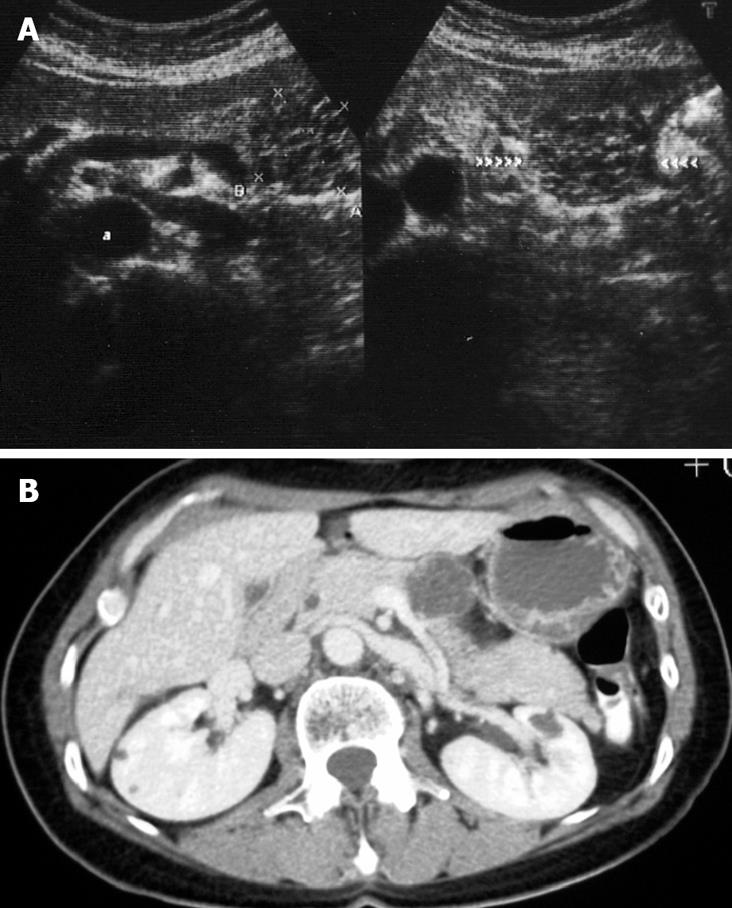
A 49-year-old women presented with increasing upper abdominal pain and nausea in November 2007. She had a past medical history of a uterine myomectomy in 1997, and a hysterectomy and left oophorectomy in 2006. On examination, she was found to be slightly tender at the epigastrium, and laboratory analyses were all within normal limits. An ultrasonography (US) and computer tomography (CT) scan revealed a well-circumscribed 35 mm polycystic lesion in the body of the pancreas, with thin septa within the lesion (Figure 1).
At laparotomy the lesion was found in the lower part of the body of the pancreas, and did not involve the main pancreatic duct. The lesion was completely excised and the main pancreatic duct was preserved. No other pathology was found within the abdomen, and the postoperative recovery was uneventful. Abdominal pain disappeared postoperatively and the patient has been doing well for the following 12 mo until now.
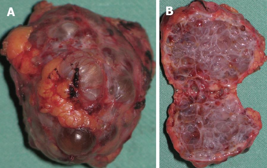
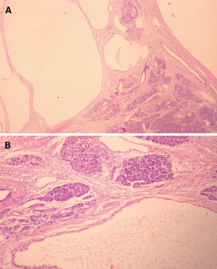
The tumour, measuring 34 mm × 32 mm × 29 mm, had a nodular, gray-blue surface and was surrounded by normal pancreatic tissue (Figure 2A). On sectioning, it had a honeycomb appearance with 1-7 mm polycystic spaces filled with murky haemorrhagic yellowish fluid (Figure 2B). Microscopically, all the sections showed a polycystic structure composed of ectatic lymphatics lined with endothelial cells (Figure 3). The cysts were separated by thin hypocellular septa similar in appearance to the thin capsule surrounding the tumour mass itself. No cell atypia was found. Immunohistochemistry showed immunoreactivity to the factor VIII-R antigen (++), CD 31 positivity (+++) and CD 34 negativity (-). The final histological diagnosis was of microcystic lymphangioma of the pancreas.
Lymphangioma of the pancreas is rare, accounting for less than 1% of lymphangiomas[4]. It occurs more frequently in females and is often located in the distal pancreas[5]. The tumour size may vary between 3 and 20 cm in diameter (average 12 cm)[6]. Patients usually present with abdominal pain[5] and an associated palpable abdominal mass[7-9], although an acute abdomen has also been described[10]. Pancreatitis, weight loss, and laboratory abnormalities are not usual disease manifestations[1]. US typically shows a polycystic tumour, and calcifications, which are typical for cystadenomas of the pancreas, are very rare[11]. On CT, the tumour is a well-circumscribed, encapsulated, water-isodense, polycystic tumour with thin septa, similar in appearance to cystadenomas, which occur far more frequently[1,12].
Differential diagnoses include pancreatic pseudocysts, mucinous and serous cystadenomas, other congenital cysts and pancreatic ductal carcinoma with cystic degeneration[1,13,14]. The final diagnosis is histological[1], with the endothelial cells showing immunohistochemical reactivity to factor VIII/R antigen, CD 31 (+) positivity[6,8] and CD 34 (-) negativity[6], as seen in our patient.
A complete surgical excision is curative[6,8,15], with incomplete excision being the only reason for recurrent disease[7]. Depending on the tumour location and size, complete excision may involve a simple marginal tumorectomy[10] or may require larger pancreatic resections with anastomoses.
| 1. | Gray G, Fried K, Iraci J. Cystic lymphangioma of the pancreas: CT and pathologic findings. Abdom Imaging. 1998;23:78-80. |
| 2. | Khandelwal M, Lichtenstein GR, Morris JB, Furth EE, Long WB. Abdominal lymphangioma masquerading as a pancreatic cystic neoplasm. J Clin Gastroenterol. 1995;20:142-144. |
| 3. | Kullendorff CM, Malmgren N. Cystic abdominal lymphangioma in children. Case report. Eur J Surg. 1993;159:499-501. |
| 4. | Leung TK, Lee CM, Shen LK, Chen YY. Differential diagnosis of cystic lymphangioma of the pancreas based on imaging features. J Formos Med Assoc. 2006;105:512-517. |
| 5. | Gui L, Bigler SA, Subramony C. Lymphangioma of the pancreas with "ovarian-like" mesenchymal stroma: a case report with emphasis on histogenesis. Arch Pathol Lab Med. 2003;127:1513-1516. |
| 6. | Paal E, Thompson LD, Heffess CS. A clinicopathologic and immunohistochemical study of ten pancreatic lymphangiomas and a review of the literature. Cancer. 1998;82:2150-2158. |
| 7. | Daltrey IR, Johnson CD. Cystic lymphangioma of the pancreas. Postgrad Med J. 1996;72:564-566. |
| 8. | Igarashi A, Maruo Y, Ito T, Ohsawa K, Serizawa A, Yabe M, Seki K, Konno H, Nakamura S. Huge cystic lymphangioma of the pancreas: report of a case. Surg Today. 2001;31:743-746. |
| 9. | Murao T, Toda K, Tomiyama Y. Lymphangioma of the pancreas. A case report with electron microscopic observations. Acta Pathol Jpn. 1987;37:503-510. |
| 10. | Itterbeek P, Vanclooster P, de Gheldere C. Cystic lymphangioma of the pancreas: an unusual cause of the acute surgical abdomen. Acta Chir Belg. 1997;97:297-298. |
| 11. | Hanelin LG, Schimmel DH. Lymphangioma of the pancreas exhibiting an unusual pattern of calcification. Radiology. 1977;122:636. |
| 12. | Koenig TR, Loyer EM, Whitman GJ, Raymond AK, Charnsangavej C. Cystic lymphangioma of the pancreas. AJR Am J Roentgenol. 2001;177:1090. |
| 13. | Schneider G, Seidel R, Altmeyer K, Remberger K, Pistorius G, Kramann B, Uder M. Lymphangioma of the pancreas and the duodenal wall: MR imaging findings. Eur Radiol. 2001;11:2232-2235. |
| 14. | Casadei R, Minni F, Selva S, Marrano N, Marrano D. Cystic lymphangioma of the pancreas: anatomoclinical, diagnostic and therapeutic considerations regarding three personal observations and review of the literature. Hepatogastroenterology. 2003;50:1681-1686. |
| 15. | Letoquart JP, Marcorelles P, Lancien G, Pompilio M, Denier P, Leveque J, Procyk S, Haffaf Y, Mambrini A. [A new case of cystic lymphangioma of the pancreas]. J Chir (Paris). 1989;126:650-658. |
Peer reviewer: Massimo Raimondo, PhD, Division of Gastroenterology and Hepatology, Mayo Clinic, 4500 San Pablo Road, Jacksonville, FL 32224, United States
S- Editor Li DL L- Editor Negro F E- Editor Yin DH


