Published online Mar 28, 2008. doi: 10.3748/wjg.14.1903
Revised: February 3, 2008
Published online: March 28, 2008
AIM: To examine the characteristics of colonic polyps, where it is difficult to distinguish adenomatous polyps from hyperplastic polyps, with the aid of acetic acid chromoendoscopy.
METHODS: Acetic acid spray was applied to colonic polyps smaller than 10 mm before complete excision. Endoscopic images were taken before and 15-30 s after the acetic acid spray. Both pre- and post-sprayed images were shown to 16 examiners, who were asked to interpret the lesions as either hyperplastic or adenomatous polyps. Regression analysis was performed to determine which factors were most likely related to diagnostic accuracy.
RESULTS: In 50 cases tested by the 16 examiners, the overall accuracy was 62.4% (499/800). Regression analysis demonstrated that surrounding colonic mucosa was the only factor that was significantly related to accuracy in discriminating adenomatous from hyperplastic polyps (P < 0.001). Accuracy was higher for polyps with linear surrounding colonic mucosa than for those with nodular surrounding colonic mucosa (P < 0.001), but was not related to the shape, location, or size of the polyp.
CONCLUSION: The accuracy of predicting histology is significantly related to the pattern of colonic mucosa surrounding the polyp. Making a histological diagnosis of colon polyps merely by acetic acid spray is helpful for colon polyps with linear, regularly patterned surrounding colonic mucosa, and less so for those with nodular, irregularly patterned surrounding colonic mucosa.
- Citation: Kim JH, Lee SY, Kim BK, Choe WH, Kwon SY, Sung IK, Park HS, Jin CJ. Importance of the surrounding colonic mucosa in distinguishing between hyperplastic and adenomatous polyps during acetic acid chromoendoscopy. World J Gastroenterol 2008; 14(12): 1903-1907
- URL: https://www.wjgnet.com/1007-9327/full/v14/i12/1903.htm
- DOI: https://dx.doi.org/10.3748/wjg.14.1903
The management of neoplastic and non-neoplastic colonic polyps is quite different[1]. Therefore, it is of great interest for a colonoscopist to distinguish them during colonoscopic examination without having to take a biopsy sample. In addition, it takes a few days to establish a histological diagnosis from a biopsy sample, and there is no assurance that the pathological result of the biopsied specimen represents the lesion as a whole[2]. The accuracy of conventional colonoscopy in distinguishing neoplastic from non-neoplastic lesions is reported to be between 68% to 84% even in the hands of experienced colonoscopists[34]. Therefore, several methods are usually required to make the distinction between hyperplastic and adenomatous colonic polyps colonoscopically, such as special techniques like high-resolution chromoendoscopy, magnifying colonoscopy, or narrow band imaging magnification[5–9]. However, these techniques are not used routinely in clinical practice for various reasons, for example. (1) lack of the appropriate equipment, (2) time restrictions, and (3) additional cost not covered by the insurance company.
Acetic acid enables a detailed examination of colonic neoplasms during colonoscopic examination by breaking the disulfide bonds of mucus, thus revealing the detail of the mucosal surface and allowing an analysis of the pit pattern of the colonic polyps[10]. Acetic acid is a cheap, efficient, safe and convenient tool[1112]. Therefore, once its accuracy in distinguishing between neoplastic and non-neoplastic polyps is established, its use might allow a one-stage colon polypectomy. Unfortunately, there are only few published data on the accuracy of acetic acid chromoendoscopy in the diagnosis of colonic polyps, and most of the previous studies were performed in conjunction with other methods such as magnifying endoscopy or with indigo carmine spraying[12].
It would be helpful to colonoscopists if the factors affecting the accuracy in distinguishing between adenomatous and hyperplastic colon polyps were established. In the present study we examined the characteristics of colonic polyps in relation to the surrounding mucosa with the aid of acetic acid chromoendoscopy.
From April to June 2006, 35 patients, in whom colonic polyps were revealed out of 299 routine colonoscopic examinations, were included in the study. Only 35 patients were included in the study since 258 examinations revealed no polyp and 6 revealed polyps only greater than 1 cm. Polyps greater than 1 cm were not included due to the higher probability of associated malignancy requiring removal regardless of the acetic acid chromoendoscopic finding. Colonoscopy was performed by one of the two colonoscopists (JH Kim and SY Lee) at the Digestive Disease Center of Konkuk University Hospital (the colonoscope Olympus CF260, Olympus Corp., Tokyo, Japan, was used). All patients provided written informed consent prior to colonoscopy. None of the 35 patients refused acetic acid chromoendoscopy. This prospective study was approved by the institutional review board of Konkuk University School of Medicine, which agreed that the study was in accordance with the ethical guidelines of the Helsinki Declaration.
When a colon polyp smaller than 1 cm was found, 5-10 mL of 1.5% acetic acid was sprayed onto the lesion from a side channel of the colonoscope. On full air inflation, several endoscopic images were taken before and 15-30 s after spraying. Once the images were taken, the polyps were completely resected either by polypectomy or by cold biopsy sampling. The resected specimens were examined by pathologists, who were unaware of the endoscopic findings.
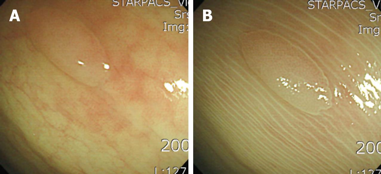
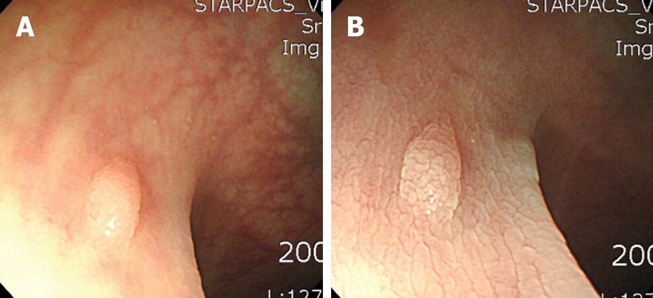
Size and location of the polyps were recorded. With respect to shape, the polyps were classified as either sessile or non-sessile. After being sprayed with acetic acid, the patterns of colonic mucosa surrounding the polyps were classified as having either (1) a linear and regular pattern (Figure 1) or (2) a nodular and irregular pattern (Figure 2).
After the final pathology report, colon polyps other than adenomatous or hyperplastic polyps were excluded from the study. In our 35 patients, a total number of 54 colonic polyps smaller than 10 mm were detected during the colonoscopic examination. However, four cases were excluded since three were inflammatory polyps and one was a leiomyoma. The endoscopic images were selected by the colonoscopist who was not scheduled for the blind test (Lee SY). Finally, endoscopic images of 50 polyps, both pre- and post-sprayed, were collected.
A total of 100 endoscopic images, pre- and post-acetic acid sprayed images of each of the 50 colonic polyps, were shown to 16 examiners (6 gastroenterologists who were not familiar with the colonic pit patterns, 5 residents, and 5 medical students) at the same time in the same place. Before the blind test, a short, 10-min lecture on colonic pit patterns, that included presentation of 24 PowerPoint slides, was given by the colonoscopist who had selected the images for examination (SY Lee). Typical images of mucosal pit patterns in hyperplastic colon polyps (star-like or papillary-like pattern) and adenomatous colon polyps (tubular or gyrus-like pattern) were shown during the lecture[13]. In addition, 10 cases of acetic acid sprayed images of colon polyps were shown to the 16 examiners. None of these images were included in the blind test and all of them were taken in Konkuk University Hospital only colonoscopically without using any other method.
The blind test was performed by showing the PowerPoint slides to the examiners. After examining two images (one pre-acetic acid sprayed image and one post-acetic acid sprayed image) of each of the cases, the examiners were required to record their interpretation as either hyperplastic or adenomatous polyps. After the blind test, the responses were collected, transcribed, and analyzed for raw data associations.
A P-value of less than 0.05 was considered statistically significant. Differences between the groups were analyzed using the chi-square tests and Student’s t-tests. Regarding age and polyp size, results were expressed as mean ± SD. Regression analysis was performed to assess the accuracy of predicting pathology (adenomatous polyp versus hyperplastic polyp). Binary logistic regression analysis was performed with the factors which colonoscopists can notice during the colonoscopic examination, i.e., (1) shape of the polyp, (2) size of the polyp, (3) location of the polyp, and (4) surrounding colonic mucosa.
In the 50 cases tested by 16 examiners, the overall accuracy was 62.4% (499/800). There was no significant difference in test scores between the gastroenterologist group (30.3 ± 2.0, mean ± SD), the resident group (32.4 ± 5.4), and the medical student group (31.0 ± 5.6). Sensitivity, specificity, positive predictive value, and negative predictive value of acetic acid spray for adenomatous polyp were 81.8%, 41.2%, 73.0%, and 53.8%, respectively.
By regression analysis, the pattern of the surrounding colonic mucosa (P < 0.001) was the only factor predicting pathology (i.e. adenomatous polyp versus hyperplastic polyp). In 34 cases (68%), the colonic mucosa surrounding the polyp was linear and regular (Figure 1), and in 16 cases (32%) nodular and irregular (Figure 2). In contrast, the accuracy of distinguishing between hyperplastic and adenomatous colon polyps was not related to the shape, location, or size of the polyp.
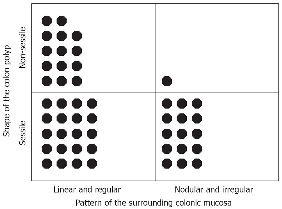
Neither age nor sex of the patient was related to the pattern of colonic mucosa surrounding the polyp (Table 1). The only related factor was the shape of the polyp (P = 0.02; Figure 3). Whereas size and location of the polyp were irrelevant.
| Linear and regular (n = 34) | Nodular and irregular (n = 16) | P value | |
| Size of the polyp (mm, mean ± SD) | 5.29 ± 2.18 | 4.75 ± 2.27 | NS |
| Shape of the polyp | 0.023 | ||
| Sessile | 20 (58.8) | 15 (93.7) | |
| Non-sessile | 14 (41.2) | 1 (6.3) | |
| Location of the polyp | NS | ||
| Cecum | 2 (5.9) | 0 (0.0) | |
| Ascending colon | 5 (14.7) | 2 (12.4) | |
| Hepatic flexure | 3 (8.8) | 0 (0.0) | |
| Transverse colon | 1 (2.9) | 1 (6.3) | |
| Splenic flexure | 0 (0.0) | 1 (6.3) | |
| Descending colon | 4 (11.8) | 1 (6.3) | |
| Sigmoid colon | 12 (35.3) | 8 (50.0) | |
| Rectum | 7 (20.6) | 3 (18.7) | |
| Pathology | NS | ||
| Hyperplastic polyp | 10 (29.4) | 7 (43.8) | |
| Adenomatous polyp | 24 (70.6) | 9 (56.2) |
To the best of our knowledge, this study is the first to evaluate the significance of the type of colonic mucosa surrounding a colon polyp for distinguishing between adenomatous and hyperplastic polyps during acetic acid chromoendoscopy. Our findings revealed that linear and regularly patterned surrounding colonic mucosa enables a higher accuracy in predicting the pathology of the associated polyp when compared to those surrounded by nodular and irregularly patterned colonic mucosa.
Although the reasons for these two different surrounding colonic mucosal patterns are unclear, the nodular pattern seems to be similar to that seen in acetic acid sprayed gastric mucosa, which is considered to be indicative of chronic mucosal damage induced either by H pylori infection or by acid irritation (unpublished data). In the present study, the pattern of the mucosa surrounding a polyp was associated with the shape of the polyp, the non-sessile type being found more frequently in linear patterned surrounding colonic mucosa. However, this should be evaluated further by a large-scale study in conjunction with pathological analysis.
Most of the previous studies on acetic acid chromo-endoscopy have examined the significance of detecting sessile polyps or analyzed polyp pit patterns[510–12]. In the present study, we did not classify colonic adenomas according to their degree of dysplasia, but simply assessed the accuracy of acetic acid chromoendoscopy, which is a cheap, easy, convenient, safe and fast procedure, in distinguishing between adenomatous and hyperplastic colon polyps. We therefore analyzed only the mucosal pit patterns, and not the microvascular pattern, which is automatically masked by spraying with acetic acid.
It has been reported that residents are able to safely and effectively screen for colorectal neoplasms with a flexible sigmoidoscope when supervised[14]. Interestingly, there was no significant difference in the blind test scores between the gastroenterologists, residents, and medical students. This indicates that the results achieved using acetic acid chromoendoscopy are easy to interpret, even for those who have no experience in gastrointestinal endoscopy. However, it also indicates that although uniform descriptions of colonic mucosal pit patterns in hyperplastic colon polyps (star-like or papillary-like pattern) and in adenomatous colon polyps (tubular or gyrus-like pattern) are useful, they are not completely visualized merely by acetic acid chromoendoscopy.
The limitation of our study is that this was the data from16 examiners, who were not familiar with the colonic pit patterns, predicted the pathology only on the basis of 2 pictures (pre- and post-sprayed images) not by full colonoscopic examination. Moreover, no additional methods such as magnifying endoscopy or indigo carmine spray were used. Therefore, the overall diagnostic accuracy for distinguishing between neoplastic and non-neoplastic lesions was lower than previous studies which were done by experienced colonoscopists[3915].
In conclusion, acetic acid chromoendoscopy can be used to distinguish between hyperplastic and adenomatous polyps without magnifying endoscopy with an accuracy of 62.4%, and the accuracy is significantly related to the pattern of colonic mucosa surrounding the polyp. In addition, we have found that making a histological diagnosis of colon polyps merely by acetic acid spray is helpful for colon polyps with linear, regularly patterned surrounding colonic mucosa, and less so for those with nodular, irregularly patterned surrounding colonic mucosa.
The management of neoplastic and non-neoplastic colonic polyps is quite different, and it is of great interest for a colonoscopist to distinguish them during colonoscopic examination without having to take a biopsy sample. Acetic acid is a cheap, efficient, safe and convenient tool which enables a detailed examination of colonic neoplasms during colonoscopic examination by breaking the disulfide bonds of mucus. Acetic acid chromoendoscopy is effective in revealing the detail of the mucosal surface and allowing an analysis of the pit pattern of the colonic polyps.
Recently, several endoscopic methods that help to make the distinction between hyperplastic and adenomatous colonic polyps colonoscopically have been introduced. Special techniques such as high-resolution chromoendoscopy, magnifying colonoscopy, or narrow band imaging magnification are being innovated.
This is the first study that evaluated the significance of the type of colonic mucosa surrounding a colon polyp for distinguishing between adenomatous and hyperplastic polyps during acetic acid chromoendoscopy. Our findings revealed that linear and regularly patterned surrounding colonic mucosa enables a higher accuracy in predicting the pathology of the associated polyp when compared to those surrounded by nodular and irregularly patterned colonic mucosa.
Through this study, we showed a new way to distinguish between neoplastic and non-neoplastic polyps by acetic acid chromoendoscopy which is a cheap, easy, convenient, safe and fast procedure. The patterns of surrounding colonic mucosa will help colonoscopists in distinguishing between adenomatous and hyperplastic colon polyps.
Acetic acid chromoendoscopy is a method done by spraying 5-10 mL of 1.5% acetic acid onto the lesion from a side channel of the colonoscope. Endoscopic images are usually taken before and 15-30 s after spraying.
This study interpreted the lesions as either hyperplastic or adenomatous polyps after acetic acid chromoendoscopy, and proved that making a histological diagnosis of colon polyps merely by acetic acid spray is helpful for colon polyps with linear, regularly patterned surrounding colonic mucosa, and less so for those with nodular, irregularly patterned surrounding colonic mucosa.
| 1. | Winawer SJ, Zauber AG, Fletcher RH, Stillman JS, O’brien MJ, Levin B, Smith RA, Lieberman DA, Burt RW, Levin TR. Guidelines for colonoscopy surveillance after polypectomy: a consensus update by the US Multi-Society Task Force on Colorectal Cancer and the American Cancer Society. CA Cancer J Clin. 2006;56:143-159; quiz 184-185. |
| 2. | Sasajima K, Kudo SE, Inoue H, Takeuchi T, Kashida H, Hidaka E, Kawachi H, Sakashita M, Tanaka J, Shiokawa A. Real-time in vivo virtual histology of colorectal lesions when using the endocytoscopy system. Gastrointest Endosc. 2006;63:1010-1017. |
| 3. | Konishi K, Kaneko K, Kurahashi T, Yamamoto T, Kushima M, Kanda A, Tajiri H, Mitamura K. A comparison of magnifying and nonmagnifying colonoscopy for diagnosis of colorectal polyps: A prospective study. Gastrointest Endosc. 2003;57:48-53. |
| 4. | Fu KI, Sano Y, Kato S, Fujii T, Nagashima F, Yoshino T, Okuno T, Yoshida S, Fujimori T. Chromoendoscopy using indigo carmine dye spraying with magnifying observation is the most reliable method for differential diagnosis between non-neoplastic and neoplastic colorectal lesions: a prospective study. Endoscopy. 2004;36:1089-1093. |
| 5. | Apel D, Jakobs R, Schilling D, Weickert U, Teichmann J, Bohrer MH, Riemann JF. Accuracy of high-resolution chromoendoscopy in prediction of histologic findings in diminutive lesions of the rectosigmoid. Gastrointest Endosc. 2006;63:824-828. |
| 6. | Kato S, Fu KI, Sano Y, Fujii T, Saito Y, Matsuda T, Koba I, Yoshida S, Fujimori T. Magnifying colonoscopy as a non-biopsy technique for differential diagnosis of non-neoplastic and neoplastic lesions. World J Gastroenterol. 2006;12:1416-1420. |
| 7. | Tanaka S, Oka S, Hirata M, Yoshida S, Kaneko I, Chayama K. Pit pattern diagnosis for colorectal neoplasia using narrow band imaging magnification. Dig Endosc. 2006;18:52-56. |
| 8. | Axelrad AM, Fleischer DE, Geller AJ, Nguyen CC, Lewis JH, Al-Kawas FH, Avigan MI, Montgomery EA, Benjamin SB. High-resolution chromoendoscopy for the diagnosis of diminutive colon polyps: implications for colon cancer screening. Gastroenterology. 1996;110:1253-1258. |
| 9. | Eisen GM, Kim CY, Fleischer DE, Kozarek RA, Carr-Locke DL, Li TC, Gostout CJ, Heller SJ, Montgomery EA, Al-Kawas FH. High-resolution chromoendoscopy for classifying colonic polyps: a multicenter study. Gastrointest Endosc. 2002;55:687-694. |
| 10. | Lambert R, Rey JF, Sankaranarayanan R. Magnification and chromoscopy with the acetic acid test. Endoscopy. 2003;35:437-445. |
| 11. | Canto MI. Acetic-acid chromoendoscopy for Barrett's esophagus: the "pros". Gastrointest Endosc. 2006;64:13-16. |
| 12. | Togashi K, Hewett DG, Whitaker DA, Hume GE, Francis L, Appleyard MN. The use of acetic acid in magnification chromocolonoscopy for pit pattern analysis of small polyps. Endoscopy. 2006;38:613-616. |
| 13. | Kudo S, Tamura S, Nakajima T, Yamano H, Kusaka H, Watanabe H. Diagnosis of colorectal tumorous lesions by magnifying endoscopy. Gastrointest Endosc. 1996;44:8-14. |
| 14. | Mullins RJ, Whitworth PW, Polk HC Jr. Screening before surgery for colon neoplasms with a flexible sigmoidoscope by surgical residents. Ann Surg. 1987;205:659-664. |
| 15. | De Palma GD, Rega M, Masone S, Persico M, Siciliano S, Addeo P, Persico G. Conventional colonoscopy and magnified chromoendoscopy for the endoscopic histological prediction of diminutive colorectal polyps: a single operator study. World J Gastroenterol. 2006;12:2402-2405. |


