Published online Feb 14, 2007. doi: 10.3748/wjg.v13.i6.978
Revised: December 25, 2006
Accepted: January 22, 2007
Published online: February 14, 2007
We report a case of sigmoid colon perforation in a patient with Crohn’s disease undergoing computed-tomographic (CT) colonography. A 70-year-old patient with Crohn’s disease with terminal ileitis and sigmoid stricture underwent CT colonography after incomplete conventional colonoscopy. During the procedure, the colon was inflated by air insufflation and the patient developed abdominal pain with radiological evidence of retroperitoneal and intraperitoneal free gas. Hartmann’s operation was performed. This case highlights that CT colonography is not risk-free. The risk of perforation may be higher in patients with inflammatory bowel disease.
- Citation: Wong SH, Wong VW, Sung JJ. Virtual colonoscopy-induced perforation in a patient with Crohn’s disease. World J Gastroenterol 2007; 13(6): 978-979
- URL: https://www.wjgnet.com/1007-9327/full/v13/i6/978.htm
- DOI: https://dx.doi.org/10.3748/wjg.v13.i6.978
Computed-tomographic (CT) colonography is a relatively non-invasive tool to examine the colon. It was once thought to be almost risk-free when compared to conventional colonoscopy. However, cases of colonic perforation during CT colonography were reported in normal subjects undergoing primary screening of colonic carcinoma. It is obvious that CT colonography carries a low but definite risk of colonic perforation. A few cases of perforation were also reported in diseased colons, as in patients suffering from ulcerative colitis and colonic carcinoma. In this article, we report a case of colonic perforation in a patient with Crohn’s disease undergoing CT colonography.
A 70-year-old gentleman suffered from Crohn’s disease for more than 10 years. He presented with chronic loose stool diarrhea. He did not have per-rectal bleeding, abdominal pain or other extra-intestinal symptoms. His body weight remained static at around 42 kg. Colonoscopy in 2005 showed a stricture at the sigmoid colon with aphthous ulcers, and a barium enema follow-through study showed terminal ileal narrowing. Tissue biopsy of the colon showed active chronic colitis with hyperplastic crypts consistent with Crohn’s disease.
Since 2004, the patient had worsened diarrhea and progressive weight loss to 40 kg. His hemoglobin dropped from 13.1 to 12.0 g/dL. Plasma C-reactive protein increased to 33.5 mg/L. He was treated with sulphasalazine and iron supplements. A regular colonoscopy was arranged to assess disease extent and to rule out colorectal cancer, but the endoscope failed to advance beyond the tight sigmoid stricture even using a pediatric colonoscope.
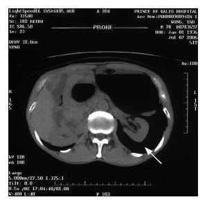
The patient was admitted to our hospital for computed tomographic (CT) colonography on 7 July 2006 for further evaluation. A 20-French Foley catheter without balloon inflation was inserted with the tip in the rectum, and air was introduced into the large bowel through manual insufflation. Immediately after air insufflation, the patient complained of lower abdominal pain. Plain CT film demonstrated large amounts of free retroperitoneal air and a small amount of intraperitoneal free gas (Figure 1).
The patient was diagnosed to have iatrogenic colonic perforation. During emergency surgery, a 3 mm perforation at the anti-mesenteric side of proximal sigmoid was noticed, and there was a tight stricture at descending colon just 1 cm proximal to the perforation. A left hemicolectomy was performed to resect the stricture and perforated segment. The distal transverse colon was brought out as end colostomy. He subsequently had uneventful recovery apart from transient perioperative atrial fibrillation.
CT colonography is a relatively non-invasive imaging technique to examine the colon. Adequate bowel preparation is essential. During the procedure, air or carbon dioxide is insufflated through a rectal catheter. Helical CT scans of the abdomen and pelvis during a breath-hold are then performed to give a 3-dimensional ‘fly-through’ reconstruction, allowing examination of the colonic surface with a simulated endoscopic view.
CT colonography has been most extensively evaluated for the detection of colonic polyps and cancer. In 1233 asymptomatic patients in the United States, the sensitivity and specificity of CT colonography in detecting adenomatous polyps of at least 10 mm in size were 92 percent and 96 percent, respectively[1]. Less commonly, CT colonography has also been utilized in patients with Crohn's disease. The greatest advantage of CT colonography lies in patients with strictures, adhesions or obstructing tumors which prevent complete colonoscopic examination. Studies have shown that CT colonography can detect thickened bowel wall and deep ulcers in patients with Crohn’s disease[2].
Since CT colonography does not involve the insertion of an endoscope, it is generally perceived as a safe procedure. The International Working Group on Virtual Colonography reviewed a total of 21 923 CT colonography procedures and reported only 2 cases of colonic perforations with a rate of 0.009%[3]. Nevertheless, it is noteworthy that most of the reported series involve people with normal colon, some undergoing primary screening only. In patients with colonic diseases, the diseased colonic wall may not accommodate a high intraluminal pressure as well as a normal colon. In addition, strictures may prevent the even distribution of the air insufflated, resulting in an increased intraluminal pressure locally. This may have accounted for the close proximity between the perforation site and stricture in our patient. There are case reports of colonic perforations in patients with ulcerative colitis[4] or carcinoma of the colon[5].
In conclusion, our case highlights that CT colonography is not risk-free. The risk of perforation may be higher in patients with inflammatory bowel disease. Prospective studies should be conducted to further evaluate the risk of CT colonography in these patients.
| 1. | Pickhardt PJ, Choi JR, Hwang I, Butler JA, Puckett ML, Hildebrandt HA, Wong RK, Nugent PA, Mysliwiec PA, Schindler WR. Computed tomographic virtual colonoscopy to screen for colorectal neoplasia in asymptomatic adults. N Engl J Med. 2003;349:2191-2200. [RCA] [PubMed] [DOI] [Full Text] [Cited by in Crossref: 1495] [Cited by in RCA: 1292] [Article Influence: 56.2] [Reference Citation Analysis (0)] |
| 2. | Tarján Z, Zágoni T, Györke T, Mester A, Karlinger K, Makó EK. Spiral CT colonography in inflammatory bowel disease. Eur J Radiol. 2000;35:193-198. [RCA] [PubMed] [DOI] [Full Text] [Cited by in Crossref: 33] [Cited by in RCA: 28] [Article Influence: 1.1] [Reference Citation Analysis (0)] |
| 3. | Pickhardt PJ. Incidence of colonic perforation at CT colonography: review of existing data and implications for screening of asymptomatic adults. Radiology. 2006;239:313-316. [RCA] [PubMed] [DOI] [Full Text] [Cited by in Crossref: 169] [Cited by in RCA: 140] [Article Influence: 7.0] [Reference Citation Analysis (0)] |
| 4. | Coady-Fariborzian L, Angel LP, Procaccino JA. Perforated colon secondary to virtual colonoscopy: report of a case. Dis Colon Rectum. 2004;47:1247-1249. [RCA] [PubMed] [DOI] [Full Text] [Cited by in Crossref: 54] [Cited by in RCA: 48] [Article Influence: 2.2] [Reference Citation Analysis (0)] |
| 5. | Kamar M, Portnoy O, Bar-Dayan A, Amitai M, Munz Y, Ayalon A, Zmora O. Actual colonic perforation in virtual colonoscopy: report of a case. Dis Colon Rectum. 2004;47:1242-1244; discussion 1244-1246;. [RCA] [PubMed] [DOI] [Full Text] [Cited by in Crossref: 56] [Cited by in RCA: 52] [Article Influence: 2.4] [Reference Citation Analysis (0)] |
S- Editor Liu Y L- Editor Zhu LH E- Editor Lu W


