INTRODUCTION
Lipomas are the most common nonepithelial benign tumors of the gastrointestinal tract. Nevertheless symptoms of a colonic lipoma are rare generally with a silent clinical course. When colonic lipomas achieve a proper size, they have manifestations such as change in bowel habits, rectal bleeding, abdominal pain or more disastrous consequences like obstruction and intussusception requiring urgent interventions. Herein, we report four patients suffering from various sizes of colonic lipoma who were treated with different theraupatic modalities.
CASE REPORT
The first patient was a 77-year old female who presented with recurrent episodes of right upper quadrant abdominal cramping. Her physical examination and previous medical history were unremarkable except for a cholecystectomy. Standard laboratory values were within normal ranges. Abdominal computed tomography (CT) revealed a regular contoured 4 cm × 3 cm mass lesion with fatty density localized in the midportion of ascending colon causing a luminal narrowing defect (Figure 1A). In double contrast enema, a filling defect of the protruded polypoid lesion was detected at the same location (Figure 1B). Colonoscopy demonstrated a mass lesion with a broad base and normal overlying mucosa (about 4 cm in diameter) adjacent to the hepatic flexura. The patient was diagnosed having a symptomatic colonic lipoma during surgery. Following colotomy, mucosa overlying the mass lesion was dissected and a tumor with a macroscopic appearance similar to lipoma was enucleated (Figure 1C). Histopathologic examination verified the diagnosis of lipoma localized in the submucosal layer and the patient remained asymptomatic through a 2-year follow-up period.
Figure 1 Computed tomography (CT) scan showing a regular contoured 4 cm x 3 cm lesion with fatty density localised in midportion of ascending colon causing a luminal narrowing defect (A), double contrast barium enema showing a filling defect of the protruded polipoid lesion at the hepatic flexura of colon (B), and appearance of the colonic submucosal lipoma during the operation (C).
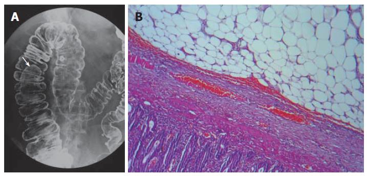
The second patient was a 41-year old male admitted to our clinic with similar complaints of the first patient. Physical and laboratory examinations were natural only with a positive fecal occult blood test. He denied any significant medical or surgical history. Abdominal CT demonstrated a regular contoured 3 cm × 2 cm mass lesion with fatty density localized in the hepatic flexura causing a filling defect which was confirmed by double contrast enema (Figure 2A). A segmental resection was performed with the diagnosis of lipoma, pathological evaluation of the specimen showed that it was a benign submucosal lipoma (Figure 2B).
Figure 2 A mass lesion located in the hepatic flexura causing filling defect shown by double contrast enema (A), resected biopsy specimen showing a submucosal benign lipoma composed of mature lipocytes by hematoxylin and eosin staining (B) (x 400).
The third patient was a 77-year old female presented with recurrent abdominal pain in the left lower quadrant for a year, bloody defecation for 6 mo and constipation for 4 wk. Physical examination revealed no pathological change except for left lower quadrant distention. Blood biochemical analysis, sedimentation rate and complete blood count were normal. Colonoscopy revealed a broad-based giant mass acquiring spontaneous hemorrhagic areas and obstructing more than 75% of the colonic lumen (Figure 3A). Punch biopsy exhibited benign colonic mucosa showing inflammatory granulation. Abdominal CT showed diffuse thickening of the sigmoid colon wall, invagination and an intraluminal mass with a size of approximately 6 cm × 7 cm having density values equal to fat (Figure 3B). An evident sigmoid redundancy and a mass lesion nearly obliterating colonic lumen were detected during surgery. A segmenter resection of the sigmoid colon was the operative procedure of choice (Figure 3C). Microscopic examination confirmed that the lesion was a submucosal lipoma with a size of 6 cm × 7 cm.
Figure 3 Colonoscopic appearance of a broad- based giant mass acquiring spontaneous hemorrhagic areas and obstructing more than 75% of the lumen of sigmoid colon (A); abdominal computed tomography scans showing the diffuse thickening of sigmoid colon wall, invagination and a distal intraluminal giant mass with fat density with a size of approximately 6 cm x 7 cm (B); macroscopic appearance of the giant sigmoid colon lipoma (C).
The fourth patient was a 44-year old male presented with tenesmus. Physical examination revealed nothing significant. A pedinculated polypoid mass lesion projecting into the lumen with an approximate diameter of 2.5 cm was discovered at the 15th cm of the rectum by colonoscopy. An endoscopic snare polypectomy and rectal biopsy were performed during the same intervention. The histopathologic examination revealed normal rectal mucosa and underlocated lipoma.
DISCUSSION
Lipoma of the gastrointestinal tract was first described by Bauer et al[1] in 1757. Lipoma is the second most common benign colonic tumor following adenomatous polyps and the incidence has been reported to range between 0.2% and 4.4%[2,3]. Colonic lipoma is more common in elderly women and tends to derive from right hemicolon with a decreasing frequency from cecum to sigmoid colon[2-5]. Approximately in 90% of cases, lipoma is defined to arise from the submucosal layer and the subserosal or intermucosal layer accounting for the remaining 10%[1,6]. They are usually solitary with varying sizes and may be sessile or pedunculated. Although the majority of these lesions are asymptomatic and detected incidentally during the examination of symptoms like abdominal pain, change in bowel habits, and rectal bleeding or in surgical specimen removed for various other reasons, on rare occasions colonic lipoma may present with massive hemorrhage, obstruction, perforation, intussusception, or prolapse[7-11]. Severity of the signs and symptoms is attributed to the size of the lesions. Lipomas larger than 2 cm in diameter may cause symptoms such as constipation, diarrhea, abdominal pain, or rectal bleeding[1,3,5]. Colicky pain may be due to intermittent intussusception whereas rectal bleeding can occur as a result of ulceration of the overlying mucosa. One of the greatest clinical significances of lipoma is its potential to be confused with colonic malignancies according to the similarity in both symptomatology, fortunately sarcomatous changes in colonic lipomas have not been reported yet[1]. Colonoscopy, CT and barium enema are considered to be the diagnostic tools, but pyhsicians should be aware that colonoscopic biopsies usually possess no histopathologic value as the lesion is beneath the normal mucosa and biopsy often can not promote diagnosis[12]. The radiographic appearance of colonic lipomas may resemble that of carcinomas. The “squeeze sign” is told to be pathognomonic for colonic lipomas, and is characterized by the elongation of a spherical filling defect during peristalsis on barium enema examination[1,13]. Despite recent diagnostic innovations in radiology, histopathologic evaluation is the gold standard in precise diagnosis. However, most colonic lipomas are asymptomatic incidental entities, the decision of removal is based on the criteria including those with suspicion of malignancy, and symptomatic lipomas. A wide range of treatment modalities have been suggested. The way of removal depends on the presentation of the case as an elective or an emergency. Surgical intervention is mandatory in surgical emergencies such as obstruction, intussusception, perforation, or very rarely massive hemorrhage. Elective endoscopic removal of submucosal lipoma up to 2 cm in diameter is reported to be safe and appropriate, however in larger lesions the risk of associated complications like uncontrolled hemorrhage and perforation makes the procedure controversial[1,14-16]. Conventional laparotomy including enucleation, colotomy and excision, and segmental colonic resection has been described as a choice of treatment as well as minilaparotomy or transanal resection of lesions mimicking rectal prolapse[1,7,16]. Minimally invasive surgeries such as laparoscopy -assisted resection under colonoscopic guidance has also been defined for selected cases[15,17].
In the literature, the general consensus on colonic lipomas is that local enucleation is an appropriate and effective procedure of choice for the management of these lesions possessing no malignant potantial. Colonoscopic removal can be safely performed for submucosal lipomas smaller than 2.5 cm in diameter, however larger or subserosal tumors associated with complication risks of obstruction, intussusception, hemorrhage and even perforation deserve extended interventions. Surgical approach should be established through the features of each case on individual basis.


