Liver Cancer Open Access
Copyright ©2005 Baishideng Publishing Group Inc. All rights reserved.
World J Gastroenterol. May 7, 2005; 11(17): 2564-2569
Published online May 7, 2005. doi: 10.3748/wjg.v11.i17.2564
Alpha-fetoprotein triggers hepatoma cells escaping from immune surveillance through altering the expression of Fas/FasL and tumor necrosis factor related apoptosis-inducing ligand and its receptor of lymphocytes and liver cancer cells
Meng-Sen Li, Qiu-Ling Ma, Qian Chen, Department of Biochemistry, Hainan Medical College, Haikou 571101, Hainan Province, China
Xin-Hua Liu, Ping-Feng Li, Guo-Guang Du, Gang Li, Department of Biochemistry and Molecular Biology, Peking University Health Science Center, Beijing 100083, China
Author contributions: All authors contributed equally to the work.
Correspondence to: Dr. Gang Li, Department of Biochemistry and Molecular Biology, Peking University Health Science Center, Beijing 100083, China. mengsenli@163.com
Telephone: +86-10-82802891
Received: June 7, 2004
Revised: June 8, 2004
Accepted: June 17, 2004
Published online: May 7, 2005

Abstract

AIM: To investigate the mechanism of α-fetoprotein (AFP) in escaping from the host immune surveillance of hepatoc-ellular carcinoma.

METHODS: AFP purified from human umbilical blood was administrated into the cultured human lymphoma Jurkat T cell line or hepatoma cell line, Bel7402 in vitro. The expression of tumor necrosis factor related apoptosis-inducing ligand (TRAIL) and its receptor (TRAILR) mRNA were analyzed by Northern blot and Western blot was used to detect the expression of Fas and Fas ligand (FasL) protein.

RESULTS: AFP (20 mg/L) could promote the expression of FasL and TRAIL, and inhibit the expression of Fas and TRAILR of Bel7402 cells. For Jurkat cell line, AFP could suppress the expression of FasL and TRAIL, and stimulate the expression of Fas and TRAILR. AFP also could synergize with Bel7402 cells to inhibit the expression of FasL protein and TRAIL mRNA in Jurkat cells. The monoclonal antibody against AFP (anti-AFP) could abolish these functions of AFP.

CONCLUSION: AFP is able to promote the expression of FasL and TRAIL in hepatoma cells and enhance the expression of Fas and TRAILR in lymphocytes. These could elicit the escape of hepatocellular carcinoma cells from the host’s lymphocytes immune surveillance.

Key Words: Alpha-fetoprotein; Hepatocellular carcinoma; Immune escape


INTRODUCTION

Though a lot of investigations were carried out on the function of α-fetoprotein (AFP), the biological role of AFP is still indistinct so far. Because the number and sequence of amino acid of AFP are very similar to those of human serum albumin (HSA), it is thought that the biological function of AFP just like HSA, plays an important role in transporting materials and regulating osmotic pressure in human blood. During the course of embryo development, the serum concentration of AFP is higher (3 g/L). The concentration of AFP falls down to a low level that it is hard to detect (<20 ng/L) when 2 years birth after. However, the expressed quantities of AFP would rise abnormally when an individual suffers from liver tumor or benign hepatic growth[1,2]. Therefore, it is reasonable to imagine that the high exp-ression of AFP is related to the proliferation of tumor or normal cells. Previous researches have shown that AFP could promote the proliferation of different types of tumor or normal cells when a sole administration or in combination with other growth factors[3,8]. Since AFP is a kind of biological macromolecule (MW: 69 ku), it is impossible to enter cells directly to regulate biological responses of the cells. Our research revealed that AFP could regulate cell growth was mediated by its receptor, which exists on the membrane of cells. Some studies also showed that AFP could inhibit immune responses mediated by its receptors[2,9,10]. The cells secrete plenty of AFP during the development of liver cancer. The potential role of AFP in the proliferation of tumor cells is still unclear though AFP has been thought as an important marker of hepatoma. It was traditionally considered that AFP was a substance accompanied with the development of liver cancer, so it was regarded as a diagnostic standard of liver cancer. However, recent studies indicated that AFP was a kind of protein with some biological activities, such as promoting the growth of cells and escaping from the host’s immune monitor of liver cancer cells in vivo. Some proteins, such as Fas as well as its natural ligand (FasL protein) and TRAIL as well as TRAILR play an important role in escaping from immune surveillance of liver cancer cells in vivo[11-14]. In the present investigation, we observed the influence of AFP on the expression of TRAIL/TRAILR or Fas/FasL and explored the possible mechanism of AFP in the growth and immune escape of liver cancer cells.

MATERIALS AND METHODS
Materials

Bel 7402 cells and Jurkat cells were presented by the Depar-tment of Cell Biology, Peking University Health Science Center (PR China). Purified AFP was purchased from Sigma (USA). Monoclonal antibody against AFP (anti-AFP) used to block AFP was offered by the Department of Biochemistry and Molecular Biology, Peking University Health Science Center (PR China). HSA and RPMI-1640 were purchased from Gibco (USA). Monoclonal antibodies against Fas and FasL were purchased from Santa Cruz (USA). Total RNA extract reagent kit and the random primer-a-gene labeling system reagent kit were products from Promage Company (Madison, WI, USA). α-32P-dCTP was bought from Yahui Biological Engineering Company (Beijing, China). TRAIL, TRAILR (DR4) and β-actin cDNA probe were presented by the Department of Endocrinology, Northwestern University (Chicago, USA). Salmon fish sperm DNA, fraction V of bovine serum albumin (BSA) and Ficoll-400 were purchased from the Jingke Chemical Reagents Company (Beijing, PR China).

Methods

Cell culture Bel7402 cells and Jurkat cells were maintained in RPMI 1640 medium supplemented with 10% fetal calf serum (FCS) at 37 °C in a humidified atmosphere of 50 mL/L CO2. Bel7402 cells were digested and scattered by using 0.25% trypsin containing 0.02% EDTA, and then continuously cultured. The cultured medium was replaced at every 24 h.

Northern blot analysis of TRAIL and TRAILR expression Cells were treated with AFP (20 mg/L), anti-AFP (40 mg/L) or AFP (20 mg/L) plus anti-AFP (40 mg/L) for 12 h. Total cellular RNA was isolated from Bel 7402 cells and Jurkat cells with the TRIzol reagent kit according to the manufacturer’s protocol. RNA (10-20 μg) was isolated by electrophoresis through a 1% formaldehyde agarose gel, and transferred (in 20×SSC) onto the nitrocellulose membranes (Millipore Corporation Bedford, MA, USA) with a standard procedure[15]. The membranes were hybridized with α-32P labeled probes and then exposed to X-ray films at -80 °C.

Western blot detected the expression of Fas and FasL Cells were treated with either AFP (20 mg/L), HSA (20 mg/L), anti-AFP (40 mg/L) or AFP (20 mg/L) plus anti-AFP (40 mg/L) for 24 h. After washing thrice with PBS (pH 7.4, 0.15 mol/L), the cells in each group were lyzed with 10 µL of lysis buffer containing 0.2% Triton X-100, 500 mmol/L NaCl, 500 mmol/L sucrose, 1.0 mmol/L EDTA, 0.15 mmol/L spermine, 0.5 mmol/L spermidine, 10 mmol/L HEPES (pH 8.0), 200 µmol/L phenylmethylsulfonyl fluoride, 2.0 mg/L leupeptin, 2.0 mg/L pepstatin, 24000 IU/L aprotinin and 7.0 mmol/L β-mercaptoethanol. Forty or twenty micrograms of proteins were subjected to sodium dodecyl sulfate-polyacrylamide gel electrophoresis (SDS-PAGE) and transferred onto the PVDF membrane for immunodetection. Molecular weight markers (Sigma) verified the correct locations of visualized bands. The membranes were blocked with 50 g/L nonfat milk in PBS-Tween, then probed with anti-Fas or anti-FasL followed by second antibody (goat anti-rabbit IgG-horse radish peroxidase). Immunoreactive proteins were detected using Western blotting chemiluminescence luminol reagent developing systems. The results of Western blot were analyzed by a standard procedure[15].

Statistical analysis

Data were analyzed by Student’s t test and expressed as mean±SD based on three independent experiments.

RESULTS
Influence of AFP on the expression of Fas and FasL protein in human Bel 7402 cells and Jurkat cells

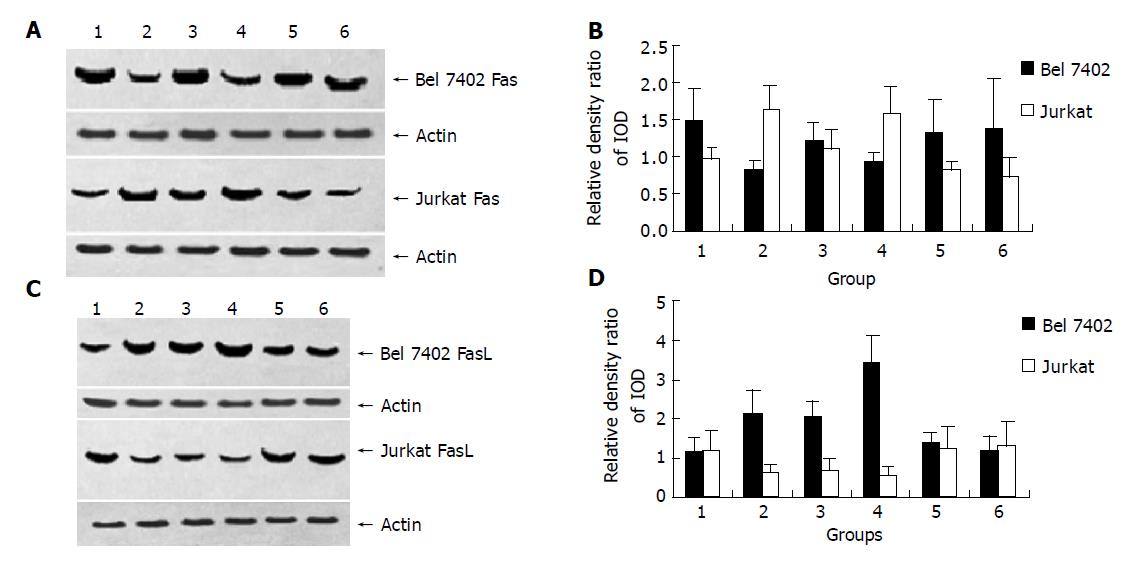
After being treated with AFP (20 mg/L), anti-AFP (40 mg/L), AFP (20 mg/L) plus anti-AFP (40 mg/L) and HSA (20 mg/L) for 24 h, AFP promoted Bel 7402 cells to express FasL and inhibit Fas protein express significantly. For Jurkat cells, AFP suppressed the expression of FasL protein, but stimulated the expression of Fas protein. When Bel7402 cells and Jurkat cells were co-incubated, AFP co-operated with Bel7402 cells to inhibit Jurkat cells to express FasL protein. HSA and anti-AFP had no significant influence on the expression of Fas and FasL protein in the two types of cells, but anti-AFP had a role in blocking the function of AFP (Figures 1 and 2).

Figure 1
Figure 1 Influence of AFP (20 mg/L), anti-AFP (40 mg/L), AFP (20 mg/L) plus anti-AFP (40 mg/L) and HSA (20 mg/L) on the expression of Fas and FasL protein in Bel7402 cells and Jurkat cells. A, C: Western blot of Fas and FasL protein of Bel7402 cells or of Jurkat cells; B, D: Quantitative analysis of IOD of Fas/IOD of actin, the columns represent mean±SD.
Figure 2
Figure 2 Effect of AFP (20 mg/L), anti-AFP (40 mg/L) and AFP (20 mg/L) plus anti-AFP (40 mg/L) on the expression of Fas and FasL protein in Bel7402 cells or in Jurkat when the cells were cultured individually or co-cultured. A and C: Western blot analysis of Fas and FasL protein of Bel7402 cells or of Jurkat cells; B and D: Quantitative analysis IOD of Fas/IOD of actin, the columns represent mean±SD.

Figure 1, Lane 1: Control group; lane 2: AFP treated groups; lane 3: Anti-AFP (40 mg/L) treated groups; lane 4: AFP (20 mg/L) plus Anti-AFP (40 mg/L) treated groups; lane 5: HSA (20 mg/L) treated groups.

Figure 2, Lane 1: Control groups (cells were cultured individually, respectively); lane 2: AFP treated groups (cells were cultured individually, respectively); lane 3: Cells were co-cultured groups; lane 4: Cells were co-cultured and treated with AFP group; lane 5: Cells were co-cultured and treated with anti-AFP group; lane 6: Cells were co-cultured and treated with AFP plus anti-AFP group.

Effects of AFP on the expression of TRAIL and TRAILR mRNA of human hepatoma Bel 7402 cells and Jurkat cells

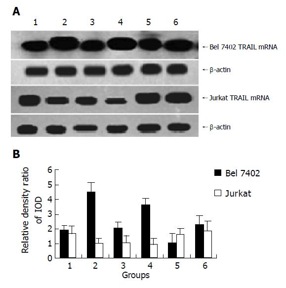
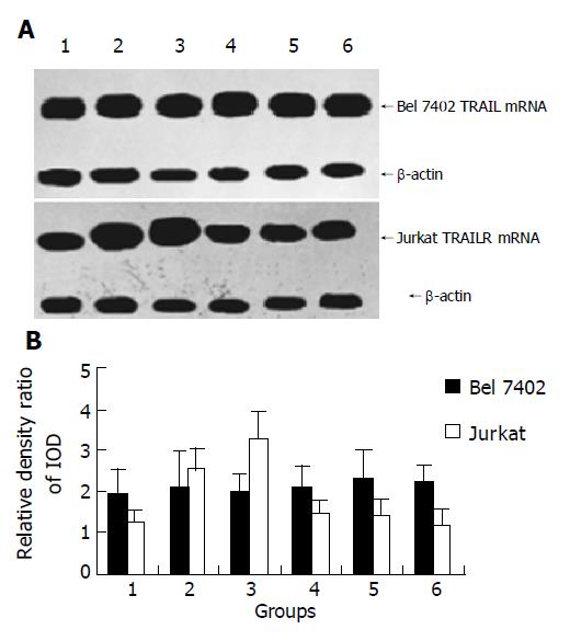
After being treated with AFP (20 mg/L), anti-AFP (40 mg/L), AFP (20 mg/L) plus anti-AFP (40 mg/L) for 12 h, it showed that AFP could enhanced the expression of TRAIL mRNA apparently, but no significant influence on the expression of TRAILR mRNA was detected in Bel7402 cells. The role of AFP in Jurkat cells indicated that APF could inhibit the expression of TRAIL mRNA and promote the expression of TRAILR mRNA. When Bel7402 cells and Jurkat cells were co-incubated, AFP functioned co-operatively with Bel 7402 cells to restrain the expression of TRAIL mRNA in Jurkat cells, AFP could not synergize with Bel 7402 cells to stimulate the expression of TRAILR mRNA in Jurkat cells. Anti-AFP had no significant influence on the expression of TRAIL mRNA and TRAILR mRNA in the two types of cells, but it could prohibit the function of AFP (Figures 3 and 4).

Figure 3
Figure 3 Effect of AFP (20 mg/L), anti-AFP (40 mg/L) and AFP (20 mg/L) plus anti-AFP (40 mg/L) on the TRAIL mRNA expression of Bel7402 cells and Jurkat cells were cultured individually or cultured together. A: Northern blot of TRAIL mRNA of Bel7402 cells and Jurkat cells; B: Quantitative analysis IOD of TRAIL/IOD of β-actin, the columns represent mean±SD.
Figure 4
Figure 4 Influence of AFP (20 mg/L), anti-AFP (40 mg/L) and AFP (20 mg/L) plus anti-AFP (40 mg/L) on the expression of TRAILR mRNA in Bel7402 cells and Jurkat cells were cultured individually or cultured together. A: Northern blot analysis of TRAILR mRNA of Bel7402 cells and Jurkat cells; B: Quantitative analysis IOD of TRAILR/IOD of β-actin, the columns represent mean±SD.

Figure 3, lane 1: Control groups (cells were cultured individually, respectively); lane 2: AFP treated groups (cells were cultured individually, respectively); lane 3: Cells were co-cultured groups; lane 4: Cells were co-cultured and treated with AFP group; lane 5: Cells were co-cultured and treated with anti-AFP group; lane 6: Cells were co-cultured and treated with AFP plus anti-AFP group.

Figure 4, Lane 1: Control groups (cells were cultured individually, respectively); lane 2: AFP treated groups (cells were cultured individually, respectively); lane 3: Cells were co-cultured groups; lane 4: Cells were co-cultured and treated with AFP group; lane 5: Cells were co-cultured and treated with anti-AFP group; lane 6: Cells were co-cultured and treated with AFP plus anti-AFP group.

DISCUSSION

It has been well known that AFP plays a role in inhibiting immunity rejection and cellular immune response during embryo development[16,17]. The synthesis and secretion of AFP always accompany liver cell growth. Researches have proved that AFP could promote the proliferation of various tumor cells, such as liver cancer cells[3,6]. For tumor cell growth in vivo, one of the mechanisms is to escape from immune surveillance. Lymphocytes play a very important role in inhibiting the proliferation of tumor cells. Previous investigations have confirmed the inhibitory effect of AFP on lymphocyte responses of cancer patients[18-20].

It has been known that escape of AFP from the immunity monitors may lead to the proliferation and survival of cancer cells. Researches have documented that AFP has a capability of regulating the growth of various cells. However, the mechanism of AFP in controlling the growth and immune escape of liver cancer cells is unclear. Formerly, it was thought that AFP could carry unsaturated fatty acids, which are essential nutrients for cell growth, AFP in this manner may promote the metabolism of unsaturated fatty acids to yield inositol triphosphate (IP3) which can affect the signal transduction of cells[21]. As a result, liver cancer cells are maintained to grow in vivo. The present research showed that AFP could promote the expression of FasL and TRAIL, and suppress the expression of Fas in hepatoma cells, but the expression of TRAILR was not significantly influenced, and an opposite role of AFP in Jurkat lymphocytes was indicated. The fact that AFP could inhibit the expression of TRAIL and FasL, and promote the expression of Fas and TRAILR when Jurkat cells and Bel 7402 cells were co-incubated, the results also indicated AFP synergism with Bel7402 cells to restrain the expression of FasL and TRAIL in Jurkat cells. FasL and Fas are a pair of systems that can induce cell death (apoptosis). Fas is defined as the death receptor and may induce the apoptotic process of cells by combining with its natural ligand FasL[22-25]. There is evidence that Bcl-2 protein displays a reciprocal pattern of expression during the development of lymphocytes. Bcl-2 family is a participant in the pathway that leads to cell apoptosis by mediating the signals from “death receptors” known as Fas on the cell surface, and Bcl-2 could affect the apoptosis of hepatocellular cancer cells through Fas[26,27], but this apoptosis-inducing effect has non-specificity of cells. Tumor cells can escape from the attack of lymphocytes by changing its Fas/FasL system[28,29]. TRAIL is mainly expressed in activated lymphocytes and can combine specifically with its receptor to induce apoptosis of tumor cells, but not for normal cells[30-33]. Lots of researches have found that the FasL/Fas system plays a very important role in promoting apoptosis and escaping from host immune surveillance of hepatocellular cancer cells[34-38]. The high expression of Fas in hepatocellular cancer cells can be beneficial to the growth of tumor cells[37,39]. Hepatocellular cancer cells could suppress the infiltrated lymphocytes to express FasL, which triggers hepatoma cells to escape from the immunity attack of lymphocytes[12,40,41]. The present results showed that AFP not only reduced Fas expression in cancer cells, which could suppress the attack of lymphocytes, but also promoted the expression of FasL in cancer cells and the expression of Fas in lymphocytes. It may be speculated that FasL of tumor cells may induce apoptosis of lymphocytes by combining its Fas. In the present study, FasL of Bel7402 cells could bind to Fas of Jurkat cells that lead the lymphocytes to apoptosis. This may be one of the mechanisms of hepatocellular cancer cells escaping from host immune surveillance.

The reason why TRAIL could induce tumor cell apoptosis is complicated. It has been reported that TRAIL may induce cell apoptosis through its five kinds of receptors[42-44]. Recently, it has been found that the effect of TRAIL was mediated through death receptors DR4 and DR5 on the membrane of tumor cells, which could combine with TRAIL to trigger caspase signal transduction pathways and induce tumor cell apoptosis[45]. Some studies have verified that TRAIL gene transferring adenoviral vector system could inhibit proliferation of human hepatoma SMMC7721 cells and induce cell apoptosis, TRAIL has been considered as an anticancer cytokine, which may generate a novel strategy for the treatment of hepatocellular carcinoma. Our data showed that AFP could enhance the expression of TRAIL, and had no obvious influence on the expression of DR4 in Bel7402 cells. On the other hand, the effect of AFP in Jurkat cells showed that it could promote the expression of DR4 and suppress the expression of TRAIL. The co-operative effect of the level of DR4 elevated by AFP in Jurkat cells and elevated TRAIL in Bel 7402 cells could facilitate apoptosis of infiltrated lymphocytes. Thus, AFP secreted from liver cancer cells could play a role in protecting itself in escaping from immune attack during the growth of liver cancer cells. In our previous studies, AFP could enhance tumor cell growth and the expression of some oncogenes[46-49]. The present study manifested that liver cancer cells persisted malignant proliferation in vivo, and secreted AFP which played a role not only in promoting cancer cells to cleavage, but also in triggering hepatoma cells to escape from host immune surveillance by altering the expression of TRAIL/TRAILR and Fas/FasL in hepatocellular cancer cells or in Jurkat lymphocytes.

Some researches showed that AFP was not necessary for the embryo development, but only required for female fertility[50]. A recent study revealed that AFP’s DNA vaccine had a distinctive antitumor immunopreventive effect on AFP-producing tumors. Some studies and our previous researches[46-49] have discovered that AFP could promote cancer cell growth by influenced cAMP and Ca2+ mediated signal transduction pathways[3,5,46-49]. AFP could cause apoptosis of cancer cells by activating caspase-3-like proteases rather than Fas[52]. These results showed that AFP had a complex biological function. It indicated that AFP could regulate the growth of tumor cells and lymphocytes by various intracellular signal pathways and at multiple regulation levels[17,52,53]. In this study, HSA as a negative control and mono-clonal antibody against AFP (anti-AFP) could not notably influence the expression of TRAIL/TRAILR and Fas/FasL in Bel7402 cells or in Jurkat cells. However, the fact that anti-AFP could block the function of AFP these indicates the high specificity of AFP in regulating the expression of TRAIL/TRAILR and Fas/FasL in the two types of cells. The present study showed that AFP could affect the expression of apoptosis related-genes of lymphocytes and hepatoma cells to maintain cancer cell growth in vivo. The precise mechanism of AFP contributes to a better understanding of the physiological effect of AFP on regulating tumor cell growth and escaping from host immune surveillance of liver cancer cells.

Footnotes
References
1.  Parasole R, Izzo F, Perrone F, Pignata S, Galati MG, Leonardi E, Castiglione F, Orlando R, Castello G, Esposito G. Prognostic value of serum biological markers in patients with hepatocellular carcinoma. Clin Cancer Res. 2001;7:3504-3509.  [PubMed]  [DOI]  [Cited in This Article: ]
2.  Deutsch HF. Chemistry and biology of alpha-fetoprotein. Adv Cancer Res. 1991;56:253-312.  [PubMed]  [DOI]  [Cited in This Article: ]  [Cited by in Crossref: 127]  [Cited by in F6Publishing: 126]  [Article Influence: 3.8]  [Reference Citation Analysis (0)]
3.  Dudich E, Semenkova L, Gorbatova E, Dudich I, Khromykh L, Tatulov E, Grechko G, Sukhikh G. Growth-regulative activity of human alpha-fetoprotein for different types of tumor and normal cells. Tumour Biol. 1998;19:30-40.  [PubMed]  [DOI]  [Cited in This Article: ]  [Cited by in Crossref: 62]  [Cited by in F6Publishing: 69]  [Article Influence: 2.9]  [Reference Citation Analysis (0)]
4.  Wang XW, Xie H. Alpha-fetoprotein enhances the proliferation of human hepatoma cells in vitro. Life Sci. 1999;64:17-23.  [PubMed]  [DOI]  [Cited in This Article: ]  [Cited by in Crossref: 73]  [Cited by in F6Publishing: 82]  [Article Influence: 3.3]  [Reference Citation Analysis (0)]
5.  Wang XW, Xu B. Stimulation of tumor-cell growth by alpha-fetoprotein. Int J Cancer. 1998;75:596-599.  [PubMed]  [DOI]  [Cited in This Article: ]  [Cited by in F6Publishing: 7]  [Reference Citation Analysis (0)]
6.  Li MS, Li PF, Zhou AR, Li G, Du GG. Growth factor like activity of α-fetoprotein in human hepatoma cell line, Bel7402 and HeLa cell (abstract). Endo 2000 the Endocrine Society 82nd Annual Meeting Toronto, Canada. 2000;P143.  [PubMed]  [DOI]  [Cited in This Article: ]
7.  Leal JA, Gangrade BK, Kiser JL, May JV, Keel BA. Human mammary tumor cell proliferation: primary role of platelet-derived growth factor and possible synergism with human alpha-fetoprotein. Steroids. 1991;56:247-251.  [PubMed]  [DOI]  [Cited in This Article: ]  [Cited by in Crossref: 15]  [Cited by in F6Publishing: 18]  [Article Influence: 0.5]  [Reference Citation Analysis (0)]
8.  Keel BA, Eddy KB, Cho S, May JV. Synergistic action of purified alpha-fetoprotein and growth factors on the proliferation of porcine granulosa cells in monolayer culture. Endocrinology. 1991;129:217-225.  [PubMed]  [DOI]  [Cited in This Article: ]  [Cited by in Crossref: 37]  [Cited by in F6Publishing: 41]  [Article Influence: 1.2]  [Reference Citation Analysis (0)]
9.  Naval J, Villacampa MJ, Goguel AF, Uriel J. Cell-type-specific receptors for alpha-fetoprotein in a mouse T-lymphoma cell line. Proc Natl Acad Sci USA. 1985;82:3301-3305.  [PubMed]  [DOI]  [Cited in This Article: ]  [Cited by in Crossref: 50]  [Cited by in F6Publishing: 55]  [Article Influence: 1.4]  [Reference Citation Analysis (0)]
10.  Torres JM, Geuskens M, Uriel J. Receptor-mediated endocytosis and recycling of alpha-fetoprotein in human B-lymphoma and T-leukemia cells. Int J Cancer. 1991;47:110-117.  [PubMed]  [DOI]  [Cited in This Article: ]  [Cited by in Crossref: 27]  [Cited by in F6Publishing: 27]  [Article Influence: 0.8]  [Reference Citation Analysis (0)]
11.  Nagao M, Nakajima Y, Hisanaga M, Kayagaki N, Kanehiro H, Aomatsu Y, Ko S, Yagita H, Yamada T, Okumura K. The alteration of Fas receptor and ligand system in hepatocellular carcinomas: how do hepatoma cells escape from the host immune surveillance in vivo? Hepatology. 1999;30:413-421.  [PubMed]  [DOI]  [Cited in This Article: ]  [Cited by in Crossref: 72]  [Cited by in F6Publishing: 82]  [Article Influence: 3.3]  [Reference Citation Analysis (0)]
12.  Taimr P, Higuchi H, Kocova E, Rippe RA, Friedman S, Gores GJ. Activated stellate cells express the TRAIL receptor-2/death receptor-5 and undergo TRAIL-mediated apoptosis. Hepatology. 2003;37:87-95.  [PubMed]  [DOI]  [Cited in This Article: ]  [Cited by in Crossref: 169]  [Cited by in F6Publishing: 165]  [Article Influence: 7.9]  [Reference Citation Analysis (0)]
13.  Shin EC, Ahn JM, Kim CH, Choi Y, Ahn YS, Kim H, Kim SJ, Park JH. IFN-gamma induces cell death in human hepatoma cells through a TRAIL/death receptor-mediated apoptotic pathway. Int J Cancer. 2001;93:262-268.  [PubMed]  [DOI]  [Cited in This Article: ]  [Cited by in Crossref: 64]  [Cited by in F6Publishing: 68]  [Article Influence: 3.0]  [Reference Citation Analysis (0)]
14.  Kaplan MJ, Lewis EE, Shelden EA, Somers E, Pavlic R, McCune WJ, Richardson BC. The apoptotic ligands TRAIL, TWEAK, and Fas ligand mediate monocyte death induced by autologous lupus T cells. J Immunol. 2002;169:6020-6029.  [PubMed]  [DOI]  [Cited in This Article: ]  [Cited by in Crossref: 132]  [Cited by in F6Publishing: 142]  [Article Influence: 6.5]  [Reference Citation Analysis (0)]
15.  Sambrook J, Fritsch EF, Maniatis T. Molecular cloning: a laboratory manual, 4nd edition. New York: Cold Spring Harbor Laboratory Press. 2001;141-160.  [PubMed]  [DOI]  [Cited in This Article: ]
16.  Semeniuk DJ, Boismenu R, Tam J, Weissenhofer W, Murgita RA. Evidence that immunosuppression is an intrinsic property of the alpha-fetoprotein molecule. Adv Exp Med Biol. 1995;383:255-269.  [PubMed]  [DOI]  [Cited in This Article: ]  [Cited by in Crossref: 30]  [Cited by in F6Publishing: 35]  [Article Influence: 1.2]  [Reference Citation Analysis (0)]
17.  Nunez EA. Biological role of alpha-fetoprotein in the endocrinological field: data and hypotheses. Tumour Biol. 1994;15:63-72.  [PubMed]  [DOI]  [Cited in This Article: ]  [Cited by in Crossref: 32]  [Cited by in F6Publishing: 34]  [Article Influence: 1.1]  [Reference Citation Analysis (0)]
18.  Chakraborty M, Mandal C. Immuno-suppressive effect of human alphafetoprotein: a cross species study. Immunol Invest. 1993;22:329-339.  [PubMed]  [DOI]  [Cited in This Article: ]  [Cited by in Crossref: 20]  [Cited by in F6Publishing: 21]  [Article Influence: 0.7]  [Reference Citation Analysis (0)]
19.  Bei R, Budillon A, Reale MG, Capuano G, Pomponi D, Budillon G, Frati L, Muraro R. Cryptic epitopes on alpha-fetoprotein induce spontaneous immune responses in hepatocellular carcinoma, liver cirrhosis, and chronic hepatitis patients. Cancer Res. 1999;59:5471-5474.  [PubMed]  [DOI]  [Cited in This Article: ]
20.  Mizejewski GJ. Alpha-fetoprotein structure and function: relevance to isoforms, epitopes, and conformational variants. Exp Biol Med (Maywood). 2001;226:377-408.  [PubMed]  [DOI]  [Cited in This Article: ]
21.  Bevan S, Wood JN. Arachidonic-acid metabolites as second messengers. Nature. 1987;328:20.  [PubMed]  [DOI]  [Cited in This Article: ]  [Cited by in Crossref: 35]  [Cited by in F6Publishing: 38]  [Article Influence: 1.0]  [Reference Citation Analysis (0)]
22.  Walczak H, Krammer PH. The CD95 (APO-1/Fas) and the TRAIL (APO-2L) apoptosis systems. Exp Cell Res. 2000;256:58-66.  [PubMed]  [DOI]  [Cited in This Article: ]  [Cited by in Crossref: 446]  [Cited by in F6Publishing: 434]  [Article Influence: 18.1]  [Reference Citation Analysis (0)]
23.  Irmler M, Thome M, Hahne M, Schneider P, Hofmann K, Steiner V, Bodmer JL, Schröter M, Burns K, Mattmann C. Inhibition of death receptor signals by cellular FLIP. Nature. 1997;388:190-195.  [PubMed]  [DOI]  [Cited in This Article: ]  [Cited by in Crossref: 1947]  [Cited by in F6Publishing: 1909]  [Article Influence: 70.7]  [Reference Citation Analysis (0)]
24.  Patel T. Immune escape in hepatocellular cancer: is a good offense the best defense? Hepatology. 1999;30:576-578.  [PubMed]  [DOI]  [Cited in This Article: ]  [Cited by in Crossref: 9]  [Cited by in F6Publishing: 10]  [Article Influence: 0.4]  [Reference Citation Analysis (0)]
25.  Bodey B, Bodey B, Siegel SE, Kaiser HE. Fas (Apo-1, CD95) receptor expression in childhood astrocytomas. Is it a marker of the major apoptotic pathway or a signaling receptor for immune escape of neoplastic cells? In Vivo. 1999;13:357-373.  [PubMed]  [DOI]  [Cited in This Article: ]
26.  Chao DT, Korsmeyer SJ. BCL-2 family: regulators of cell death. Annu Rev Immunol. 1998;16:395-419.  [PubMed]  [DOI]  [Cited in This Article: ]  [Cited by in Crossref: 1249]  [Cited by in F6Publishing: 1263]  [Article Influence: 48.6]  [Reference Citation Analysis (0)]
27.  Yin XM, Wang K, Gross A, Zhao Y, Zinkel S, Klocke B, Roth KA, Korsmeyer SJ. Bid-deficient mice are resistant to Fas-induced hepatocellular apoptosis. Nature. 1999;400:886-891.  [PubMed]  [DOI]  [Cited in This Article: ]  [Cited by in Crossref: 795]  [Cited by in F6Publishing: 770]  [Article Influence: 30.8]  [Reference Citation Analysis (0)]
28.  Reichmann E. The biological role of the Fas/FasL system during tumor formation and progression. Semin Cancer Biol. 2002;12:309-315.  [PubMed]  [DOI]  [Cited in This Article: ]  [Cited by in Crossref: 80]  [Cited by in F6Publishing: 85]  [Article Influence: 3.9]  [Reference Citation Analysis (0)]
29.  Hahne M, Rimoldi D, Schröter M, Romero P, Schreier M, French LE, Schneider P, Bornand T, Fontana A, Lienard D. Melanoma cell expression of Fas(Apo-1/CD95) ligand: implications for tumor immune escape. Science. 1996;274:1363-1366.  [PubMed]  [DOI]  [Cited in This Article: ]  [Cited by in Crossref: 942]  [Cited by in F6Publishing: 982]  [Article Influence: 35.1]  [Reference Citation Analysis (0)]
30.  Wiley SR, Schooley K, Smolak PJ, Din WS, Huang CP, Nicholl JK, Sutherland GR, Smith TD, Rauch C, Smith CA. Identification and characterization of a new member of the TNF family that induces apoptosis. Immunity. 1995;3:673-682.  [PubMed]  [DOI]  [Cited in This Article: ]  [Cited by in Crossref: 2187]  [Cited by in F6Publishing: 2185]  [Article Influence: 75.3]  [Reference Citation Analysis (0)]
31.  Renshaw SA, Parmar JS, Singleton V, Rowe SJ, Dockrell DH, Dower SK, Bingle CD, Chilvers ER, Whyte MK. Acceleration of human neutrophil apoptosis by TRAIL. J Immunol. 2003;170:1027-1033.  [PubMed]  [DOI]  [Cited in This Article: ]  [Cited by in Crossref: 130]  [Cited by in F6Publishing: 140]  [Article Influence: 6.7]  [Reference Citation Analysis (0)]
32.  Smyth MJ, Takeda K, Hayakawa Y, Peschon JJ, van den Brink MR, Yagita H. Nature's TRAIL--on a path to cancer immunotherapy. Immunity. 2003;18:1-6.  [PubMed]  [DOI]  [Cited in This Article: ]  [Cited by in Crossref: 250]  [Cited by in F6Publishing: 259]  [Article Influence: 12.3]  [Reference Citation Analysis (0)]
33.  Gura T. How TRAIL kills cancer cells, but not normal cells. Science. 1997;277:768.  [PubMed]  [DOI]  [Cited in This Article: ]  [Cited by in Crossref: 131]  [Cited by in F6Publishing: 141]  [Article Influence: 5.2]  [Reference Citation Analysis (0)]
34.  Jiang S, Song MJ, Shin EC, Lee MO, Kim SJ, Park JH. Apoptosis in human hepatoma cell lines by chemotherapeutic drugs via Fas-dependent and Fas-independent pathways. Hepatology. 1999;29:101-110.  [PubMed]  [DOI]  [Cited in This Article: ]  [Cited by in Crossref: 68]  [Cited by in F6Publishing: 72]  [Article Influence: 2.9]  [Reference Citation Analysis (0)]
35.  Nakamoto Y, Kaneko S, Buttner SW, Matsushita E, Kobayashi K. Inhibition of peripheral blood lymphocyte apoptosis by soluble fas ligand in patients with hepatocellular carcinoma. Oncol Rep. 1999;6:733-739.  [PubMed]  [DOI]  [Cited in This Article: ]
36.  Ito Y, Monden M, Takeda T, Eguchi H, Umeshita K, Nagano H, Nakamori S, Dono K, Sakon M, Nakamura M. The status of Fas and Fas ligand expression can predict recurrence of hepatocellular carcinoma. Br J Cancer. 2000;82:1211-1217.  [PubMed]  [DOI]  [Cited in This Article: ]
37.  Chang YC, Xu YH. Expression of Bcl-2 inhibited Fas-mediated apoptosis in human hepatocellular carcinoma BEL-7404 cells. Cell Res. 2000;10:233-242.  [PubMed]  [DOI]  [Cited in This Article: ]  [Cited by in Crossref: 22]  [Cited by in F6Publishing: 24]  [Article Influence: 1.0]  [Reference Citation Analysis (0)]
38.  Lee SH, Shin MS, Lee HS, Bae JH, Lee HK, Kim HS, Kim SY, Jang JJ, Joo M, Kang YK. Expression of Fas and Fas-related molecules in human hepatocellular carcinoma. Hum Pathol. 2001;32:250-256.  [PubMed]  [DOI]  [Cited in This Article: ]  [Cited by in Crossref: 71]  [Cited by in F6Publishing: 78]  [Article Influence: 3.4]  [Reference Citation Analysis (0)]
39.  Okano H, Shiraki K, Inoue H, Kawakita T, Saitou Y, Enokimura N, Yamamoto N, Sugimoto K, Murata K, Nakano T. Fas stimulation activates NF-kappaB in SK-Hep1 hepatocellular carcinoma cells. Oncol Rep. 2003;10:1145-1148.  [PubMed]  [DOI]  [Cited in This Article: ]
40.  Yuen MF, Hughes RD, Heneghan MA, Langley PG, Norris S. Expression of Fas antigen (CD95) in peripheral blood lymphocytes and in liver-infiltrating, cytotoxic lymphocytes in patients with hepatocellular carcinoma. Cancer. 2001;92:2136-2141.  [PubMed]  [DOI]  [Cited in This Article: ]  [Cited by in F6Publishing: 1]  [Reference Citation Analysis (0)]
41.  Fukuzawa K, Takahashi K, Furuta K, Tagaya T, Ishikawa T, Wada K, Omoto Y, Koji T, Kakumu S. Expression of fas/fas ligand (fasL) and its involvement in infiltrating lymphocytes in hepatocellular carcinoma (HCC). J Gastroenterol. 2001;36:681-688.  [PubMed]  [DOI]  [Cited in This Article: ]  [Cited by in Crossref: 38]  [Cited by in F6Publishing: 45]  [Article Influence: 2.0]  [Reference Citation Analysis (0)]
42.  Pan G, O'Rourke K, Chinnaiyan AM, Gentz R, Ebner R, Ni J, Dixit VM. The receptor for the cytotoxic ligand TRAIL. Science. 1997;276:111-113.  [PubMed]  [DOI]  [Cited in This Article: ]  [Cited by in Crossref: 1333]  [Cited by in F6Publishing: 1335]  [Article Influence: 49.4]  [Reference Citation Analysis (0)]
43.  Golstein P. Cell death: TRAIL and its receptors. Curr Biol. 1997;7:R750-R753.  [PubMed]  [DOI]  [Cited in This Article: ]  [Cited by in Crossref: 144]  [Cited by in F6Publishing: 151]  [Article Influence: 5.6]  [Reference Citation Analysis (0)]
44.  Mongkolsapaya J, Cowper AE, Xu XN, Morris G, McMichael AJ, Bell JI, Screaton GR. Lymphocyte inhibitor of TRAIL (TNF-related apoptosis-inducing ligand): a new receptor protecting lymphocytes from the death ligand TRAIL. J Immunol. 1998;160:3-6.  [PubMed]  [DOI]  [Cited in This Article: ]
45.  Schmaltz C, Alpdogan O, Kappel BJ, Muriglan SJ, Rotolo JA, Ongchin J, Willis LM, Greenberg AS, Eng JM, Crawford JM. T cells require TRAIL for optimal graft-versus-tumor activity. Nat Med. 2002;8:1433-1437.  [PubMed]  [DOI]  [Cited in This Article: ]  [Cited by in Crossref: 113]  [Cited by in F6Publishing: 113]  [Article Influence: 5.1]  [Reference Citation Analysis (0)]
46.  Li MS, Li PF, Li G, Du GG. Enhancement of proliferation of HeLa cells by the alpha-fetoprotein. ShengWu HuaXue Yu ShengWu WuLi XueBao (Shanghai). 2002;34:769-774.  [PubMed]  [DOI]  [Cited in This Article: ]
47.  Li MS, Li PF, Yang FY, He SP, Du GG, Li G. The intracellular mechanism of alpha-fetoprotein promoting the proliferation of NIH 3T3 cells. Cell Res. 2002;12:151-156.  [PubMed]  [DOI]  [Cited in This Article: ]  [Cited by in Crossref: 59]  [Cited by in F6Publishing: 66]  [Article Influence: 3.0]  [Reference Citation Analysis (0)]
48.  Li MS, Li PF, Du GG, L G. The enhancement effects of alpha-fetoprotein on the expression on N-ras and p53 and p21ras in HeLa cells. Chin J Biochem Mol Biol. 2002;18:750-754.  [PubMed]  [DOI]  [Cited in This Article: ]
49.  Li MS, Li PF, He SP, Du GG, Li G. The properties and mediated cAMP-PKA signal transduction pathway of alpha-fetoprotein receptor. Chin J Biochem Mol Biol. 2004;20:101-106.  [PubMed]  [DOI]  [Cited in This Article: ]
50.  Gabant P, Forrester L, Nichols J, Van Reeth T, De Mees C, Pajack B, Watt A, Smitz J, Alexandre H, Szpirer C. Alpha-fetoprotein, the major fetal serum protein, is not essential for embryonic development but is required for female fertility. Proc Natl Acad Sci USA. 2002;99:12865-12870.  [PubMed]  [DOI]  [Cited in This Article: ]  [Cited by in Crossref: 76]  [Cited by in F6Publishing: 78]  [Article Influence: 3.5]  [Reference Citation Analysis (0)]
51.  Dudich E, Semenkova L, Dudich I, Gorbatova E, Tochtamisheva N, Tatulov E, Nikolaeva M, Sukhikh G. alpha-fetoprotein causes apoptosis in tumor cells via a pathway independent of CD95, TNFR1 and TNFR2 through activation of caspase-3-like proteases. Eur J Biochem. 1999;266:750-761.  [PubMed]  [DOI]  [Cited in This Article: ]  [Cited by in Crossref: 43]  [Cited by in F6Publishing: 47]  [Article Influence: 1.9]  [Reference Citation Analysis (0)]
52.  Semenkova L, Dudich E, Dudich I, Tokhtamisheva N, Tatulov E, Okruzhnov Y, Garcia-Foncillas J, Palop-Cubillo JA, Korpela T. Alpha-fetoprotein positively regulates cytochrome c-mediated caspase activation and apoptosome complex formation. Eur J Biochem. 2003;270:4388-4399.  [PubMed]  [DOI]  [Cited in This Article: ]  [Cited by in Crossref: 32]  [Cited by in F6Publishing: 35]  [Article Influence: 1.7]  [Reference Citation Analysis (0)]
53.  Hanke P, Rabe C, Serwe M, Böhm S, Pagenstecher C, Sauerbruch T, Caselmann WH. Cirrhotic patients with or without hepatocellular carcinoma harbour AFP-specific T-lymphocytes that can be activated in vitro by human alpha-fetoprotein. Scand J Gastroenterol. 2002;37:949-955.  [PubMed]  [DOI]  [Cited in This Article: ]  [Cited by in Crossref: 40]  [Cited by in F6Publishing: 42]  [Article Influence: 1.9]  [Reference Citation Analysis (0)]