Published online Mar 28, 2005. doi: 10.3748/wjg.v11.i12.1884
Revised: October 20, 2004
Accepted: November 19, 2004
Published online: March 28, 2005
A diagnosis of small-bowel perforation, caused by a sharp or pointed foreign body, is rarely made preoperatively because the clinical symptoms are usually nonspecific and can mimic other surgical conditions, such as appendicitis and diverticulitis. We report the case of a 62-year-old woman who experienced severe pain in the right iliac fossa and fever for about five days before arrival at our hospital. The presumptive diagnosis was acute purulent appendicitis and an emergency appendectomy was planned. Swelling and erythema were noted in a segment of the small bowel in the lower right abdomen. A tiny pointed object was found penetrating the inflamed portion of the bowel, which proved to be a sharp fish bone (gray snapper). The bone was removed, followed by segmental resection of the terminal ileum and ascending colon. The postoperative course was uneventful.
- Citation: Hsu SD, Chan DC, Liu YC. Small-bowel perforation caused by fish bone. World J Gastroenterol 2005; 11(12): 1884-1885
- URL: https://www.wjgnet.com/1007-9327/full/v11/i12/1884.htm
- DOI: https://dx.doi.org/10.3748/wjg.v11.i12.1884
A variety of foreign bodies are seen on abdominal radiographs in emergency departments. Most foreign-body ingestion is accidental, but there may be contributory factors such as mental disorder, bulimia, alcoholism, and prison incarceration. When foreign bodies are ingested, they usually pass spontaneously through the entire alimentary tract and out in the feces. Perforation of the gastrointestinal tract is a well-recognized complication of foreign-body ingestion and the ileum is the most common site of perforation[1]. We report the case of a fish bone perforating the distal ileum, resulting in a clinical presentation mimicking acute appendicitis.
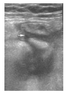
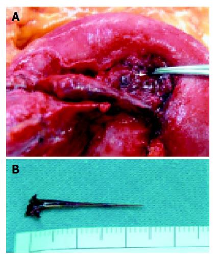
A 62-year-old woman, with no previous abdominal complaints, was admitted to our emergency department with acute abdominal pain in the lower right quadrant for the preceding five days. The pain was initially generalized, but later localized to the lower right quadrant, worsening in intensity. There was no nausea, vomiting, or diarrhea. Physical examination revealed a body temperature of 38.7 °C. An abdominal examination showed localized tenderness in the lower right quadrant with rebound and voluntary guarding. Laboratory tests indicated an elevated white cell count of 16500 with 81.9% neutrophils, elevated C-reactive protein (123 mg/L), and normal serum amylase (33 U/L). Urinalysis was normal. Other laboratory data were within normal limits. A plain X-ray of the abdomen showed local ileus in the lower right abdomen. Sonography of whole abdomen revealed fluid accumulated over the lower right quadrant (Figure 1). The presumptive diagnosis was acute purulent appendicitis and an emergency appendectomy was planned. Para-median exploratory laparotomy revealed turbid peritoneal fluid at the right paracolic gutter. However, the appendix was normal on inspection. The loop of the ileum in the right lower abdomen was swollen and showed evidence of an erythematous change. On examination of the small bowel, a tiny sharp object was seen penetrating the wall in the inflamed region. The foreign body was grasped, pulled out directly from the bowel, and removed through the abdomen (Figures 2A, B). Due to severe inflammatory changes and gangrene of the terminal ileum, segmental resection of the terminal ileum and ascending colon was performed. End-to-side iliac-colonic anastomosis was also performed, with subsequent irrigation of the purulent fluid and cleaning of the pelvic area. A closed-wound suction drainage tube was placed in the pelvis. The final pathological diagnosis was small-bowel perforation caused by a foreign body (fish bone) with extra-mural abscess formation and fibropurulent serositis. The patient had an uneventful postoperative course and was discharged eight days after the procedure.
Foreign-body ingestion is a common clinical problem presenting in emergency departments. Perforation of the gastrointestinal tract by ingested foreign bodies is uncommon and less than 1% of ingested foreign bodies perforate the bowel. The types of foreign bodies ingested depend on the dietary habits of the relevant countries. Madrona et al[1], reported that chicken bones are the most common foreign bodies causing gastrointestinal tract perforation. Fish bones are the most commonly ingested foreign bodies in Hong Kong[2]. The ileum is the most common site of perforation. Clinical presentations vary, depending on the site of perforation and the extent of peritonitis. Bowel perforation by foreign bodies can mimic acute appendicitis and should be considered in differential diagnoses. Clinically, patients often do not recall ingesting the foreign body, which makes the clinical diagnosis more challenging, and a correct diagnosis is frequently delayed. Several paraclinical investigations, such as small-bowel series, ultrasonography, and computed tomography scans, may lead to the correct diagnosis, but in most patients, the diagnosis is not confirmed until the surgical intervention has been performed.
These foreign bodies may lodge anywhere in the gastrointestinal tract, from the level of the esophagus to the rectum. Retrieval of a foreign body that has lodged in the stomach using a flexible overtube has been reported[3]. Ten to twenty percent of objects must be removed endoscopically, and about 1% requires surgery [4].
In recent years, laparoscopy has been increasingly recognized as a procedure offering precise, visual assessment of the intra-abdominal condition, allowing consequent prompt intervention. Massa et al[5], suggested that laparoscopy should be performed routinely in cases of acute lower-quadrant abdominal pain of suspected appendiceal origin. If acute appendicitis is not the cause of the abdominal pain, laparoscopy allows further examination of the entire abdomen to identify other intraperitoneal diseases that cause similar symptoms, which can then be treated simultaneously.
Although intestinal perforation occurs infrequently after foreign-body ingestion, this complication must always be considered.
Science Editor Li WZ Language Editor Elsevier HK
| 1. | Pinero Madrona A, Fernández Hernández JA, Carrasco Prats M, Riquelme Riquelme J, Parrila Paricio P. Intestinal perforation by foreign bodies. Eur J Surg. 2000;166:307-309. [RCA] [PubMed] [DOI] [Full Text] [Cited by in Crossref: 109] [Cited by in RCA: 135] [Article Influence: 5.2] [Reference Citation Analysis (0)] |
| 2. | Chu KM, Choi HK, Tuen HH, Law SY, Branicki FJ, Wong J. A prospective randomized trial comparing the use of the flexible gastroscope versus the bronchoscope in the management of foreign body ingestion. Gastrointest Endosc. 1998;47:23-27. [RCA] [PubMed] [DOI] [Full Text] [Cited by in Crossref: 35] [Cited by in RCA: 26] [Article Influence: 0.9] [Reference Citation Analysis (0)] |
| 3. | Chang JJ, Yen CL. Endoscopic retrieval of multiple fragmented gastric bamboo chopsticks by using a flexible overtube. World J Gastroenterol. 2004;10:769-770. [PubMed] |
| 4. | Webb WA. Management of foreign bodies of the upper gastrointestinal tract. Gastroenterology. 1988;94:204-216. [PubMed] |
| 5. | Mazza D, Fabiani P, Casaccia M, Baldini E, Gugenheim J, Mouiel J. A rare laparoscopic diagnosis in acute abdominal pain: torsion of epiploic appendix. Surg Laparosc Endosc. 1997;7:456-458. [RCA] [PubMed] [DOI] [Full Text] [Cited by in Crossref: 11] [Cited by in RCA: 10] [Article Influence: 0.4] [Reference Citation Analysis (0)] |


