Published online Feb 1, 2004. doi: 10.3748/wjg.v10.i3.342
Revised: January 4, 2004
Accepted: January 11, 2004
Published online: February 1, 2004
AIM: To clone and express the human colon mast cell carboxypeptidase (MC-CP) gene.
METHODS: Total RNA was extracted from colon tissue, and the cDNA encoding human colon mast cell carboxypeptidase was amplified by reverse-transcription PCR (RT-PCR). The product cDNA was subcloned into the prokaryotic expression vector pMAL-c2x and eukaryotic expression vector pPIC9K to construct prokaryotic expression vector pMAL/human MC-CP (hMC-CP) and eukaryotic pPIC9K/hMC-CP. The recombinant fusion protein expressed in E.coli was induced with IPTG and purified by amylose affinity chromatography. After digestion with factor Xa, recombinant hMC-CP was purified by heparin agarose chromatography. The recombinant hMC-CP expressed in Pichia pastoris (P.pastoris) was induced with methanol and analyzed by SDS-PAGE, Western blot, N-terminal amino acid sequencing and enzyme assay.
RESULTS: The cDNA encoding the human colon mast cell carboxypeptidase was cloned, which had five nucleotide variations compared with skin MC-CP cDNA. The recombinant hMC-CP protein expressed in E.coli was purified with amylose affinity chromatography and heparin agarose chromatography. SDS-PAGE and Western blot analysis showed that the recombinant protein expressed by E. coli had a molecular weight of 36 kDa and reacted to the anti-native hMC-CP monoclonal antibody (CA5). The N-terminal amino acid sequence confirmed further the product was hMC-CP. E. coli generated hMC-CP showed a very low level of enzymatic activity, but P. pastoris produced hMC-CP had a relatively high enzymatic activity towards a synthetic substrate hippuryl-L-phenylalanine.
CONCLUSION: The cDNA encoding human colon mast cell carboxypeptidase can be successfully cloned and expressed in E.coli and P. pastoris, which will contribute greatly to the functional study on hMC-CP.
- Citation: Chen ZQ, He SH. Cloning and expression of human colon mast cell carboxypeptidase. World J Gastroenterol 2004; 10(3): 342-347
- URL: https://www.wjgnet.com/1007-9327/full/v10/i3/342.htm
- DOI: https://dx.doi.org/10.3748/wjg.v10.i3.342
Mast cells and their inflammatory mediators have been implicated to play a pivotal role in intestinal diseases such as inflammatory bowel disease[1-7], collagenous colitis[8,9], intestinal anaphylaxis[10,11] and irritable bowel syndrome[11,12]. Mast cell neutral proteases constitute more than 50% granule proteins in mast cells. They are tryptase, MC-CP, chymase, and a cathepsin G-like protease[13-16]. Upon degranulation, these neutral proteases are released and carry out numerous functions in tissues nearby or distant as pro-inflammatory mediators. Recently it was found that mast cell products tryptase and histamine might play an important role in the amplification of degranulation signals in human[17-21].
hMC-CP is a distinctive carboxypeptidase, which is exclusively located in MCTC mast cells, possesses pancreatic carboxypeptidase A (CPA)-like activity, but has a closer amino acid sequence identical to carboxypeptidase B (CPB)[13,22]. The evolution analysis demonstrated that MC-CP originated from a gene duplication along the pancreatic CPB lineage rather than along the pancreatic CPA lineage[22,23]. Although hMC-CP has been detected in skin, lung, and intestinal tissues with immunohistochemistry[13,22,24], and hMC-CP genes from skin and lung were cloned[22,23,25], hMC-CP gene from intestinal tissue is still unknown.
Investigations on the structures and functions of human tryptase and chymase have made impressive progress and a number of potent functions of these two mast cell proteases were found in last decade. These include induction of microvascular leakage in skin of guinea pig[26], stimulation of inflammatory cell accumulation in peritoneum of mouse[27,28] and modulation of mast cell degranulation[29,30], indicating that they may be key mediators of allergic inflammation and promising targets for diagnosis and therapeutic intervention[31-35]. However, little is known about the function of hMC-CP except for its ability to cleave angiotension I[13]. This could be resulted from lack of sufficient amount of hMC-CP. In the current study, a procedure for cloning and expression of hMC-CP was developed and enzymatically active human intestinal recombinant MC-CP was produced.
RNA extract kit, total RNA purification system, multi-copy Pichia expression kit were purchased from Invitrogen (Carlsbad, CA, USA). First strand cDNA synthesis kit, restriction endonucleases, T4 DNA ligase, the expression vector pMAL-c2x, E.coli hosts TB1 and amylose resin were obtained from Biolabs (Beverly, MA,USA). Antibiotics, isopropyl thio-β-D-galctopyranoside (IPTG), heparin agarose, extr-Avidin peroxidase, biotinlated sheep anti-mouse IgG, for E.coli growth medium and P. Pichia growth medium were from Sigma (Saint Louis, MO, USA). Qiaquick gel extraction kit and Taq polymerase were from Qiagen (Hilden, Germany). Protein molecular weight markers were from Bio-Rad (Hercules, CA, USA). A monoclonal antibody against human mast cell carboxypeptidase (CA5) was donated by University of Southampton, UK. All other chemicals were of analytical grade.
Human colon tissue was obtained from patients with carcinoma of colon at colectomy. Only macroscopically normal tissue was used for the study. The specimens were kept in liquid nitrogen until use.
Total cellular RNA was extracted from normal colon tissues according to the manufacturer’s protocol. The purity of RNA was confirmed by formaldehyde denaturing agarose gel electrophoresis, and the concentration of RNA was determined with a spectrophotometer (DU640, Beckman).
cDNAs were generated from total RNA by using the ProtoScriptTM first strand cDNA synthesis kit. A total of 10 μL of RNA (1 μg), 2 μL of oligo (dT) primer and 4 μL of 2.5 mM dNTP were heated at 70 °C for 5 min. Reverse transcription was performed for 1 h at 42 °C in a solution (20 μL of total volume) containing 1 μL of 25 U/μL M-MuLV reverse transcriptase. The reaction was terminated by incubating the mixture at 95 °C for 5 min, and placed on ice immediately.
Based on the published DNA sequence of human skin mast cell carboxypeptidase[22], a pair of primers (P1: 5’-GCTATGAGGCTCATCCTGCCTGT-3’; P2: 5’-GCTTTAGGAAGTATGCTTGAGGATATAC-3’) were used to amplify hMC-CP cDNA. A hot-star PCR protocol was followed under the condition: at 95 °C for 15 min prior to amplification, then at 94 °C for 30 s, at 57 °C for 30 s, and at 72 °C for 1 min. The amplification was carried out for 30 cycles, followed by incubation at 72 °C for 10 min. The PCR products were analyzed with 1% agarose gel electrophoresis, and recovered with a Qiaquick gel extraction kit. The purified PCR product was cloned to pGEM-T Easy vector, forming a new plasmid pGEM/hMC-CP. The ligation mixtures were transformed into E.coli TB1. The positive recombinant clones were seeded on LB/agar plates with 100 μg/mL ampicillin, and the clones were further determined by PCR and DNA sequencing using a DNA sequencer (ABI 377 PRISM).
To express hMC-CP in E. coli, an expression plasmid comprising the expression vector pMAL-C2x and hMC-CP cDNA was constructed. For this construction, a pair of specific primers (P3: 5’-GCTGAATTCATCGAGGGAAGGATCCCAGGCAGGCACAGCTAC-3’; P4: -GCTCTGCAGTTAGGAAGTATGCTTGAGGATATAC-3’) were designed and used to amplify the coding region of the mature hMC-CP. The forward primer contained the recognition sequences for EcoR I, coding sequences for Factor Xa rEcognition sequence and the N-terminal region of the mature hMC-CP, and the reverse primer contained the rEcognition sequences for Pst I, and the coding sequence of the C-terminal region of the mature hMC-CP. pGEM/hMC-CP was used as the template for PCR. The resulting PCR fragments and pMAL-c2x plasmid were digested with EcoR I and Pst I. The fragments of interests were recovered from agarose gel, purified and ligated by T4 DNA ligase, which resulted in the expression plasmid pMAL/hMC-CP. The ligation mixtures were used to transform E.coli TB1 cells. The positive recombinant products were selected on LB agar plates with 100 μg/mL ampicillin, and confirmed by PCR and DNA sequencing.
To express hMC-CP in pichia pastoris, aNother expression plasmid comprising the expression vector pPIC9K and hMC-CP cDNA was constructed. For this construction, a pair of specific primers (P5: 5’-GCTGAATTCATCCCAGGCAGGCACAGCTAC-3’; P6: -TACGCGGCCGCTTAGGAAGTATGCTTGAGGATATAC-3’) were designed and used to amplify the coding region of the mature hMC-CP. The forward primer contained the recognition sequences for EcoR I, and the reverse primer contained the recognition sequences for Pst I. pGEM/hMC-CP was used as the template for PCR. The resulting PCR fragments and pPIC9K plasmid were digested with EcoR I and Not I. The fragments of interests were recovered from agarose gel, purified and ligated by T4 DNA ligase, which resulted in the expression plasmid pPIC9K/hMC-CP. The ligation mixtures were used to transform E.coli TB1 cells. The positive recombinant products were selected on LB agar plates with 100 μg/mL ampicillin. The nucleotide sequences of cDNA insert and flanking sequence were verified. The expression plasmid pPIC9K/hMC-CP was linearized by digestion with Bgl II. Competent cells of P. pastoris GS115 were prepared for electroporation with the linearized plasmid pPIC9K/hMC-CP. The electroporation was performed in a 2 mm gap cuvette at 2.0 kV, 25 μF, and 200 Ω using a gene-pulser (Bio-Rad). Transformants were screened for a His+ pheNotype on minimal dextrose (MD) agar plates. MD and minimal methanol (MM) plates were used to identify MutS clones. YPD plates containing Geneticin at a final concentration of 0.25, 0.5, 0.75, 2.0, 3.0, 4.0 mg/mL were used to screen multiple inserts for further expression.
E.coli TB1 cells harboring the expression plasmid pMAL/hMC-CP were inoculated into LB medium containing 100 μg/mL ampicillin overnight at 37 °C in an orbital shaker (220 rpm). IPTG was added to a final concentration of 0.3 mM before the culture mixture was transferred to a 23 °C air shaker.
For the expression of hMC-CP in P. pastoris, a single colony of GS115 harboring the expression plasmid pPIC9K/hMC-CP was inoculated into 200 mL of buffered minimal glycerol complex medium (BMGY), and grew at 30 °C until the culture reached an A600 = 2.0. Cultured cells were harvested by centrifugation and transferred to 1/10 of the original culture volume of buffered minimal methanol complex medium (BMMY), then grew at 30 °C. Methanol was added to a final concentration of 0.5% (v/v) every 24 h to maintain induction.
At 24 h after induction, the bacterial cells were harvested by centrifugation at 5000 g for 10 min at 4 °C. The pellet was resuspended in 50 mL ice-cold cells lysis buffer (20 mM Tris, 200 mM NaCl, 0.01% Triton X-100) at pH8.0, then sonicated 6 times for 10 s (300 w) at 30 s intervals. The clarified cell extract was obtained by centrifugation at 20000 g for 20 min, at 4 °C.
Amylose resin was used for purification of the fusion protein, and equilibrated with the running buffer (20 mM Tris, 200 mM NaCl, pH8.0), then the cell extract was loaded onto the column at a flow rate. The fusion protein was eluted from column with a buffer containing 10 mM maltose. In order to obtain the recombinant hMC-CP, the fusion protein was cleaved with factor Xa in 20 mM Tris, pH8.0, containing 100 mM NaCl, 2 mM CaCl2. The digestion was performed at 23 °C for 3 h. The above cleavage mixture was applied to heparin agarose in an equilibration buffer (20 mM Tris, 200 mM NaCl, pH8.0), and eluted from heparin agarose by the elution buffer containing 20 mM Tris, 2 M NaCl, pH8. The fractions containing hMC-CP were collected and stored at -80 °C. The procedures above were mainly performed at 4 °C.
SDS-PAGE was performed on a 15% polyacrylamide gel. The gel was then stained with 0.25% Coomassie brilliant blue R-250 or transferred to polyvinylidene fluoride (PVDF) membranes for Western blotting. The membranes were incubated for 1 h at room temperature in PBS containing 4% BSA and 0.02% Tween-20 in order to prevent nonspecific binding. After incubated with CA5, biotinylated sheep anti-mouse antibody followed by extr-avidin peroxidase was added to the strips. The immunoreactive protein was visualized by DAB.
Protein was sequenced by automated Edman degradation on a model 491A protein sequencer (Applied Biosystem). Purified protein was applied to a SDS-PAGE. After blotting, the polyvinylidene difluoride membranes were stained with Coomassie brilliant blue R-250, the protein bands of interest were cut out for N-terminal amino acids sequence determination.
Protein concentration was determined using the method of Bradford with the protein assay dye reagent concentrator (Bio-Rad) and bovine serum albumin (BSA) was used as a standard protein.
In this study, the hMC-CP activity was measured spectrophometrically by hydrolyzing a substrate of synthesis peptide of hippuryl-L-phenylalanine[13]. The rate of hydrolysis of hippuryl-L-phenylalanine was determined by measuring the increase in absorbance at 254 nm. The assay mixtures contained 1 mM substrate in 0.05 M Tris-HCl, pH7.5, 0.5 M NaCl.△A254/minute from the initial linear portion of the curve was determined. Unit definition: One unit hydrolyzes one micromole of hippuryl-L-phenylalanine per minute at pH7.5 and 25 °C. Bovine pancreatic CPA (51 U/mg, Sigma) was used as positive control.
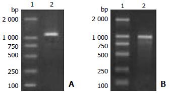
RT-PCR was performed with total RNA template extracted from human colon tissues. The PCR product showed a single band about 1250 bp on 1% agarose gel (Figure 1A).
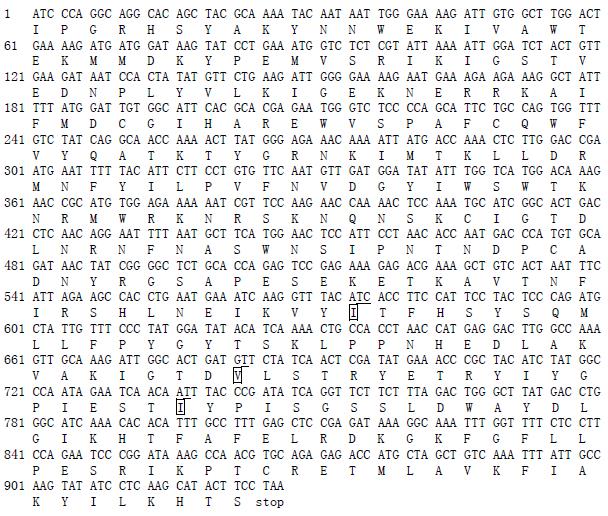
DNA sequencing revealed that the human colon MC-CP cDNA had 1254 bp. The DNA sequence and deduced amino acid sequence of human colon MC-CP are shown in Figure 2. The DNA sequence of human colon MC-CP cDNA was identical to the skin MC-CP except for five nucleotides at the positions 109, 575, 576, 708, 737: C→G, C→T, T→C, A→C, A→T (nucleotide numbering starts from the first codon of mature enzyme). Only the fourth (A1035→C) of the nucleotide differences described above did Not represent an altered amino acid residue in the putative protein products. The other 4 nucleotide variations caused 3 amino acid substitutions at the positions 146, 301, 355. The human colon MC-CP had Gly146, Ile301 and Ile355 residues, whereas the skin MC-CP had Arg146, Thr146 and Asn355 residues. In contrast, the colon MC-CP cDNA was 100% identical to the lung MC-CP cDNA.
A 924 bp of PCR product was obtained following amplification of the coding region of the mature hMC-CP (Figure 1B). DNA sequencing showed that the recombinant pMAL/hMC-CP and pPIC9K/hMC-CP plasmids had the correct open reading frame coding for 308 amino acids mature polypeptide and no substitutions were introduced by PCR.
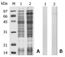
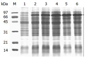
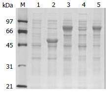
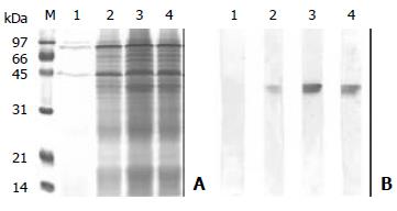
As shown in Figure 3A, a high level of expression of an induced protein of about 80 kD was achieved after the E.coli harbouring expression plasmid pMAL/hMC-CP, which was in agreement with the expected molecular mass of the fusion protein MBP (45 kDa) and hMC-CP (36 kDa). Figure 3B showed a band at about 80 kDa (expected in E.coli cells with IPTG induction) reacted to CA5, suggesting that the recombinant protein had a good immunological activity. The best expression of the recombinant protein after IPTG induction was at 23 °C for 16 h (Figure 4). The recombinant products generated by the above procedures were mainly insoluble inclusion body with a small proportion of the soluble recombinant proteins (Figure 5).
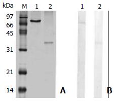
In P. pastoris expression, 2 colonies resistant to 4.0 mg/ml Geneticin were screened and used for the expression of recombinant protein. There was a substantial quantity of recombinant proteins in cell-free supernatant, and SDS-PAGE showed a major band of approximately 37 kDa (Figure 6A), which reacted to CA5 on Western blot (Figure 6B).
Maltose-binding protein (MBP) was used as a fusion partner to provide a “tag” which could be used for the subsequent purification. The yield of the recombinant fusion protein was 12 mg/L of bacterial culture. The purified fusion protein showed a single protein band of approximately 80 kDa on SDS-PAGE.
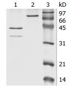
After the fusion protein cleavage, SDS-PAGE analysis showed that the fusion protein was completely cleaved by factor Xa (Figure 7). The cleavage mixtures were loaded to heparin agarose, and the target protein showed one band about 36 kDa on SDS-PAGE, which was corresponding to the molecular weight of the native hMC-CP published previously (Figure 8A). About 1.2 mg pure recombinant protein was obtained from 5 mg fusion protein following the above procedures. The Western blot showed that this 36 kDa protein band strongly reacted to CA5 (Figure 8B), suggesting that the recombinant protein had a good immunology activity. The N-terminal sequence of the purified recombinant protein expressed in E.coli was IPGRHSYAKY, and no additional amino acids were found at the N-terminus.
The purified recombinant hMC-CP expressed in E.coli had a very low level of enzymatic activity. In contrast, enzymatic activity in cell-free supernatant of P. pastoris culture was 11.7 U/mg secreted protein.
A cDNA of human colon hMC-CP was cloned and active enzyme was expressed in the current study, which will offer an essential tool for investigating the functions of hMC-CP, a zinc containing metalloexopeptidase.
Our result revealed that the human colon MC-CP cDNA comprised 1251 bp, which agreed with the skin and lung mast cell carboxypeptidase[22,23,26]. The hMC-CP was predicted to be translated as a 417 amio acid preproenzyme which includes a 15 amino acid signal peptide, a 94 amino acid activation peptide and 308 amino acid mature mast cell carboxypeptidase. When comparison of the DNA sequence of human colon MC-CP cDNA with skin MC-CP cDNA, five variations were found which caused 3 amino acid substitutions, but there was Not any difference between the human colon and skin MC-CP. The meaning of these variations between tissues in man requires more investigations.
Since the role of hMC-CP in man remains unclear and human mast cells contain large amount of MC-CP, there is a pressing need to investigate the functions of this enzyme. One of the difficulties in investigating the potential functions of MC-CP over the years was that it was uneasy to obtain a substantial quantity of the active enzyme. Purification of MC-CP from human tissues was not only hard to perform, but also difficult to collect enough tissues for purification. Therefore, development of an efficient heterologous expression system for the production of recombinant hMC-CP is an alternative for obtaining a sufficient quantity of hMC-CP. There are a number of options for heterologous recombinant expressions, among them E.coli expression system is the most convenient and frequently used, therefore, E.coli expression system was used to express hMC-CP. The pMAL-C2x plasmid[36,37], a vector that allows the fusion of the target protein N-terminus to the MBP tag, made the purification of recombinant proteins much easier.
The extra residue(s) is often added to the C-terminus or N-terminus of recombinant protein. In this study, a pair of specific primers were designed. The upstream primer contained the sequences for a factor Xa recognition site just before the sequence for N-terminus of hMC-CP, and the downstream primer contained a terminator. The PCR product was inserted into the expression vector pMAL-c2x, which yielded the recombinant protein without extra residues after the fusion protein was cut with factor Xa. The result of N-terminal amino acid sequencing also showed that the N-terminus of recombinant hMC-CP had no extra residue.
After induction with IPTG, the recombinant protein was expressed in E.coli, with a molecular weight of about 80 kDa. This was in agreement with the expected molecular mass of the fusion proteins MBP (45 kD) and HMC-CP (36 kD). It was reported that the target gene fused to bacterial gene could improve the expression level and increase the solubility of recombinant proteins[37]. The expression vector pMAL-2x containing malE gene of E.coli encoding MBP was used for fusion expression. The target gene was inserted downstream from the malE gene, which resulted in the expression of hMC-CP fused to MBP. The solubility of recombinant proteins generally could be increased when the cell culture temperature decreased[38]. In our case, although the culture temperature was reduced to 23 °C, insoluble recombinant proteins were still the major products. Since purification of recombinant proteins from inclusion body was a complicated process, we only used soluble products to isolate active recombinant hMC-CP.
Fusion protein was purified with one–step affinity chromatography with maltose. Once the fusion protein was isolated, it was necessary to remove the tag. In this study, the linker sequence recognized by factor Xa was designed between the MBP and target protein, because there were no such sequences in MBP and hMC-CP. After the fusion protein cleavage, usually ion exchange chromatography and hydroxyapatite chromatography were used in separating the protein of interest from MBP[36,39]. But in this study, the recombinant protein was purified by heparin agarose chromatography as MC-MBP could tightly bind to heparin[40]. In comparison with ion exchange chromatography and hydroxyapatite chromatography, heparin agarose chromatography was simpler and more convenient. Approximately 1.2 mg target protein was obtained from 5 mg fusion protein following the established procedures. N-terminal amino acid sequencing showed that the first 10 amino acids of the recombinant hMC-CP were in good agreement with the human skin and lung MC-CP. Western blotting analysis showed that the recombinant protein had the similar immuno-reactivity with its natural counterpart, indicating that the recombinant hMC-CP could be used as an antigen to produce a specific antibody.
Our studies revealed that the purified recombinant hMC-CP expressed in E.coli had a very low level of enzymatic activity to substrate hippuryl-L-phenylalanine. It might be possible that the E. coli expression system is a prokaryotic expression system, which can not carry out post-translation modifications. In order to obtain higher levels of enzymatic activity of recombinant hMC-CP, we used P. pastoris to express hMC-CP. The enzymatic assay showed that the hMC-CP expressed in P.asptoris had a relatively high activity (11.7 U/mg secreted protein) towards hippuryl-L-phenylalanine. It is possible that P.asptoris is an eukaryotic expression system, which has the ability to perform eukaryotic post-translational modifications, such as glycosylation, disulfide bond formation and proteolytic processing[41]. Our result showed that the supernatant of P.pastoris culture had the highest enzymatic activity on the second day after induction by methanol, the enzymatic activity would decrease when induction time increased. It is possible that the secreted recombinant protein was degraded with the increase of induction time.
In conclusion, cDNA encoding human colon MC-CP can be cloned and expressed in E.coli and P.asptoris. The expression of recombinant hMC-CP can facilitate its functional study including its role in intestinal diseases.
Edited by Wang XL
| 1. | He SH. Key role of mast cells and their major secretory products in inflammatory bowel disease. World J Gastroenterol. 2004;10:309-318. [PubMed] |
| 2. | Crowe SE, Luthra GK, Perdue MH. Mast cell mediated ion transport in intestine from patients with and without inflammatory bowel disease. Gut. 1997;41:785-792. [RCA] [PubMed] [DOI] [Full Text] [Cited by in Crossref: 66] [Cited by in RCA: 66] [Article Influence: 2.3] [Reference Citation Analysis (0)] |
| 3. | Raithel M, Winterkamp S, Pacurar A, Ulrich P, Hochberger J, Hahn EG. Release of mast cell tryptase from human colorectal mucosa in inflammatory bowel disease. Scand J Gastroenterol. 2001;36:174-179. [RCA] [PubMed] [DOI] [Full Text] [Cited by in Crossref: 96] [Cited by in RCA: 101] [Article Influence: 4.0] [Reference Citation Analysis (0)] |
| 4. | Stoyanova II, Gulubova MV. Mast cells and inflammatory mediators in chronic ulcerative colitis. Acta Histochem. 2002;104:185-192. [RCA] [PubMed] [DOI] [Full Text] [Cited by in Crossref: 97] [Cited by in RCA: 103] [Article Influence: 4.3] [Reference Citation Analysis (1)] |
| 5. | Sasaki Y, Tanaka M, Kudo H. Differentiation between ulcerative colitis and Crohn's disease by a quantitative immunohistochemical evaluation of T lymphocytes, neutrophils, histiocytes and mast cells. Pathol Int. 2002;52:277-285. [RCA] [PubMed] [DOI] [Full Text] [Cited by in Crossref: 26] [Cited by in RCA: 30] [Article Influence: 1.3] [Reference Citation Analysis (1)] |
| 6. | Furusu H, Murase K, Nishida Y, Isomoto H, Takeshima F, Mizuta Y, Hewlett BR, Riddell RH, Kohno S. Accumulation of mast cells and macrophages in focal active gastritis of patients with Crohn's disease. Hepatogastroenterology. 2002;49:639-643. [PubMed] |
| 7. | Gelbmann CM, Mestermann S, Gross V, Köllinger M, Schölmerich J, Falk W. Strictures in Crohn's disease are characterised by an accumulation of mast cells colocalised with laminin but not with fibronectin or vitronectin. Gut. 1999;45:210-217. [RCA] [PubMed] [DOI] [Full Text] [Cited by in Crossref: 119] [Cited by in RCA: 122] [Article Influence: 4.5] [Reference Citation Analysis (0)] |
| 8. | Nishida Y, Murase K, Isomoto H, Furusu H, Mizuta Y, Riddell RH, Kohno S. Different distribution of mast cells and macrophages in colonic mucosa of patients with collagenous colitis and inflammatory bowel disease. Hepatogastroenterology. 2002;49:678-682. [PubMed] |
| 9. | Schwab D, Raithel M, Hahn EG. Evidence for mast cell activation in collagenous colitis. Inflamm Res. 1998;47 Suppl 1:S64-S65. [RCA] [PubMed] [DOI] [Full Text] [Cited by in Crossref: 15] [Cited by in RCA: 16] [Article Influence: 0.6] [Reference Citation Analysis (0)] |
| 10. | Stenton GR, Vliagoftis H, Befus AD. Role of intestinal mast cells in modulating gastrointestinal pathophysiology. Ann Allergy Asthma Immunol. 1998;81:1-11; quiz 12-5. [RCA] [PubMed] [DOI] [Full Text] [Cited by in Crossref: 65] [Cited by in RCA: 62] [Article Influence: 2.2] [Reference Citation Analysis (0)] |
| 11. | Gui XY. Mast cells: a possible link between psychological stress, enteric infection, food allergy and gut hypersensitivity in the irritable bowel syndrome. J Gastroenterol Hepatol. 1998;13:980-989. [RCA] [PubMed] [DOI] [Full Text] [Cited by in Crossref: 48] [Cited by in RCA: 45] [Article Influence: 1.6] [Reference Citation Analysis (0)] |
| 12. | O'Sullivan M, Clayton N, Breslin NP, Harman I, Bountra C, McLaren A, O'Morain CA. Increased mast cells in the irritable bowel syndrome. Neurogastroenterol Motil. 2000;12:449-457. [RCA] [PubMed] [DOI] [Full Text] [Cited by in Crossref: 340] [Cited by in RCA: 341] [Article Influence: 13.1] [Reference Citation Analysis (0)] |
| 13. | Goldstein SM, Kaempfer CE, Kealey JT, Wintroub BU. Human mast cell carboxypeptidase. Purification and characterization. J Clin Invest. 1989;83:1630-1636. [RCA] [PubMed] [DOI] [Full Text] [Cited by in Crossref: 66] [Cited by in RCA: 59] [Article Influence: 1.6] [Reference Citation Analysis (0)] |
| 14. | Welle M. Development, significance, and heterogeneity of mast cells with particular regard to the mast cell-specific proteases chymase and tryptase. J Leukoc Biol. 1997;61:233-245. [PubMed] |
| 15. | Schechter NM, Choi JK, Slavin DA, Deresienski DT, Sayama S, Dong G, Lavker RM, Proud D, Lazarus GS. Identification of a chymotrypsin-like proteinase in human mast cells. J Immunol. 1986;137:962-970. [PubMed] |
| 16. | Schechter NM, Irani AM, Sprows JL, Abernethy J, Wintroub B, Schwartz LB. Identification of a cathepsin G-like proteinase in the MCTC type of human mast cell. J Immunol. 1990;145:2652-2661. [PubMed] |
| 17. | He SH, Xie H, He YS. Induction of tryptase and histamine release from human colon mast cells by IgE dependent or independent mechanisms. World J Gastroenterol. 2004;10:319-322. [PubMed] |
| 18. | He SH, Xie H. Modulation of tryptase secretion from human colon mast cells by histamine. World J Gastroenterol. 2004;10:323-326. [PubMed] |
| 19. | He SH, Xie H. Inhibition of tryptase release from human colon mast cells by protease inhibitors. World J Gastroenterol. 2004;10:332-336. [PubMed] |
| 20. | He SH, He YS, Xie H. Activation of human colon mast cells through proteinase activated receptor-2. World J Gastroenterol. 2004;10:327-331. [PubMed] |
| 21. | He SH, Xie H. Modulation of histamine release from human colon mast cells by protease inhibitors. World J Gastroenterol. 2004;10:337-341. [PubMed] |
| 22. | Natsuaki M, Stewart CB, Vanderslice P, Schwartz LB, Natsuaki M, Wintroub BU, Rutter WJ, Goldstein SM. Human skin mast cell carboxypeptidase: functional characterization, cDNA cloning, and genealogy. J Invest Dermatol. 1992;99:138-145. [RCA] [PubMed] [DOI] [Full Text] [Cited by in Crossref: 15] [Cited by in RCA: 14] [Article Influence: 0.4] [Reference Citation Analysis (0)] |
| 23. | Reynolds DS, Gurley DS, Austen KF. Cloning and characterization of the novel gene for mast cell carboxypeptidase A. J Clin Invest. 1992;89:273-282. [RCA] [PubMed] [DOI] [Full Text] [Cited by in Crossref: 34] [Cited by in RCA: 30] [Article Influence: 0.9] [Reference Citation Analysis (0)] |
| 24. | Irani AM, Goldstein SM, Wintroub BU, Bradford T, Schwartz LB. Human mast cell carboxypeptidase. Selective localization to MCTC cells. J Immunol. 1991;147:247-253. [PubMed] |
| 25. | Reynolds DS, Gurley DS, Stevens RL, Sugarbaker DJ, Austen KF, Serafin WE. Cloning of cDNAs that encode human mast cell carboxypeptidase A, and comparison of the protein with mouse mast cell carboxypeptidase A and rat pancreatic carboxypeptidases. Proc Natl Acad Sci USA. 1989;86:9480-9484. [RCA] [PubMed] [DOI] [Full Text] [Cited by in Crossref: 68] [Cited by in RCA: 67] [Article Influence: 1.8] [Reference Citation Analysis (0)] |
| 26. | He S, Walls AF. The induction of a prolonged increase in microvascular permeability by human mast cell chymase. Eur J Pharmacol. 1998;352:91-98. [RCA] [PubMed] [DOI] [Full Text] [Cited by in Crossref: 43] [Cited by in RCA: 40] [Article Influence: 1.4] [Reference Citation Analysis (0)] |
| 27. | He S, Peng Q, Walls AF. Potent induction of a neutrophil and eosinophil-rich infiltrate in vivo by human mast cell tryptase: selective enhancement of eosinophil recruitment by histamine. J Immunol. 1997;159:6216-6225. [PubMed] |
| 28. | He S, Walls AF. Human mast cell chymase induces the accumulation of neutrophils, eosinophils and other inflammatory cells in vivo. Br J Pharmacol. 1998;125:1491-1500. [RCA] [PubMed] [DOI] [Full Text] [Cited by in Crossref: 118] [Cited by in RCA: 120] [Article Influence: 4.3] [Reference Citation Analysis (0)] |
| 29. | He S, Gaça MD, Walls AF. A role for tryptase in the activation of human mast cells: modulation of histamine release by tryptase and inhibitors of tryptase. J Pharmacol Exp Ther. 1998;286:289-297. [PubMed] |
| 30. | He S, Gaça MD, Walls AF. The activation of synovial mast cells: modulation of histamine release by tryptase and chymase and their inhibitors. Eur J Pharmacol. 2001;412:223-229. [RCA] [PubMed] [DOI] [Full Text] [Cited by in Crossref: 29] [Cited by in RCA: 23] [Article Influence: 0.9] [Reference Citation Analysis (0)] |
| 31. | Tomimori Y, Tsuruoka N, Fukami H, Saito K, Horikawa C, Saito M, Muto T, Sugiura N, Yamashiro K, Sumida M. Role of mast cell chymase in allergen-induced biphasic skin reaction. Biochem Pharmacol. 2002;64:1187. [RCA] [PubMed] [DOI] [Full Text] [Cited by in Crossref: 26] [Cited by in RCA: 22] [Article Influence: 0.9] [Reference Citation Analysis (0)] |
| 32. | Dell'Italia LJ, Husain A. Dissecting the role of chymase in angiotensin II formation and heart and blood vessel diseases. Curr Opin Cardiol. 2002;17:374-379. [RCA] [PubMed] [DOI] [Full Text] [Cited by in Crossref: 65] [Cited by in RCA: 67] [Article Influence: 2.8] [Reference Citation Analysis (0)] |
| 33. | Tomimori Y, Muto T, Fukami H, Saito K, Horikawa C, Tsuruoka N, Saito M, Sugiura N, Yamashiro K, Sumida M. Chymase participates in chronic dermatitis by inducing eosinophil infiltration. Lab Invest. 2002;82:789-794. [RCA] [PubMed] [DOI] [Full Text] [Cited by in Crossref: 50] [Cited by in RCA: 46] [Article Influence: 1.9] [Reference Citation Analysis (0)] |
| 34. | Leskinen MJ, Lindstedt KA, Wang Y, Kovanen PT. Mast cell chymase induces smooth muscle cell apoptosis by a mechanism involving fibronectin degradation and disruption of focal adhesions. Arterioscler Thromb Vasc Biol. 2003;23:238-243. [RCA] [PubMed] [DOI] [Full Text] [Cited by in Crossref: 88] [Cited by in RCA: 96] [Article Influence: 4.2] [Reference Citation Analysis (0)] |
| 35. | Buckley MG, Variend S, Walls AF. Elevated serum concentrations of beta-tryptase, but not alpha-tryptase, in Sudden Infant Death Syndrome (SIDS). An investigation of anaphylactic mechanisms. Clin Exp Allergy. 2001;31:1696-1704. [RCA] [PubMed] [DOI] [Full Text] [Cited by in Crossref: 45] [Cited by in RCA: 41] [Article Influence: 1.6] [Reference Citation Analysis (0)] |
| 36. | Riggs P. Expression and purification of recombinant proteins by fusion to maltose-binding protein. Mol Biotechnol. 2000;15:51-63. [RCA] [PubMed] [DOI] [Full Text] [Cited by in Crossref: 89] [Cited by in RCA: 79] [Article Influence: 3.0] [Reference Citation Analysis (0)] |
| 37. | Lazos SA, Tsiftsoglou AS. Production and purification of recombinant human cytokines (rhIL-4, rhGM-CSF, and rhIL-1beta) from genetically engineered E. coli cells bearing pMAL expression vector constructs. J Protein Chem. 1998;17:517. [PubMed] |
| 38. | Schein CH. Optimizing protein folding to the native state in bacteria. Curr Opin Biotechnol. 1991;2:746-750. [RCA] [PubMed] [DOI] [Full Text] [Cited by in Crossref: 50] [Cited by in RCA: 42] [Article Influence: 1.2] [Reference Citation Analysis (0)] |
| 39. | Ding Y, Qin L, Kotenko SV, Pestka S, Bromberg JS. A single amino acid determines the immunostimulatory activity of interleukin 10. J Exp Med. 2000;191:213-224. [RCA] [PubMed] [DOI] [Full Text] [Full Text (PDF)] [Cited by in Crossref: 96] [Cited by in RCA: 94] [Article Influence: 3.6] [Reference Citation Analysis (0)] |
| 40. | Goldstein SM, Leong J, Schwartz LB, Cooke D. Protease composition of exocytosed human skin mast cell protease-proteoglycan complexes. Tryptase resides in a complex distinct from chymase and carboxypeptidase. J Immunol. 1992;148:2475-2482. [PubMed] |
| 41. | Cereghino JL, Cregg JM. Heterologous protein expression in the methylotrophic yeast Pichia pastoris. FEMS Microbiol Rev. 2000;24:45-66. [RCA] [PubMed] [DOI] [Full Text] [Cited by in Crossref: 1396] [Cited by in RCA: 1376] [Article Influence: 52.9] [Reference Citation Analysis (0)] |


