INTRODUCTION
Apoptosis, or programmed cell death, is characterized by a series of morphological features such as chromatin condensation, nuclear fragmentation, and the appearance of membrane-enclosed apoptotic bodies, etc.[1,2], most of which are caused by the activation of caspases, a family of aspartate-specific cysteine proteases[3,4]. Caspases are constitutively present in cells as inactive zymogens and require proteolytic cleavage into the catalytic active heterodimer[5]. All activated caspases are comprised of a large subunit with Mr 17000-20000 that contains a redox-sensitive cysteine at the active site and a small subunit with Mr 10000 approximately. According to their function and sequence of activation, caspases can be grouped into three categories: Caspases that function primarily in cytokine maturation (such as caspases-1, -4, and -5); initiator caspases involved in the early steps of apoptotic signaling (caspases-8, -9, and -10) and effector proteases in the execution phase of apoptosis (caspases-3, -6, and -7). The initiator caspases lead to the activation of executioner caspases following characteristic apoptotic stimuli. The substrates of executioner caspases include many proteins, the cleavage of which causes the characteristic morphology of apoptosis[6,7]. Because apoptosis is a regulated cellular process, it offers some opportunities for therapeutic intervention[8,9]. Indeed, eliminating caspase activity, either through mutation or the use of small pharmacological inhibitors, will slow down or even prevent apoptosis.
Ribozymes are small RNA molecules that hybridize to the target RNA around the cleavage site, and catalyze site-specific cleavage of substrate. Cleaved mRNA is rapidly degraded, allowing ribozyme to react with new targets. Various ribozymes have been used to down-regulate gene expression in a sequence-specific manner. Among them the hammerhead ribozyme, composed of only 30 to 40 nucleotides, is the smallest and simplest one, and has been used extensively to down-regulate gene expression[10-13].
Caspase-3 and caspase-7 are both important executioner caspases[14]. Expression pattern of caspase-7 is distinguishable from that of caspase-3, suggesting that they may regulate apoptosis in disparate tissues, although they share strong homology. In the past years, many researches have been concentrated on interfering with the activity or expression of caspase-3 so as to intervene in apoptosis[15-17]. Following its activation, caspase-7 was translocated into the microsomal and mitochondrial fractions, where it was responsible for the cleavage of specific substrates in those distinct subcellular compartments[18]. Recent results suggested that as one of important executioner caspases, caspase-7 is also essential in apoptotic signaling[19,20]. In this study, we designed and synthesized two hammerhead ribozymes against mouse caspase-7 at sites 333 and 394 (named Rz333 and Rz394), studied their in vitro transcription and cleavage activity, and selected the one that could site-specifically cleave caspase-7 mRNA as a hopeful gene therapy tool for apoptosis-related diseases.
MATERIALS AND METHODS
Materials
TRIzol reagent was product of Gibco. RT-PCR kit, T4 DNA ligase and restriction endonucleases were Takara products. In vitro transcription kit and pGEM-T vector were purchased from Promega. pBSK neo-U6’ was kindly donated by Dr. Youxin Jin. Plasmid extraction kit and gel purification kit were products of Shanghai Sangon Biotechnology Co.
Methods
Target mRNA secondary structure analysis and design of anti-caspase-7 ribozymes Caspase-7 gene sequence of BALB/c mouse searched in GenBank (gi: 6680849) was analyzed, and its secondary structure was simulated in computer. Ribozymes against caspase-7 mRNA were designed by software and DNA sequences encoding ribozymes were synthesized. A restriction site of Xba I was introduced into 5’ end of sense strand. And a BamH I restriction site was introduced into the 5’ end of antisense strand. Two DNA strands were mixed at equal molar ratio after synthesis, and annealed by cooling to room temperature naturally after placed in boiling water for 2 min, then double strand DNA encoding ribozymes was obtained.
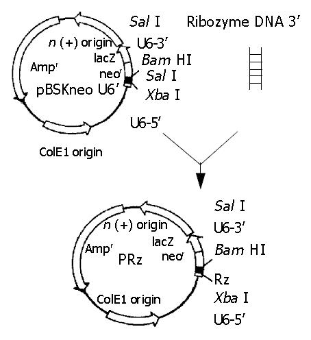
Ribozyme gene cloning Double-strand ribozyme DNAs designed by computer were inserted into pBSKneo U6’ with T4 DNA ligase after pBSKneo U6’ was cleaved by BamH I and Xba I (Figure 1). The reconstructed plasmids containing ribozyme genes were identified by Sal I digestion and sequencing.
Figure 1 Ribozyme gene cloning.
Construction of caspase-7 expression vector Total RNA was extracted with TRIzol reagent from Balb/c mouse liver. Caspase-7 gene segment (841 bp) was amplified by RT-PCR with specific primers. The segment was cloned into pGEM-T vector downstream from T7 promoter, and the transformed clones selectively grew at 37 °C overnight in LB plate (Ampicillin resistance) containing IPTG and X-gal on plate’s surface. Clones in blue were selected for sequencing. Caspase-7 primers were: P1 5’-GGATCCGAACGATGACCGATGATCAG-3’, P2 5’-AAGCTTGTGAGCATGGACACCATAC-3’.
In vitro transcription of ribozyme and caspase-7 cDNAs Constructed caspase-7 plasmid was linearized with Hind III. Reconstructed ribozyme plasmid was considered as PCR template and a pair of primers containing T7 promotor was designed. P1 5’-TCTAGAGTAATACGACTCACTATAGGGCCTTCGGCAGCACATATAC-3’, P2 5’-TATGGAACGCTTCAGGAT-3’ (The italic is T7 promotor sequence). Both caspase-7 linearized plasmid and pRz PCR product were phenol-chloroform extracted, and ethanol precipitated. In vitro transcription was performed using T7 RNA polymerase. Transcript of caspase-7 labeled with α-32P-UTP was heated (2 min, 95 °C) before loading and electrophoresed on 6% polyacrylamide gel denatured by 8 mol/L urea. Transcript band was observed after autoradiography. In vitro transcription of ribozyme was not labeled with isotope. After electrophoresis on 60 g/L polyacrylamide gel, the transcript bands were observed under ultraviolet. All transcript bands in gel, labeled with isotope or not, were cut and dipped into nucleic acid (0.5 mol/L NH4Ac, 1 mmol/L EDTA, 1 g/L SDS), precipitated in ethanol and dissolved in DEPC-H2O. Concentrations of substrate and ribozyme were calculated through cpm counting and A260 measurement, respectively.
In vitro ribozyme cleavage assays Ribozymes and caspase-7 mRNA were incubated at equal molar ratio at 37 °C for 90 min in a system of 5-10 µL containing 50 mmol/L Tris-HCl pH7.5, 20 mmol/L MgCl2, 20 mmol/L NaCl and 2 mmol/L EDTA. The products after cleavage were electrophoresed on 10% polyacrylamide gel, and the cleavage results were analyzed by autoradiography. Cleavage efficiency may be estimated according to cpm of both substrates (S) and cleaved products (P).
Cleavage ratio = [P/(S + P)] × 100%
RESULTS
Design of ribozymes
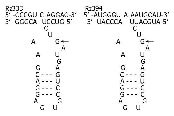
According to the results of computer simulation, triplet GUC at site 333 and GUA at site 394 of caspase-7 mRNA were chosen as the cleavage sites of ribozyme. So ribozymes targeting site 333 and site 394 (named Rz333 and Rz394) were designed to cleave the target mRNA. Both designed ribozymes were comprised of a catalytic core and two flanking sequences (Figure 2).
Figure 2 Sequences, structures and targets of ribozymes against caspase-7.
Hammerhead ribozymes (bottom strand) binding to their targets (top strand) to form a typical three-stem struc-ture that leads to the cleavage of caspase-7 RNA at the targets of GUC (Rz333) and GUA (Rz394).
Identification of reconstructed plasmid containing ribozyme
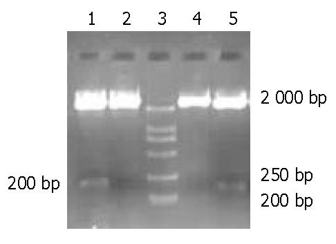
Reconstructed plasmids pU6Rz333 and pU6Rz394 were both incubated with SalI at 37 °C for 2 h and then electrophoresed on 12 g/L agarose gel (Figure 3). Clones without 200 bp segment were sent for sequencing, and the results indicated that clones 2, 4 were correctly constructed.
Figure 3 Restriction enzyme cleavage of reconstructed ribozyme plasmid.
Lanes 1, 2: pU6Rz333 digested by Sal I; lanes 4, 5: pU6Rz394 digested by Sal I; lane 3: Marker (DL2000).
Caspase-7 DNA and its clone
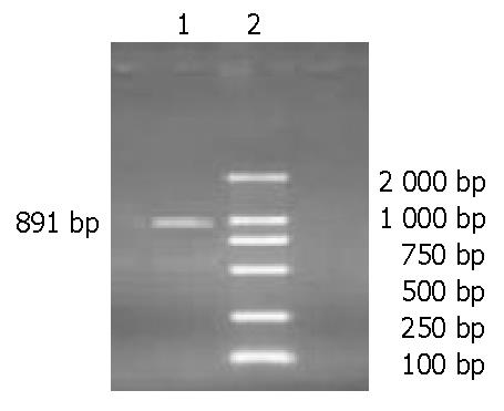
Extracted total RNA was amplified by RT-PCR and a 891-bp caspase-7 DNA segment was seen on 20 g/L agarose gel (Figure 4). It was cloned into pGEM-T vector, and white and blue monoclones were seen in LB medium after incubated at 37 °C overnight. Blue clones were selected and sent for sequencing.
Figure 4 Agarose gel electrophoresis of RT-PCR products.
Lane 1: A 891-bp caspase-7 gene segment; lane 2: Marker (DL2000).
In vitro transcription of target mRNA and ribozymes
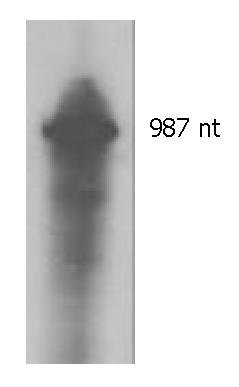
Transcript of target mRNA was electrophoresed on 60 g/L polyacrylamide gel. After autoradiography, a black band of 987 nt was observed, including 891-nt caspase-7 mRNA and 96-nt U6 segment (Figure 5). In vitro transcripts of the two ribozymes were not labeled with isotope. The results of transcription were analyzed under ultraviolet. Black bands of 47 nt (Rz333) and 50 nt (Rz394) could be seen.
Figure 5 In vitro transcription of target RNA.
The transcript is 987 nt.
In vitro cleavage of ribozymes
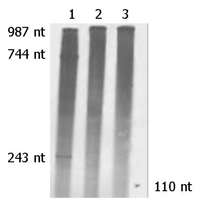
Products of cleavage assays were electrophoresed on 100 g/L polyacrylamide gel, and analyzed by autoradiography. Rz333 cleaved caspase-7 mRNA and produced two cleaved segments of 243 nt and 744 nt, with a cleavage efficiency of 67.98%. Rz394 could not cleave target RNA (Figure 6).
Figure 6 In vitro cleavage experiments of anti-caspase-7 ribozymes.
Lane 1: Caspase-7 mRNA was mixed with Rz333 and two segments of 243 nt and 744 nt produced by cleavage reaction. Lane 2: Caspase-7 mRNA was mixed with Rz394 and no segment was seen after cleavage. Lane 3: Caspase-7 mRNA was mixed with neither Rz333 nor Rz394. The 110-nt marker is indicated by a short arrow.
DISCUSSION
RNA catalysis was first described by Altman and Cech with the discovery of RNase P and the group I intron, respectively[21,22]. This makes RNA the only molecule with an information-carrying capacity and inherent catalytic activity. So far, several natural ribozyme motifs have been identified and their physical structure, biological and biochemical properties have been the subject of reviews[23,24]. In general, however, natural ribozymes have been divided into groups based on their specialized catalytic properties. As the smallest one, the hammerhead ribozyme, with its capability of self-cleavage of a particular phosphodiester bond, has been studied extensively to understand the structure-activity relationship[25-29]. It provides a very valuable tool for genetic therapy through its RNA-mediated inhibition of gene expression. Hammerhead ribozyme has been used extensively to down-regulate cellular and viral gene expression[30,31], and recognized as a novel molecular therapeutic drug in the treatment of cancer or viral infection.
Apoptosis is essential to the normal development of multicellular organisms as well as physiologic cell turnover. In pathologic states, a failure to undergo apoptosis may cause abnormal cell overgrowth and malignancy, while excessive apoptosis may lead to organ injury. Apoptosis involves the activation of the caspases, which are hallmarks of apoptosis. Central to the execution phase of apoptosis are the two closely related caspase-3 and caspase-7, which share common substrate specificity and structure. Many cellular proteins are cleaved during the execution phase of apoptosis at a DXXD motif by the effector caspases-3 and -7. Caspase-3 has been extensively studied as gene therapeutic target. Caspase-7, as another crucial executioner caspase, has been chosen to be the gene therapy target of ribozyme.
In our experiment, we only discovered two sites (333 and 394) in caspase-7 gene suitable for hammerhead ribozymes to cleave, so Rz333 and Rz394 were synthesized and cloned. Caspase-7 gene segment (891 bp) was amplified by RT-PCR from total RNA of mouse liver, and was cloned into an expression plasmid. Ribozymes and substrate were both gained by in vitro transcription. The results of cleavage assays indicated that Rz333 could catalyze site-specific cleavage of caspase-7 mRNA, and produced 243-nt and 744-nt cleaved segments, but Rz394 could not cleave the substrate. This might be because the secondary structures of ribozymes and substrate simulated by computer could not reflect the real situation. Rz333 could site-specifically cleave caspase-7 mRNA with a cleavage efficiency of 67.98%. It may prevent apoptosis solely or in association with anti-caspase-3 ribozyme synthesized and selected in our previous study. Rz333 may become a candidate for gene therapy of apoptosis-related diseases.


