Published online Oct 1, 1995. doi: 10.3748/wjg.v1.i1.25
Revised: June 10, 1995
Accepted: August 20, 1995
Published online: October 1, 1995
AIM: To study the changes of antral endocrine cells in Helicobacter pylori (Hp) infection, and to observe the relation between Hp infection and the number of gastrin (G) cells and somatostatin (D) cells.
METHODS: Sixty-five cases, 18 with Hp infection and 47 without Hp, were analyzed by endoscopy and immunohistochemical staining of the antral mucosal biopsies using antibodies against chromogranin A, gastrin, somatostatin, and bombesin. The positive cells were quantitatively studied by an image analyzer.
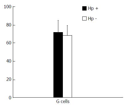
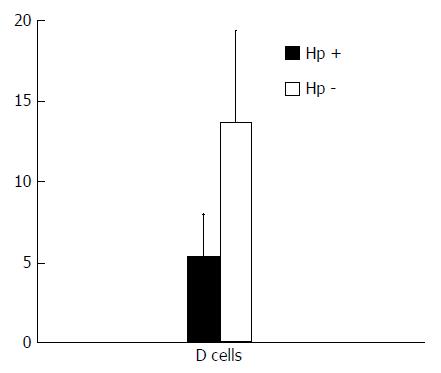
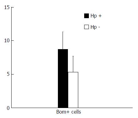
RESULTS: In the Hp infection group, the results were following: 71.28 G cells/mm2, 5.32 D cells/mm2, 8.68 bombesin positive cells/mm2, and the G/D cell ratio was 13.40. By contrast, in the group without Hp infection, the number of G cells was 67.75/mm2 while the number of D cells and bombesin positive cells were 13.65/mm2 and 5.31/mm2, respectively, with the ratio of G/D cells of 5.05. The difference in the number of D cells and the G/D cell ratio was statistically significant between the two groups (P < 0.05).
CONCLUSION: The gastrin increase in patients with Hp infection may be due to the decrease in D cells and somatostatin secretion.
- Citation: Yu JY, Wang JL, Yao L, Zheng JY, Hu M. The changes of antral endocrine cells in Helicobacter pylori infection. World J Gastroenterol 1995; 1(1): 25-26
- URL: https://www.wjgnet.com/1007-9327/full/v1/i1/25.htm
- DOI: https://dx.doi.org/10.3748/wjg.v1.i1.25
It has been suggested that Helicobacter pylori (Hp) infection induces changes in the antral endocrine cells (EC). The relationship between EC and stomach diseases has been reported. The present study aimed to examine the influence of Hp infection on the antral EC using immunohistochemical staining.
Sixty-five gastroscopic antral biopsy specimens including the entire height of the antral mucosa from the superficial epithelium to the muscularis mucosa were collected. The blocks were fixed in 10% formalin and embedded in paraffin. Serial sections (10) were cut from each block (5 μm). Hematoxylin and eosin staining was carried out. Hp was confirmed with the Warthin Starry silver staining.
The cellular localizations of endocrine substances were detected by biotin avidin (BA kit, Academy of Military Medical Sciences). The primary antibodies included anti-Chromogranin A (CGA, M869 DAKO, working dilution 1:400), anti-gastrin (GAT, A568, 1:300), anti-somatostatin (SS, A566, 1:400), and anti-bombesin (BOM, CA-08-210, Cambridge Research Biochemical Limited England, 1:20000). All antibodies were polyclonal. Peroxidase was revealed using diaminobenzidine tetrahydrochloride. Specificity of the immunoreaction was controlled by incubating consecutive sections with nonimmune serum, instead of the primary antiserum, or with the specific antiserum pre-absorbed with an excess of the respective antigens.
The EC in gastric antral mucosa was quantitatively analyzed with GmbHg image analyzer using 20 × objective. Care was taken to examine the same area in consecutive sections stained with anti-Chromogranin A and hormonal antibodies immunostaining. The number of the different types of EC in measured mucosa was counted and shown by the number of EC per square millimeter. Statistical analysis: The results were shown as x-± s of each EC. Measurements were compared with the unpaired two-tailed Student’s t test.
Table 1 shows the clinicopathological characteristics of 65 cases. The number of peptic ulcers in the Hp positive group was 8/19 (42.1%) and in the Hp negative group was 3/46 (6.5%) (P < 0.05). In the Hp positive group, 15.8% (3/19) of cases had chronic atrophic gastritis; the Hp negative group had 10.9% (5/46) of chronic atrophic gastritis (P > 0.05).
| Characteristics | Hp positive | Hp negative |
| Cases (n) | 19 | 46 |
| Age (Yr) | ||
| Median (age range in Yr) | 51 (32-67) | 48 (34-65) |
| Sex (F/M) | 14/4 | 36/11 |
| Pathological diagnosis | ||
| CSG | 8 | 38 |
| CAG | 3 | 5 |
| PU | 8 | 3 |
The mean numbers of G cells in the Hp positive and Hp negative groups are shown in Figure 1. The difference was not statistically significant (P > 0.05). Figure 2 shows the number of somatostatin positive cell (D cell) in the Hp positive group (5.32 ± 1.42/mm2) and Hp negative group 1 (3.65 ± 3.56/mm2) (P < 0.05). Figure 3 shows the number of bombesin positive cells in the two groups (Hp positive, 12.7 ± 2.6/mm2; Hp negative, 5.31 ± 2.31/mm2) (P < 0.05).
Hp infection is a cause of gastritis and peptic ulcer[1]. The antral content of progastrin and its products are increased in Hp infected patients[2]. Our study showed a more intense immunostaining of G cells, using quantitative image analysis, in the presence of Hp than in uninfected patients (P < 0.05). The ratio of G/D cell was significantly different (P < 0.05). Gastrin release was normally inhibited by somatostatin. The decrease of D cells led to decreased inhibition in the G cells by somatostatin. Our results are in agreement with some reports[3,4] which showed a significant rise in somatostatin mRNA and somatostatin immunoreactive cell density after the eradication of Hp.
The stomach is rich in endocrine cells of various kinds. When it is infected with Hp, the changes in endocrine cells, including number, structure, and function are complex. This study showed an increase in the bombesin positive cells of antrum during Hp infection. Exaggeration of gastrin release followed bombesin stimulation[5]. Gastrin release can also occur as a result of γ interferon and interleukin 2 stimulation in a dog antrum[6]. Tumor necrosis factor, found in high concentration in the inflamed antrum, has been shown to increase gastrin gene transcription.
In conclusion, the increase of gastrin in patients with Hp infection may be due to the decrease in D cell and somatostatin secretion. Further investigation is required to elucidate the relations between EC in gastric mucosa and Hp infection.
| 1. | Moss S, Calam J. Helicobacter pylori and peptic ulcers: the present position. Gut. 1992;33:289-292. [RCA] [PubMed] [DOI] [Full Text] [Cited by in Crossref: 98] [Cited by in RCA: 98] [Article Influence: 2.9] [Reference Citation Analysis (0)] |
| 2. | Beardshall K, Moss S, Gill J, Levi S, Ghosh P, Playford RJ, Calam J. Suppression of Helicobacter pylori reduces gastrin releasing peptide stimulated gastrin release in duodenal ulcer patients. Gut. 1992;33:601-603. [RCA] [PubMed] [DOI] [Full Text] [Cited by in Crossref: 39] [Cited by in RCA: 45] [Article Influence: 1.3] [Reference Citation Analysis (0)] |
| 3. | Odum L, Petersen HD, Andersen IB, Hansen BF, Rehfeld JF. Gastrin and somatostatin in Helicobacter pylori infected antral mucosa. Gut. 1994;35:615-618. [RCA] [PubMed] [DOI] [Full Text] [Cited by in Crossref: 43] [Cited by in RCA: 52] [Article Influence: 1.6] [Reference Citation Analysis (0)] |
| 4. | Moss SF, Legon S, Bishop AE, Polak JM, Calam J. Effect of Helicobacter pylori on gastric somatostatin in duodenal ulcer disease. Lancet. 1992;340:930-932. [RCA] [PubMed] [DOI] [Full Text] [Cited by in Crossref: 204] [Cited by in RCA: 178] [Article Influence: 5.2] [Reference Citation Analysis (0)] |
| 5. | Graham KY, Lew GM, Lechago J. Antral G Cell and cell numbers in Helicobacter pylori infection of Hp eradication. Gastroenterology. 1993;104:A90. |
| 6. | Savino LR, Sacent C, Midolo P. Antral gastrin (G) and somatostatin (S) cell in Helicobacter pylori (Hp) subjects: restitution of D cell number with eradication. Gastroenterology. 1993;104:A1046. |
Original title: China National Journal of New Gastroenterology (1995-1997) renamed World Journal of Gastroenterology (1998-).
S- Editor: Filipodia L- Editor: Jennifer E- Editor: Zhang FF


