Copyright
©The Author(s) 2003.
World J Gastroenterol. Sep 15, 2003; 9(9): 1954-1958
Published online Sep 15, 2003. doi: 10.3748/wjg.v9.i9.1954
Published online Sep 15, 2003. doi: 10.3748/wjg.v9.i9.1954
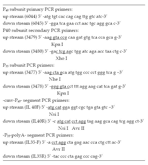
Figure 1 Primers for amplifying human interleukin 12.
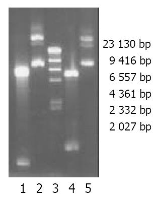
Figure 2 Electrophoresis of P (-)/P35, P (+)/P40 and their bands after restrictive endonuclease enzyme digestion in a 0.
8% aga-rose gel. 1. P (-)/P35 plasmid digested by Kpn I and Nhe I. 2. P (-)/P35 plasmid. 3. Marker (λDNA/Hind III). 4. P (+)/P40 plasmid. 5. of P (+)/P40 plasmid cut by Kpn I and Xho I.
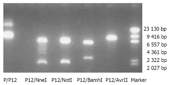
Figure 3 Electrophoresis of recombinant expression plasmid P (+)/IL-12 and the bands after restriction enzyme digestion in a 0.
8% agarose gel (Marker: λDNA/Hind III).
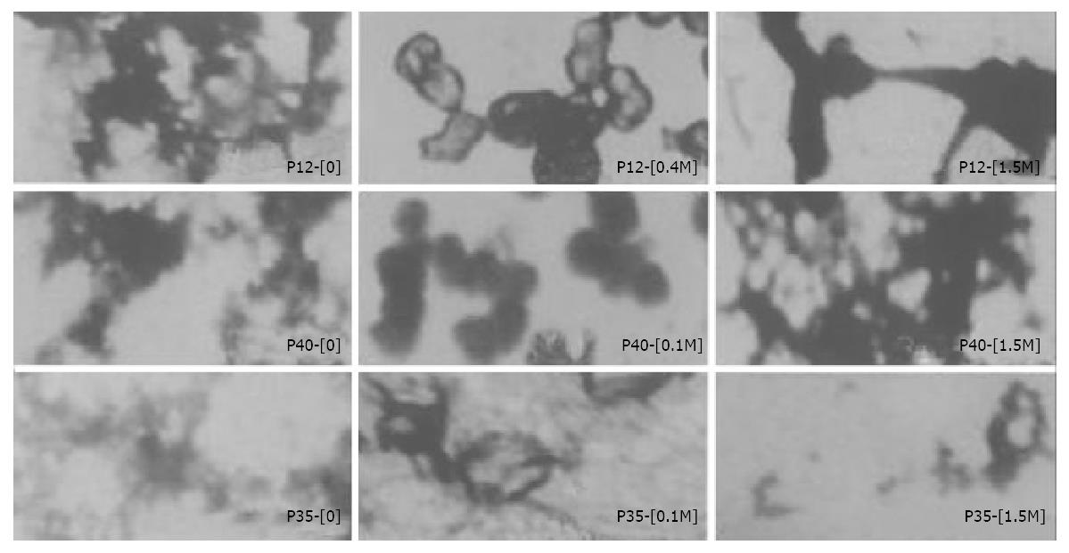
Figure 4 Differently structural features of targeting gene drugs (ASOR-PLL-DNA complexes) at various concentrations of adjuvant under transmission electron microscope.
(The bar equals 100 nm, amplified 40000 times).
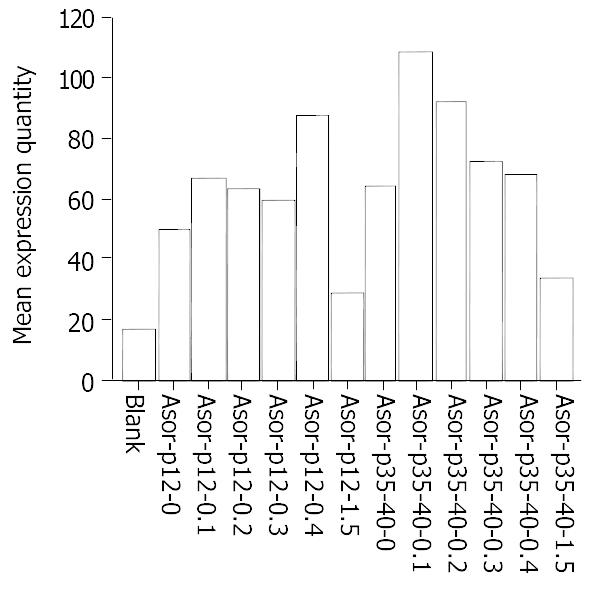
Figure 5 ELISA results of hIL-12 expressed in cell supernatant 48 hours after targeting gene drugs at various adjuvant concentrations were transfected into HepG2.
-
Citation: Yang DY, Lu FG, Tang XX, Zhao SP, Ouyang CH, Wu XP, Liu XW, Wu XY. Study on relationship between expression level and molecular conformations of gene drugs targeting to hepatoma cells
in vitro . World J Gastroenterol 2003; 9(9): 1954-1958 - URL: https://www.wjgnet.com/1007-9327/full/v9/i9/1954.htm
- DOI: https://dx.doi.org/10.3748/wjg.v9.i9.1954


