©The Author(s) 2003.
World J Gastroenterol. Aug 15, 2003; 9(8): 1762-1766
Published online Aug 15, 2003. doi: 10.3748/wjg.v9.i8.1762
Published online Aug 15, 2003. doi: 10.3748/wjg.v9.i8.1762
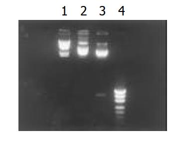
Figure 1 Restriction endonuclease EcoRI, HindIII digests of PcagA.
1. λDNA HindIIImarker, 2. PcagA, 3. Fragement di-gested by EcoRI-HindIII, 4. PUC18/MSPI marker.
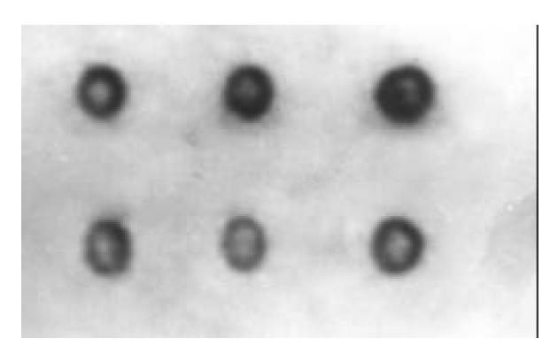
Figure 2 Colony hybridization detection of cagA.
The positive hybridization dot.
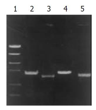
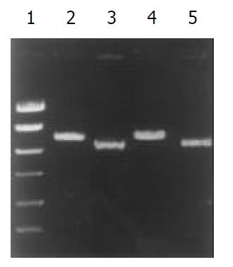
Figure 3 PCR typing of vacA signal sequence.
1.PCR marker, 2. Type s2, 3. Type s1, 4. Standard strain 86313, 5. Standard strain 60 190.
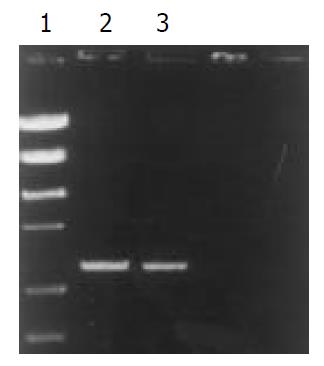
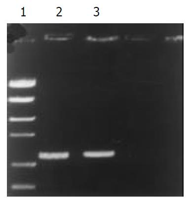
Figure 4 PCR typing of vacA s1a.
1. PCR marker, 2. Clinical isolates, 3. Standard strain 84 183.
Figure 5 PCR typing of vacA mid-region.
1. PCR marker, 2. Clinical isolates, 3. Standard strain 84 183.
Figure 6 PCR typing of vacA s1a.
1. PCR marker, 2.Type m2, 3. Type m1, 4. Standard strain 86 313, 5. Standard strain 60 190.
-
Citation: Qiao W, Hu JL, Xiao B, Wu KC, Peng DR, Atherton JC, Xue H. cagA and vacA genotype of
Helicobacter pylori associated with gastric diseases in Xi’an area. World J Gastroenterol 2003; 9(8): 1762-1766 - URL: https://www.wjgnet.com/1007-9327/full/v9/i8/1762.htm
- DOI: https://dx.doi.org/10.3748/wjg.v9.i8.1762


