Copyright
©The Author(s) 2003.
World J Gastroenterol. Jun 15, 2003; 9(6): 1352-1355
Published online Jun 15, 2003. doi: 10.3748/wjg.v9.i6.1352
Published online Jun 15, 2003. doi: 10.3748/wjg.v9.i6.1352
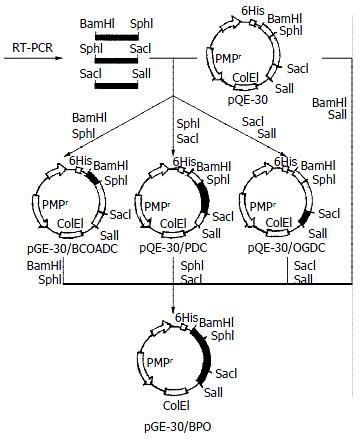
Figure 1 Construction protocol of recombinant plasmids.
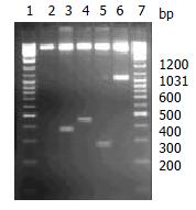
Figure 2 Segment analysis of recombinant plasmids by restriction endonuclease digestion.
1, 7. Markers; 2. pQE-30 (Bamh1); 3. pQE-30/BCOADC-E2 (Bamh1+Sph1); 4. pQE-30/PDC-E2 (Sph1+Sac1); 5. p30/OGDC-E2 (Sac1+Sal1); 6. pQE-30/BPO (Bamh1+Sal1).
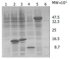
Figure 3 Expression products of recombinant plasmids detected by SDS-PAGE stained with Coomassie Brilliant Blue R-250.
Lane 1: pQE-30 (control); Lane 2: pQE-30/BCOADC-E2; Lane 3: pQE-30/PDC-E2; Lane 4: pQE-30/OGDC-E2; Lane 5: pQE-30/BPO; Lane 6: protein marker.
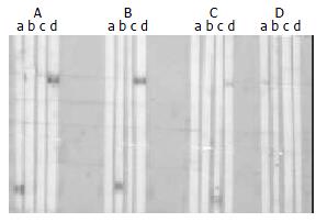
Figure 4 Immunoreactivity of sera against recombinant proteins.
Three M2 antibody positive sera (A, B, C) and a M2 antibody negative serum (D) were probed with SDS-PAGE-separated recombinant proteins of BCOADC-E2 (lane a), PDC-E2(lane b), OGDC-E2 (lane c), BPO (lane d).
- Citation: Jiang XH, Zhong RQ, Yu SQ, Hu Y, Li WW, Kong XT. Construction and expression of a humanized M2 autoantigen trimer and its application in the diagnosis of primary biliary cirrhosis. World J Gastroenterol 2003; 9(6): 1352-1355
- URL: https://www.wjgnet.com/1007-9327/full/v9/i6/1352.htm
- DOI: https://dx.doi.org/10.3748/wjg.v9.i6.1352


