©The Author(s) 2003.
World J Gastroenterol. May 15, 2003; 9(5): 946-950
Published online May 15, 2003. doi: 10.3748/wjg.v9.i5.946
Published online May 15, 2003. doi: 10.3748/wjg.v9.i5.946
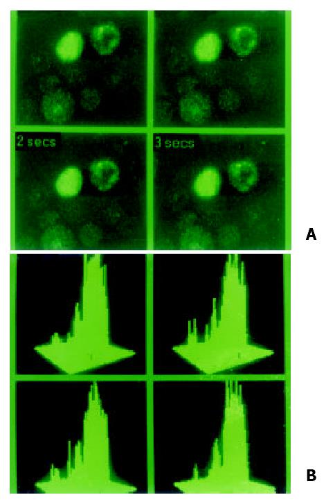
Figure 1 Special indicator Fluo-3 AM was loading at 37 °C for 90 min in hepatocarcinoma cell lines in order to examine localization and concentration of intracellular calcium in MRC-1024 laser scanning confocal microscope imaging system.
(A). In normal hepatocyte cell line QZG, stronger signal of calcium with lower peak released. (B). Concentration of intracellular calcium in HHCC was 108.37 nmol/L.
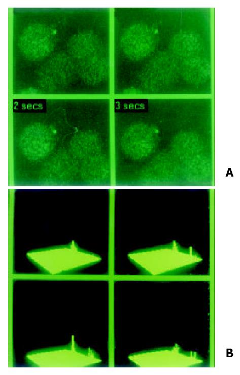
Figure 2 Special indicator Fluo-3 AM was loading at 37 °C for 90 min in hepatocarcinoma cell lines in order to examine localization and concentration of intracellular calcium in MRC-1024 laser scanning confocal microscope imaging system.
(A). In hepatocarcinoma cell line HHCC, slight signal of calcium with lower peak was observed. (B). Concentration of intracellular calcium in HHCC was 35.13 nmol/L.
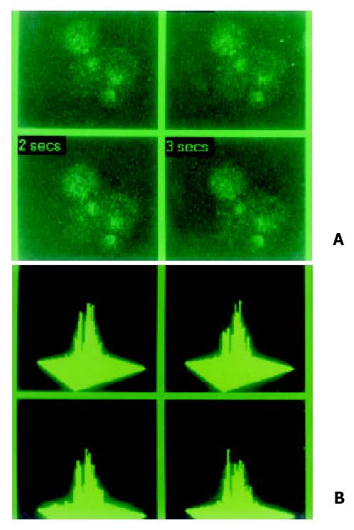
Figure 3 Special indicator Fluo-3 AM was loading at 37 °C for 90 min in hepatocarcinoma cell lines in order to examine localizatio and concentration of intracellular calcium in MRC-1024 laser scanning confocal microscope imaging system.
(A). Loading by Fluo-3 AM, dark signal of calcium and lower peak was also absent in hepatocarcinoma cell line SMMC-7721. (B). Concentration of intracellular calcium was 47.08 nmol/L.
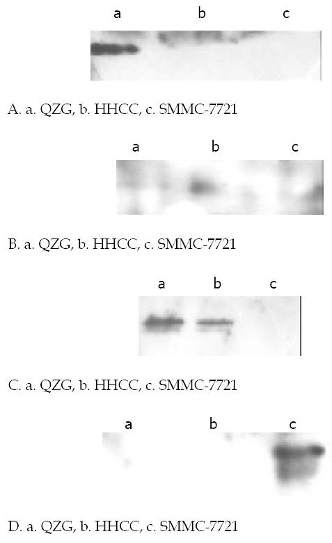
Figure 4 Western blot analysis of phosphorylation on tyrosine of CX32, CX43 proteins in various cells lines.
Cells were harvested and lysed, equal amount of cell lystates were resolved of SDS-PAGE, transferred to NC membranes, and then probed with anti-phophorylation tyrosine mAb 4G10. (A). Expressions of CX32 proteins in cell lines with special anti-CX32 mAb. CX32 showed high immunoblot signal only in normal hepatocyte cell line QZG with very slight signal in hepatocarcinoma cell lines HHCC, SMMC-7721. (B). Expressions of CX43 proteins in cell lines with special anti-CX43 mAb. CX43 apperared in both QZG and SMMC-7721 cells but no in HHCC. (C). Phosphorylation on tyrosine of CX43 with special anti-phosphrylation tyrosine 4G10, unphosphorylation appeared in QZG cells even they showed high level expression of CX32, CX43. (D). Phosphorylated tyrosine of CX43 protein was detected in SMMC-7721 cells.
- Citation: Ma XD, Ma X, Sui YF, Wang WL, Wang CM. Signal transduction of gap junctional genes, connexin32, connexin43 in human hepatocarcinogenesis. World J Gastroenterol 2003; 9(5): 946-950
- URL: https://www.wjgnet.com/1007-9327/full/v9/i5/946.htm
- DOI: https://dx.doi.org/10.3748/wjg.v9.i5.946


