Copyright
©The Author(s) 2003.
World J Gastroenterol. May 15, 2003; 9(5): 1038-1044
Published online May 15, 2003. doi: 10.3748/wjg.v9.i5.1038
Published online May 15, 2003. doi: 10.3748/wjg.v9.i5.1038
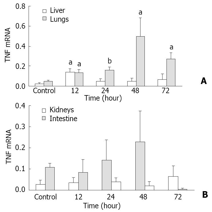
Figure 1 Semiquantitive analysis of tumor necrosis factor-α (TNF-α) mRNA in various organs after thermal injury.
Values are reported as the ratio of TNF-α to β-actin signals. aP < 0.05 and bP < 0.01 as compared to the control values.
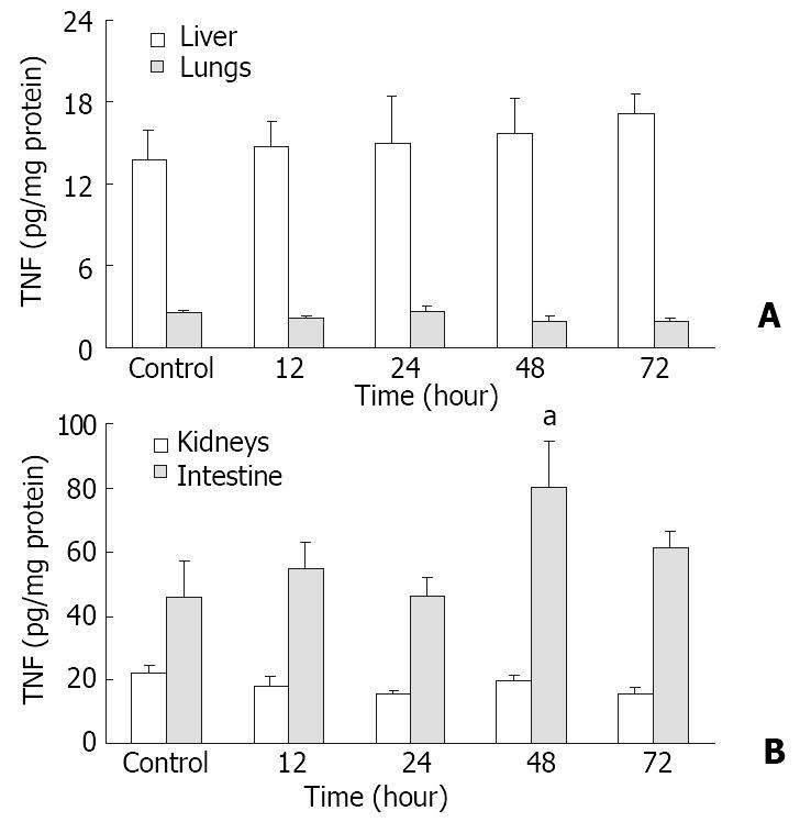
Figure 2 TNF-α protein levels in various organs after thermal injury.
Values are reported as pg/mg protein. aP < 0.05 as com-pared to the control values.
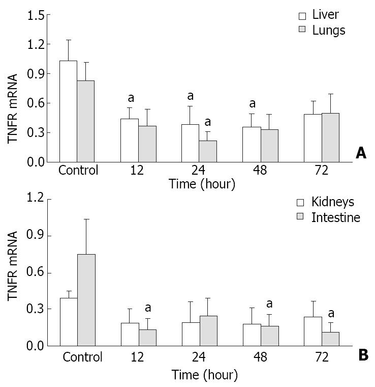
Figure 3 Semiquantitive analysis of tumor necrosis factor-α receptor-I (TNFR-I) mRNA in observed organs after thermal injury.
Values are reported as the ratio of TNFR-I to β-actin signals. aP < 0.05 as compared to the control values.
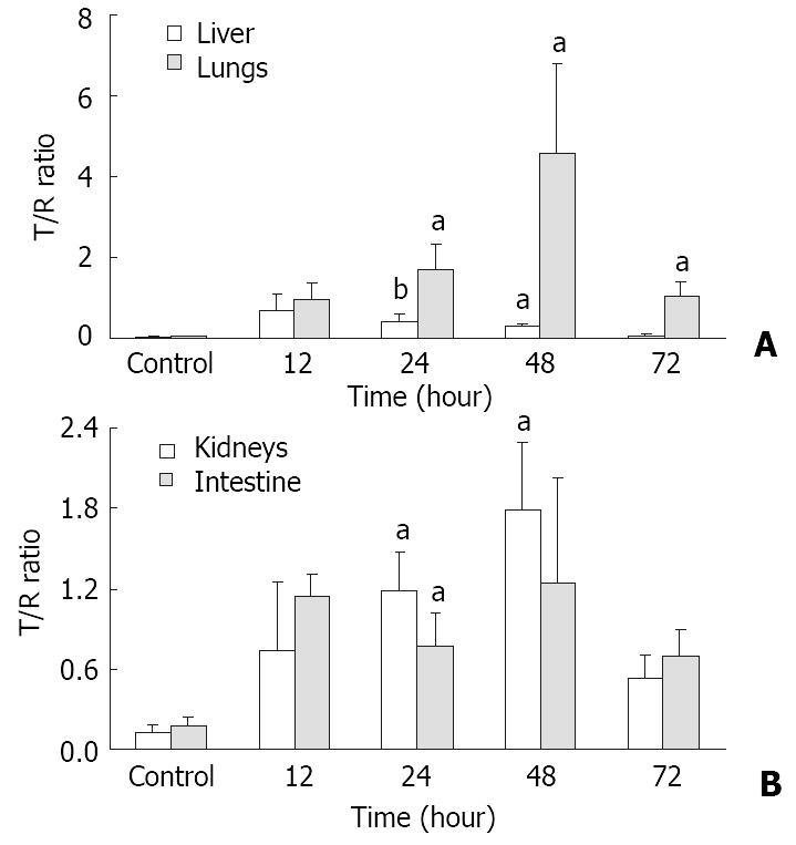
Figure 4 mRNA expression ratio of tumor necrosis factor-α to TNFR-I (T/R ratio) in observed organs after thermal injury.
aP < 0.05 and bP < 0.01 as compared to the control values.
- Citation: Fang WH, Yao YM, Shi ZG, Yu Y, Wu Y, Lu LR, Sheng ZY. The mRNA expression patterns of tumor necrosis factor-α and TNFR-I in some vital organs after thermal injury. World J Gastroenterol 2003; 9(5): 1038-1044
- URL: https://www.wjgnet.com/1007-9327/full/v9/i5/1038.htm
- DOI: https://dx.doi.org/10.3748/wjg.v9.i5.1038


