©The Author(s) 2003.
World J Gastroenterol. Apr 15, 2003; 9(4): 800-803
Published online Apr 15, 2003. doi: 10.3748/wjg.v9.i4.800
Published online Apr 15, 2003. doi: 10.3748/wjg.v9.i4.800
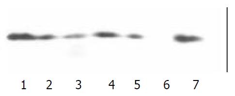
Figure 1 The binding of PH domain to PKC by Western blot with the antibody to PKC.
Lane 1, 2, 3, 4 and 5 were the GST-PH domains of IRS-1, GRF/c, β-ARK1, PLCγ/n and Btk, respectively; lane 6 was the GST; lane 7 was lysate of Jurkat cells.
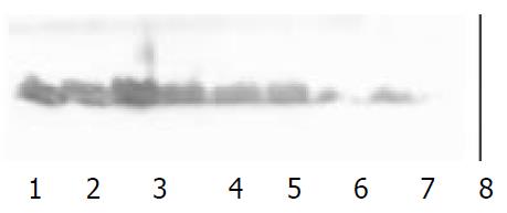
Figure 2 The expressing of GRK2 by Western blot with the antibody to GRK2.
Lane 1, 2, 3, 4 were the results of Western blot in PC-3, MDCK, 7901 and Jurkat cell line respectively. Lane 5,6 was GST.
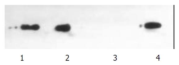
Figure 3 After stimulation of epinephrine, Co-immunopre-cipitation was performed with anti-GRK2 and anti-PKC in co-precipitation and detection of PKC and GRK2.
Lane 1 was PKC detected in precipitate of GRK antibody, lane 2 was GRK2 de-tected in precipitate of PKC antibody. Lane 3 indicated the PKC did not bind to GRK2 in the intact cells without stimulation of epinephrine , lane 4 was the lysate of Jurkat cell.
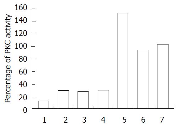
Figure 4 Effects of the PH domain on PKC activity measured by protein kinase C Assay System.
The volume was percent-age of PKC activity. The data were representation of two experiments. Lanes 1, 2, 3, 4 and 5 were GST-PH domain of IRS-1, GRF/c, β-ARK1, PLC γ/n and Btk, respectively; lane 6 wasthe GST; lane 7 was lysate of Jurkat cells.
- Citation: Yang XL, Zhang YL, Lai ZS, Xing FY, Liu YH. Pleckstrin homology domain of G protein-coupled receptor kinase-2 binds to PKC and affects the activity of PKC kinase. World J Gastroenterol 2003; 9(4): 800-803
- URL: https://www.wjgnet.com/1007-9327/full/v9/i4/800.htm
- DOI: https://dx.doi.org/10.3748/wjg.v9.i4.800


