Copyright
©The Author(s) 2003.
World J Gastroenterol. Apr 15, 2003; 9(4): 795-799
Published online Apr 15, 2003. doi: 10.3748/wjg.v9.i4.795
Published online Apr 15, 2003. doi: 10.3748/wjg.v9.i4.795
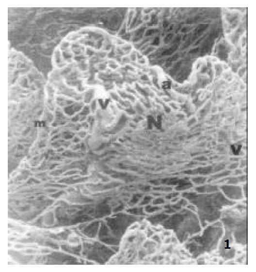
Figure 1 The microvascular architecture of adult rat jejunal villi, showing the villous arteriole (a), the villous capillary network (N), the villous venule (v), the marginal capillary (m) × 150.
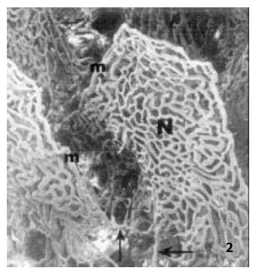
Figure 2 The microvascular architecture of adult rat jejunal villi, showing the marginal capillary (m) connecting to the basal part of its adjacent villous plexus (↑) × 130.
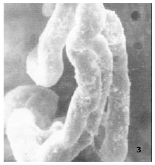
Figure 3 The microvascular architecture of newborn rat jejunal villi, × 400.
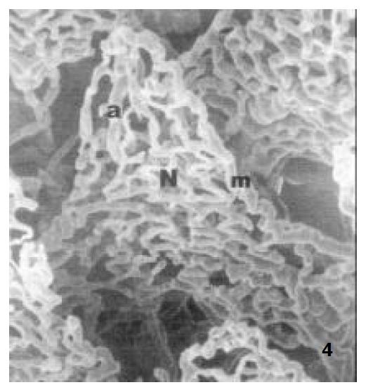
Figure 4 The microvascular architecture of ablactation rat jejunal villi, showing the villous arteriole (a), The villous capillary network (N), the marginal capillary (m) × 300.
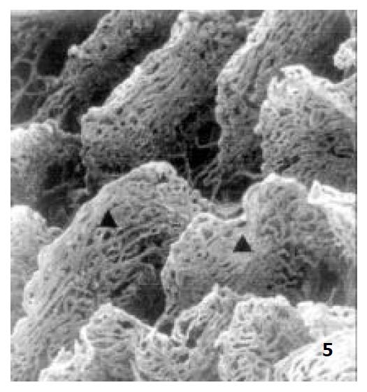
Figure 5 The microvascular architecture of aged rat jejunal villi × 100.
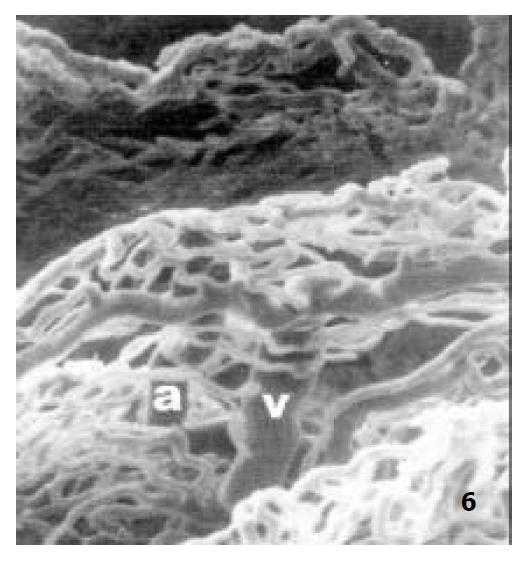
Figure 6 The microvascular architecture of aged rat jejunal villi, showing the villous arteriole (a), the villous venule (v) × 250.
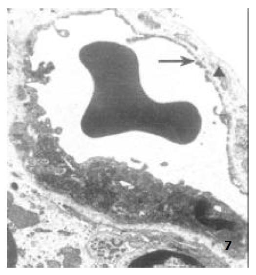
Figure 7 The capillary endothelium of ablactation rats, showing the endothelial hole (↑) and the basal membrane (▲) × 8000.
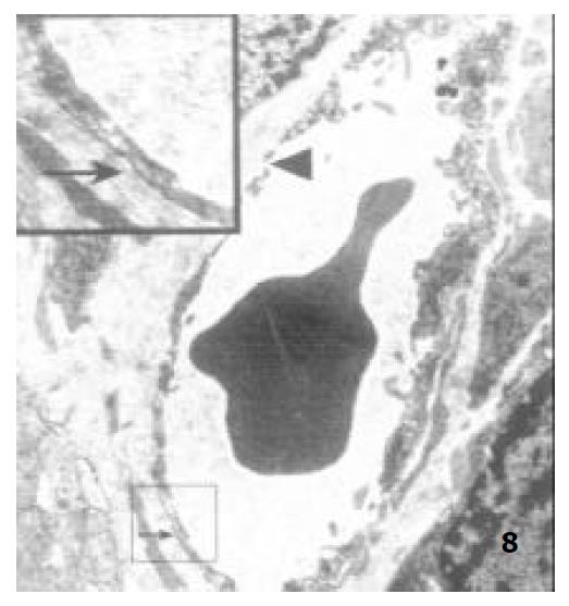
Figure 8 The capillary endothelium of adult rats, showing the basal membrane (↑) and the endothelial hole (▲) × 8000.
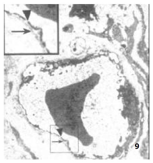
Figure 9 The capillary endothelium of aged rats, showing the basal membrane (↑) and the endothelial hole (▲) × 8000.
- Citation: Chen YM, Zhang JS, Duan XL. Changes of microvascular architecture, ultrastructure and permeability of rat jejunal villi at different ages. World J Gastroenterol 2003; 9(4): 795-799
- URL: https://www.wjgnet.com/1007-9327/full/v9/i4/795.htm
- DOI: https://dx.doi.org/10.3748/wjg.v9.i4.795


