Copyright
©The Author(s) 2003.
World J Gastroenterol. Apr 15, 2003; 9(4): 678-682
Published online Apr 15, 2003. doi: 10.3748/wjg.v9.i4.678
Published online Apr 15, 2003. doi: 10.3748/wjg.v9.i4.678
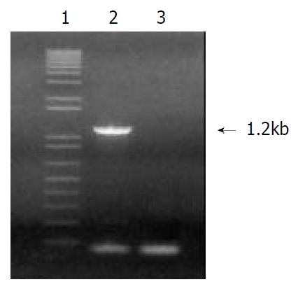
Figure 1 PCR amplified products of HCA587.
(Lanes 1: 1 kb DNA marker; lane 2: 1.2 kb fragment of HCA587; lane 3: nega-tive control).
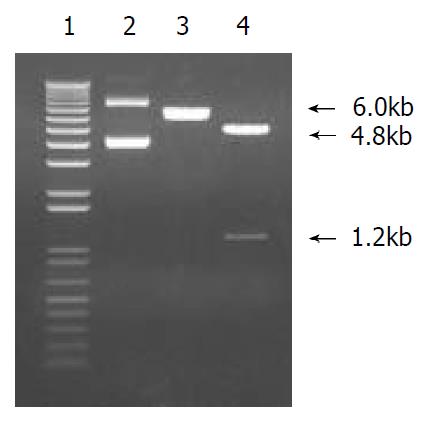
Figure 2 Recombinant pFasBac Htb-587 vector digested by BamH I and Hind III.
(Lanes 1: 1 kb DNA marker; lane 2: recom-binant plasmid; lane 3: digested by BamH I; lane 4: digested by BamH I + Hind III).
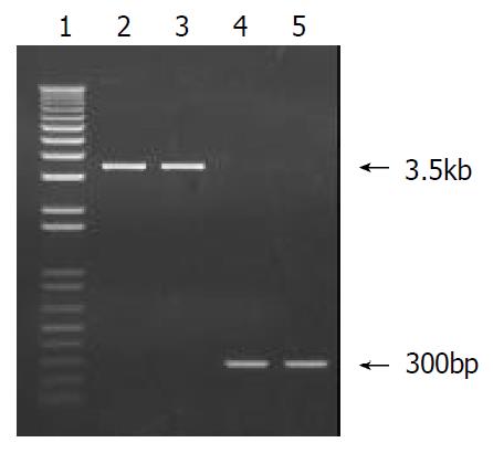
Figure 3 PCR identification of recombinant Bacmid-587.
(lane 1: DNA marker; lane 2-3: recombinant Bacmid-587; lane 4-5: blank bacmid).
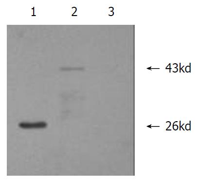
Figure 4 Western blot analysis of recombinant protein ex-pression in sf9 insect cells after transfection.
(lane 1: positive control; lane 2: Sf9 cell transfected with bacmid-587; lane 3: blank Sf9 cells).
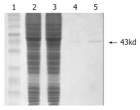
Figure 5 SDS-PAGE analysis of recombinant HCA587.
(lane 1: protein marker; lane 2: sf9 lysate; lane 3: infected sf9 lysate; lane 4-5: purified HCA587 protein).
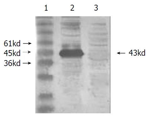
Figure 6 Western blot of recombinant HCA587 reacting with positive serum.
(lane 1: protein marker; lane 2: positive serum of HCC; lane 3: negative control).
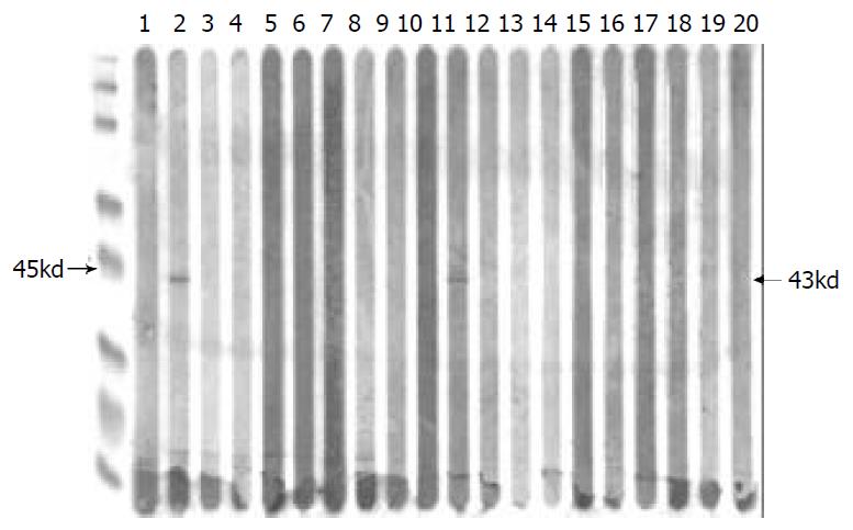
Figure 7 Serological analysis of HCC patients with recombi-nant protein HCA587.
(lane 2 and 11: positive; others: negative; left: protein marker).
- Citation: Li B, Wu HY, Qian XP, Li Y, Chen WF. Expression, purification and serological analysis of hepatocellular carcinoma associated antigen HCA587 in insect cells. World J Gastroenterol 2003; 9(4): 678-682
- URL: https://www.wjgnet.com/1007-9327/full/v9/i4/678.htm
- DOI: https://dx.doi.org/10.3748/wjg.v9.i4.678


