Copyright
©The Author(s) 2003.
World J Gastroenterol. Feb 15, 2003; 9(2): 381-384
Published online Feb 15, 2003. doi: 10.3748/wjg.v9.i2.381
Published online Feb 15, 2003. doi: 10.3748/wjg.v9.i2.381
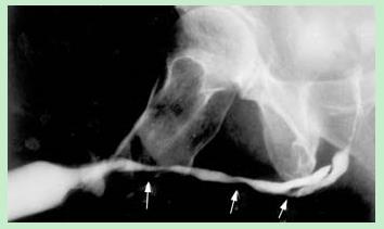
Figure 1 Retrograde urethrogram.
Small arrows indicate stricture, and big arrow indicates stricture and pseudopath.
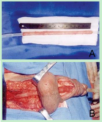
Figure 2 A tubularized free colonic mucosa over 22 F fenestrated silicone tube; B Reconstructive urethra with colonic mucosa.
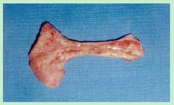
Figure 3 Colonic mucosa-replaced urethra is macroscopically difficult to distinguish from bladder mucosa.
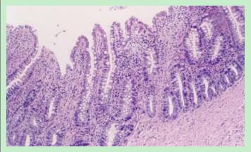
Figure 4 Cross-section of urethra.
Urethral wall is lined by the glandular epithelium (Hematoxylin and eosin stain, × 100).
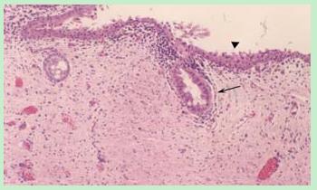
Figure 5 Longitudinal section of urethra.
Arrowhead indicates urinary epithelium, arrows indicate atrophic glandular body (Hematoxylin and eosin stain, × 100).
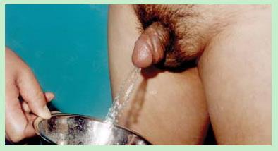
Figure 6 Patient have good urinary streams.
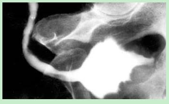
Figure 7 Cystourethrography reveals patent neourethra.
- Citation: Xu YM, Qiao Y, Sa YL, Zhang J, Zhang HZ, Zhang XR, Wu DL, Chen R. One-stage urethral reconstruction using colonic mucosa graft: An experimental and clinical study. World J Gastroenterol 2003; 9(2): 381-384
- URL: https://www.wjgnet.com/1007-9327/full/v9/i2/381.htm
- DOI: https://dx.doi.org/10.3748/wjg.v9.i2.381


