©The Author(s) 2003.
World J Gastroenterol. Feb 15, 2003; 9(2): 324-328
Published online Feb 15, 2003. doi: 10.3748/wjg.v9.i2.324
Published online Feb 15, 2003. doi: 10.3748/wjg.v9.i2.324
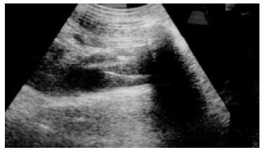
Figure 1 Intrahepatic portal vein, RHSIVC of swine and the modified RUPS-100 set wedging against the anterior-lateral wall of RHSIVC are clearly demonstrated on sonography.
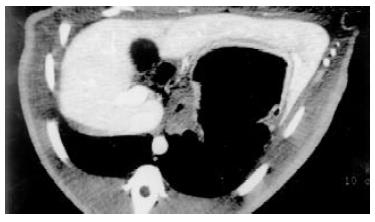
Figure 2 Sectional abdominal enhanced CT scan of swine before DIPS demonstrates the swine's intrahepatic portal vein and retrohepatic segment of IVC clearly.
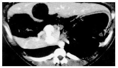
Figure 3 No hematoma beneath the hepatic integument or intraperitoneal bleeding is dectected on sectional abdominal enhanced CT imaging of swine after DIPS.
The self-expandable home-made stent is accurately deployed in the shunt connecting the intrahepatic portal vein and RHSIVC.
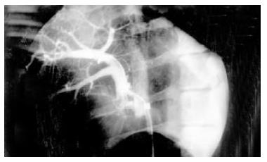
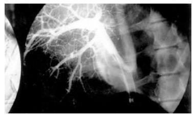
Figure 4 After intrahepatic portal vein successfully accessed via retrohepatic segment of IVC approach, portography was performed with a 5-F pigtail catheter.
Swine's anterior and posterior branch of right portal vein is clearly demonstrated.
Figure 5 The DIPS shunt is lined by an 8 mm-diamter, 6 cm-long home-made metal bare stent.
The stent is fully dilated and accurately connected the swine's RHSIVC and anterior branch of right portal vein. Swine's heptaic perfusion is well and animal's IVC was patent. No extravasation of contrast medium can be detected.
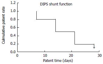
Figure 6 Median patency time of 10 DIPS shunt is 14 d.
7, 14, 21 and 28 d after DIPS procedure the cumulative patency rate of shunt is 100%, 80%, 50% and 20% respectively.
- Citation: Luo JJ, Yan ZP, Zhou KR, Qian S. Direct intrahepatic portacaval shunt: An experimental study. World J Gastroenterol 2003; 9(2): 324-328
- URL: https://www.wjgnet.com/1007-9327/full/v9/i2/324.htm
- DOI: https://dx.doi.org/10.3748/wjg.v9.i2.324


