Copyright
©The Author(s) 2003.
World J Gastroenterol. Feb 15, 2003; 9(2): 238-241
Published online Feb 15, 2003. doi: 10.3748/wjg.v9.i2.238
Published online Feb 15, 2003. doi: 10.3748/wjg.v9.i2.238
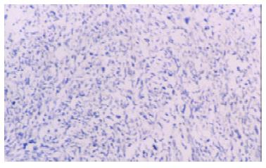
Figure 1 The expression of 1A6 protein in sarcoma.
The cells were negative staining. Immunohistochemical stain, × 100.
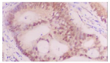
Figure 2 The expression of 1A6 protein in intestinal type gastric carcinoma.
The tumor cells showed a nuclear staining. Immunohistochemical stain, × 400.
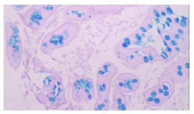
Figure 3 Type I intestinal metaplasia.
The mucins in cells were stained blue. HID/AB/PAS stain, × 200.
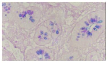
Figure 4 Type II intestinal metaplasia.
Themucins in cells were stained blue and magenta. HID/AB/PAS stain, × 200.
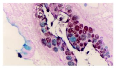
Figure 5 Type III intestinal metaplasia.
The mucins in cells were stained blue and brown. HID/AB/PAS stain, × 200.
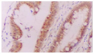
Figure 6 The expression of 1A6 protein in type III intestinal metaplasia.
The metaplastic cells showed a nuclear and plasmatic staining. Immunohistochemical stain, × 200.
- Citation: Liu YQ, Zhao H, Ning T, Ke Y, Li JY. Expression of 1A6 gene and its correlation with intestinal gastric carcinoma. World J Gastroenterol 2003; 9(2): 238-241
- URL: https://www.wjgnet.com/1007-9327/full/v9/i2/238.htm
- DOI: https://dx.doi.org/10.3748/wjg.v9.i2.238


