©The Author(s) 2003.
World J Gastroenterol. Dec 15, 2003; 9(12): 2880-2882
Published online Dec 15, 2003. doi: 10.3748/wjg.v9.i12.2880
Published online Dec 15, 2003. doi: 10.3748/wjg.v9.i12.2880
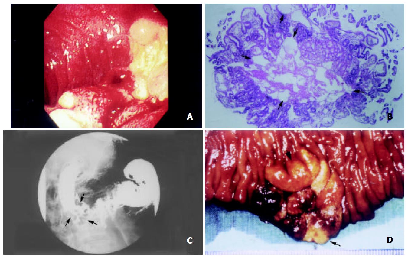
Figure 1 (A) Panendoscopy revealed a 3 cm irregular elevated lesion with whitish spots on the surface and a central crater with blood-coating in the second portion of duodenum.
(B) Histology of the endoscopic biopsy showed prominent dilatation of intramucosal and submucosal lymphatic ducts (arrows), a picture of intestinal lymphangiectasia (Hematoxylin and eosin, ×40). (C) Upper gastrointestinal series demonstrated a cluster of polypoid filling defects, 3×3 cm in size, in the second portion of the duode-num (arrows). (D) Gross appearance of the surgical specimen revealed a sessile polypoid lesion, 3 cm in diameter, in the second portion of the duodenum (arrows).
- Citation: Chen CP, Chao Y, Li CP, Lo WC, Wu CW, Tsay SH, Lee RC, Chang FY. Surgical resection of duodenal lymphangiectasia: A case report. World J Gastroenterol 2003; 9(12): 2880-2882
- URL: https://www.wjgnet.com/1007-9327/full/v9/i12/2880.htm
- DOI: https://dx.doi.org/10.3748/wjg.v9.i12.2880


