©The Author(s) 2003.
World J Gastroenterol. Dec 15, 2003; 9(12): 2715-2719
Published online Dec 15, 2003. doi: 10.3748/wjg.v9.i12.2715
Published online Dec 15, 2003. doi: 10.3748/wjg.v9.i12.2715
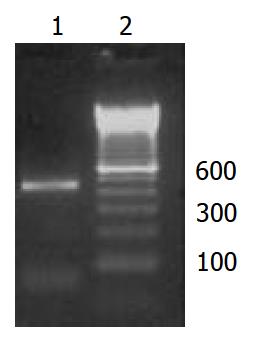
Figure 1 Amplification of hnmMLCK cDNA by PCR .
1. prod-ucts of PCR amplification, 2. the 100 bp ladder.
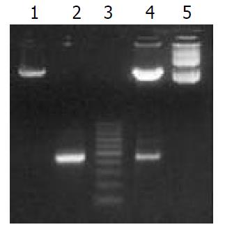
Figure 2 Analysis of human MLCK recombinant plasmids with restriction endonucleases mapping.
1. pBKcmv/EcoRI-HindIII, 2. PCR products/EcoRI-HindIII, 3. the 100 bp DNA ladder, 4. pBK-hnmMLCK/EcoRI-HindIII, 5. pBK-hnmMLCK.
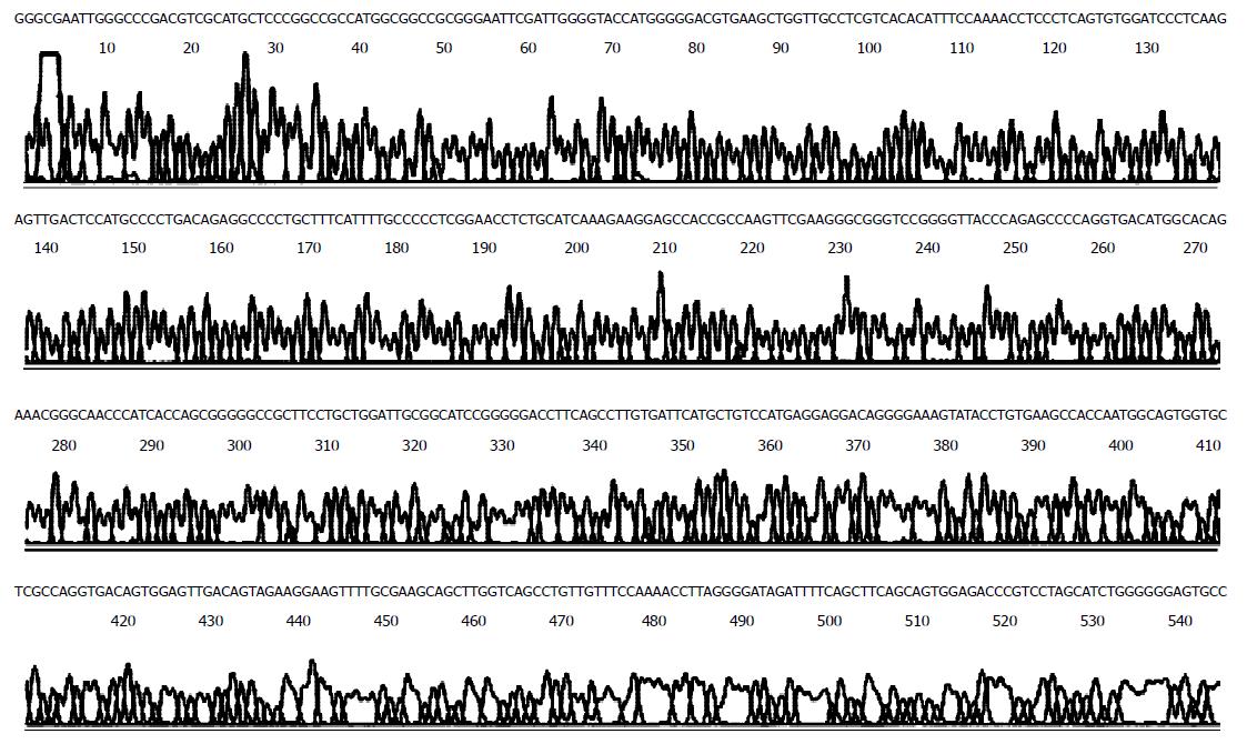
Figure 3 DNA sequences of pBK-hnmMLCK were detected by ABI377 auto analytical instrument.
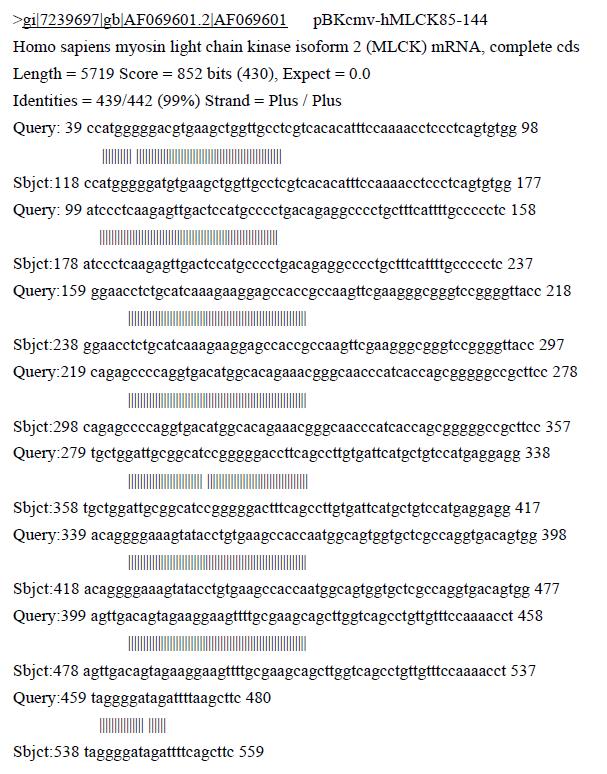
Figure 4 Comparison between sequencing results of hnmMLCK DNA and those published by Genbank.
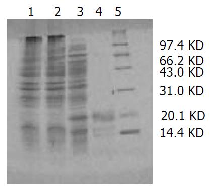
Figure 5 Analysis of pBK-hnmMLCK with SDS-PADE.
1. pBKcmv in XL1-blue, 2. pBK-hnmMLCK in XL1-blue before induced, 3. pBK-hnmMLCK in XL1-blue after induced, 4. pu-rified expression protein, 5. protein markers.
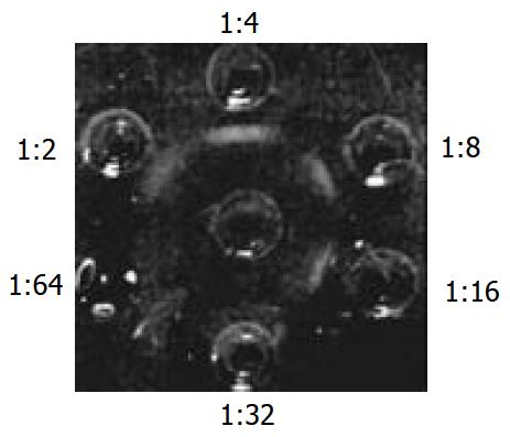
Figure 6 Antiserum titer detected by immuno-double-diffusion.
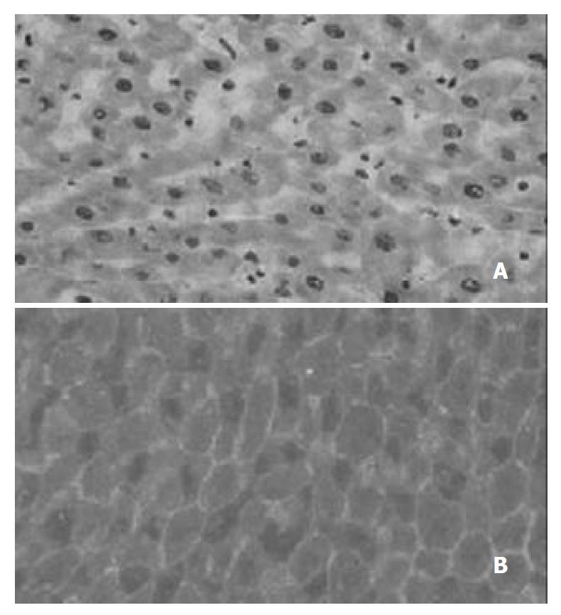
Figure 7 MLCK distribution in hepatocytes of New Zealand rabbits.
A: H and E staining, B: MLCK distribution.
- Citation: Zhu HQ, Wang Y, Hu RL, Ren B, Zhou Q, Jiang ZK, Gui SY. Distribution and expression of non-muscle myosin light chain kinase in rabbit livers. World J Gastroenterol 2003; 9(12): 2715-2719
- URL: https://www.wjgnet.com/1007-9327/full/v9/i12/2715.htm
- DOI: https://dx.doi.org/10.3748/wjg.v9.i12.2715


