©The Author(s) 2003.
World J Gastroenterol. Jan 15, 2003; 9(1): 94-98
Published online Jan 15, 2003. doi: 10.3748/wjg.v9.i1.94
Published online Jan 15, 2003. doi: 10.3748/wjg.v9.i1.94
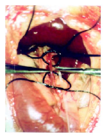
Figure 1 Catheterization of the gastroduodenal artery in vivo
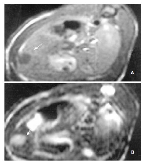
Figure 2 MR images of HCC before the application of Plcg-Mitomycin-microspheres: (a) T1-weighted image (400/14 ms) showed a hypointense pattern in the left lateral lobe of the liver, (b) T2-weighted image (3000/96 ms) showed a hyperintense pattern in the liver.
Necrosis, hemorrhage and metastase of the tumor were not observed.
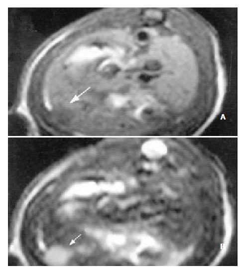
Figure 3 MR images of HCC after the application of Plcg-Mitomycin-microspheres: (a) MRI T1WI (400/14 ms); (b) MRI T2WI (3000/96 ms).
The tumor volume after the application of Plcg-Mitomycin-microspheres was not changed compared to that before the application.
- Citation: Qian J, Truebenbach J, Graepler F, Pereira P, Huppert P, Eul T, Wiemann G, Claussen C. Application of poly-lactide-co-glycolide-microspheres in the transarterial chemoembolization in an animal model of hepatocellular carcinoma. World J Gastroenterol 2003; 9(1): 94-98
- URL: https://www.wjgnet.com/1007-9327/full/v9/i1/94.htm
- DOI: https://dx.doi.org/10.3748/wjg.v9.i1.94


