Copyright
©The Author(s) 2002.
World J Gastroenterol. Dec 15, 2002; 8(6): 982-986
Published online Dec 15, 2002. doi: 10.3748/wjg.v8.i6.982
Published online Dec 15, 2002. doi: 10.3748/wjg.v8.i6.982
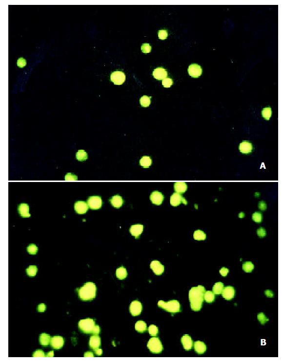
Figure 1 SGC-7901 cells stained with DAPI.
A: UT control; B: VES at 20 mg·L-1
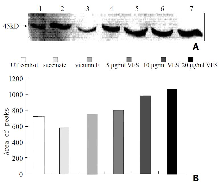
Figure 2 The expression of Fas protein in SGC-7901 cells follow-ing treatment of VES for 24 h.
Lane1: Molecular weight marker; Lane2: UT control; Lane3: succinate; Lane4: vitamin E; Lane5: VES at 5 mg·L-1; Lane6: VES at 10 mg·L-1; Lane7: VES at 20 mg·L-1.
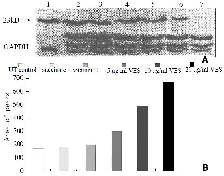
Figure 3 The expression of FADD protein in SGC-7901 cells fol-lowing treatment of VES for 24 h.
Lane1: Molecular weight marker; Lane2: VES at 20 mg·L-1; Lane3: VES at 10 mg·L-1; Lane4: VES at 5 mg·L-1; Lane5: vitamin E; Lane6: succinate; Lane7: UT control.
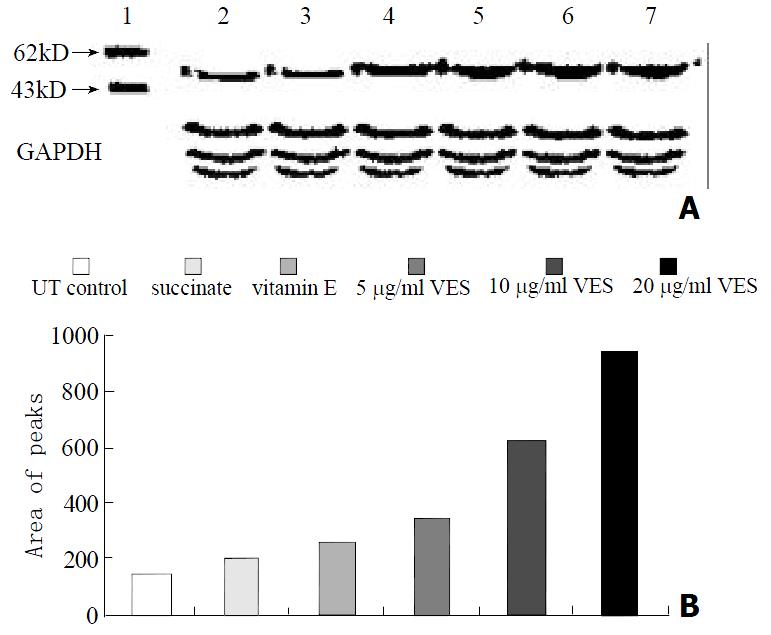
Figure 4 The expression of caspase-8 protein in SGC-7901 cells fol-lowing treatment of VES for 24 h.
Lane1: Molecular weight marker; Lane2: UT control; Lane3: succinate; Lane4: vitamin E; Lane5: VES at 5 mg·L-1; Lane6: VES at 10 mg·L-1; Lane7: VES at 20 mg·L-1.
Figure 5 The expression of FADD protein when Fas antisense was transfected into SGC-7901 cells following treatment of VES for 24 h.
- Citation: Wu K, Li Y, Zhao Y, Shan YJ, Xia W, Yu WP, Zhao L. Roles of Fas signaling pathway in vitamin E succinate-induced apoptosis in human gastric cancer SGC-7901 cells. World J Gastroenterol 2002; 8(6): 982-986
- URL: https://www.wjgnet.com/1007-9327/full/v8/i6/982.htm
- DOI: https://dx.doi.org/10.3748/wjg.v8.i6.982


