Copyright
©The Author(s) 2002.
World J Gastroenterol. Dec 15, 2002; 8(6): 1040-1044
Published online Dec 15, 2002. doi: 10.3748/wjg.v8.i6.1040
Published online Dec 15, 2002. doi: 10.3748/wjg.v8.i6.1040
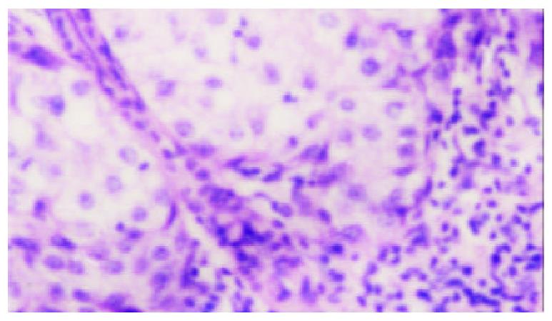
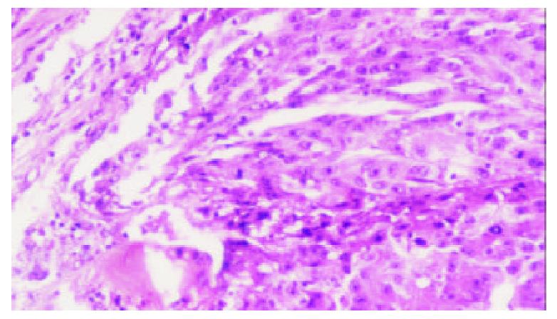
Figure 1 Massive lymphocytes infiltrating the interface be-tween HCC and surroundings (HE, ×100)
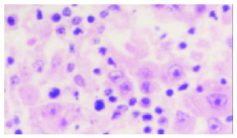
Figure 2 Many lymphocytes infiltrating tumor stroma (HE, × 100)
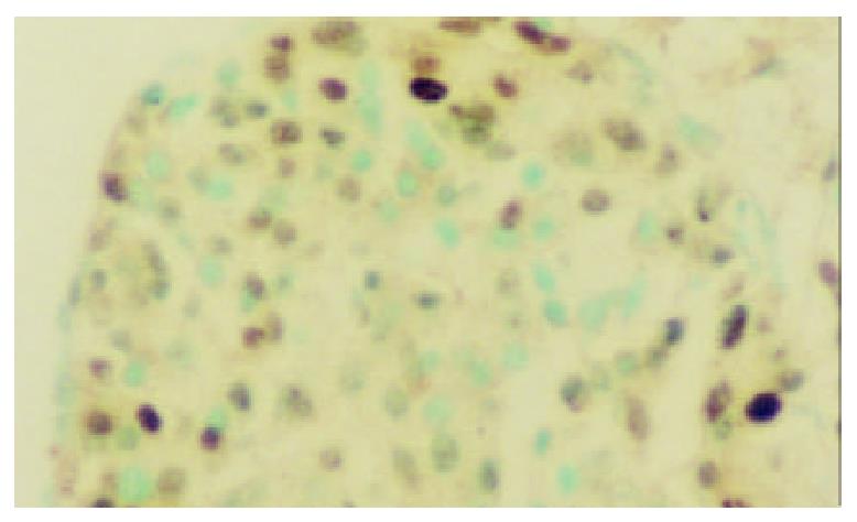
Figure 3 PCNA positive cells mainly distributed in the peripheral area of tumor nest (PCNA immunohistochemical staining, × 200)
Figure 4 HCC invasion in capsule (HE, × 100)
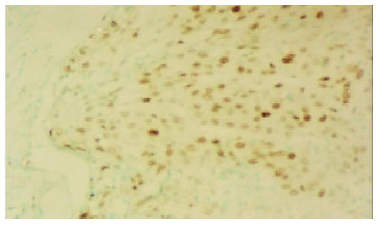
Figure 5 PCNA positive cells (the continuous section of Figure 4, PCNA immunohistochemical staining, × 100)
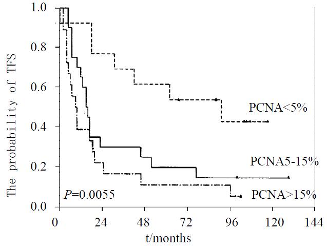
Figure 6 The Kaplan-Meier curves of different PCNA-LI groups
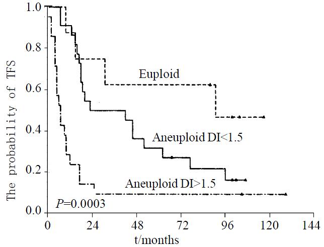
Figure 7 The Kaplan-Meier curves of different DI groups
- Citation: Zeng WJ, Liu GY, Xu J, Zhou XD, Zhang YE, Zhang N. Pathological characteristics, PCNA labeling index and DNA index in prognostic evaluation of patients with moderately differentiated hepatocellular carcinoma. World J Gastroenterol 2002; 8(6): 1040-1044
- URL: https://www.wjgnet.com/1007-9327/full/v8/i6/1040.htm
- DOI: https://dx.doi.org/10.3748/wjg.v8.i6.1040


