©The Author(s) 2002.
World J Gastroenterol. Oct 15, 2002; 8(5): 956-960
Published online Oct 15, 2002. doi: 10.3748/wjg.v8.i5.956
Published online Oct 15, 2002. doi: 10.3748/wjg.v8.i5.956
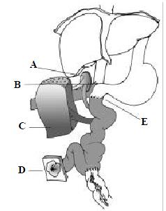
Figure 1 Small-bowel and auxiliary liver allograft.
A. Carrel patch containing the origin of the superior mesenteric artery and the coeliac artery is anastomosed to the recipient's aorta; B. Anastomosis of end of the donor infrahepatic vena cava to the side of recipient's vena cava; C. The reduced liver; D. Ilesotomy; E. Anastomosis of donor jejunum to the recipient's duodenum
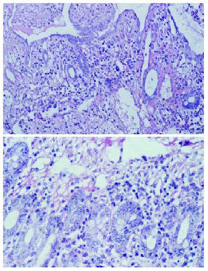
Figure 2 Photomicrographs showing acute rejection with lymphocytic crytitis on the 7 th postoperative day, the mucosal destruction and necrosis were not observed (H&E, A, × 200; B.
× 400).
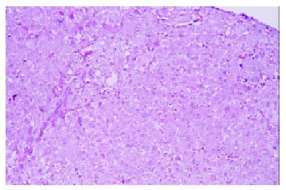
Figure 3 Liver biopsy specimens on the 9th postoperative day showed normal appearance of the allograft (× 200), no inflammatory infiltrate in the portal tract.
- Citation: Zhang WJ, Liu DG, Ye QF, Sha B, Zhen FJ, Guo H, Xia SS. Combined small bowel and reduced auxiliary liver transplantation: Case report. World J Gastroenterol 2002; 8(5): 956-960
- URL: https://www.wjgnet.com/1007-9327/full/v8/i5/956.htm
- DOI: https://dx.doi.org/10.3748/wjg.v8.i5.956


