Copyright
©The Author(s) 2002.
World J Gastroenterol. Aug 15, 2002; 8(4): 679-685
Published online Aug 15, 2002. doi: 10.3748/wjg.v8.i4.679
Published online Aug 15, 2002. doi: 10.3748/wjg.v8.i4.679
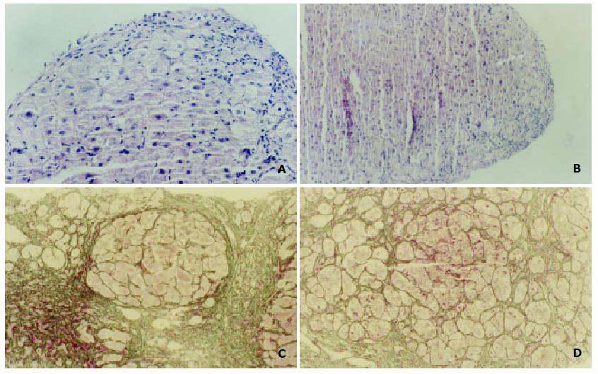
Figure 1 Histological changes of liver biopsy specimens before and after treatment in IFN-γ group: A: First liver biopsy (G3, HE stain, × 100) before treatment; B: Second liver biopsy (G3, HE stain, × 100) after treatment; C: First liver biopsy (S4, collagen stain, × 100) before treatment; D: Second liver biopsy (S3, GS stain, × 100) after treatment.
Figure 2 Histological changes of liver biopsy specimens before and after treatment in SA-B group: A: First liver biopsy (G3, HE stain, × 100) before treatment; B: Second liver biopsy (G3, HE stain, × 100) after treatment; C: First liver biopsy (S4, collagen stain, × 100) before treatment; D: Second liver biopsy (S3, collgen stain, .
× 100) after treatment.
- Citation: Liu P, Hu YY, Liu C, Zhu DY, Xue HM, Xu ZQ, Xu LM, Liu CH, Gu HT, Zhang ZQ. Clinical observation of salvianolic acid B in treatment of liver fibrosis in chronic hepatitis B. World J Gastroenterol 2002; 8(4): 679-685
- URL: https://www.wjgnet.com/1007-9327/full/v8/i4/679.htm
- DOI: https://dx.doi.org/10.3748/wjg.v8.i4.679


