Copyright
©The Author(s) 2002.
World J Gastroenterol. Aug 15, 2002; 8(4): 658-662
Published online Aug 15, 2002. doi: 10.3748/wjg.v8.i4.658
Published online Aug 15, 2002. doi: 10.3748/wjg.v8.i4.658
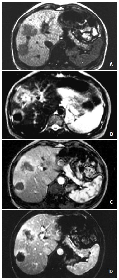
Figure 1 HCC after TACE.
A: T1WI shows two hypointense lesions in the right anterior and posterior lobe; B: T2WI shows that the central coagulative necrosis of the right posterior lesion is hypointense and the peripheral residuals of viable tumors is hyperintense. The right anterior lesion is inhomogeneous intensity with liquefied necrosis (central higher hyperintensity), coagulative necrosis (punctual hypointensity) and peripheral residuals of viable tumors (peripheral hyperintensity); C: FMPSPGR dynamic contrast early phase scanning shows that both of liquefied necrosis and coagulative necrosis have no enhancement and peripheral residuals of viable tumors enhanced; D: The peripheral residuals of viable tumors have permanent enhancement at the dynamic contrast late phase.
- Citation: Yan FH, Zhou KR, Cheng JM, Wang JH, Yan ZP, Da RR, Fan J, Ji Y. Role and limitation of FMPSPGR dynamic contrast scanning in the follow-up of patients with hepatocellular carcinoma treated by TACE. World J Gastroenterol 2002; 8(4): 658-662
- URL: https://www.wjgnet.com/1007-9327/full/v8/i4/658.htm
- DOI: https://dx.doi.org/10.3748/wjg.v8.i4.658


