Copyright
©The Author(s) 2002.
World J Gastroenterol. Aug 15, 2002; 8(4): 619-623
Published online Aug 15, 2002. doi: 10.3748/wjg.v8.i4.619
Published online Aug 15, 2002. doi: 10.3748/wjg.v8.i4.619
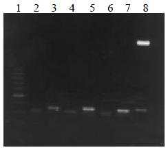
Figure 1 VH and VL DNA amplified by RT-PCR.
Lane 1: 100 bp DNA ladder (100-1000 bp); lane 2, 4, 6: VL DNA; lane 3, 5, 7: VH DNA; lane 8: VH DNA marker (2.7 kb, 350 bp)
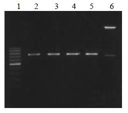
Figure 2 ScFv DNA amplified by RT-PCR.
Lane 1: 100 bp DNA ladder (100-1000 bp); lane 2, 3, 4, 5: MC5, MC3, MGD1, MGD1 ScFv DNA; lane 6 ScFv DNA marker (2.7 kb, 750 bp)
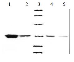
Figure 3 Western bloting of soluble MC3-ScFv clones.
1, 2, 4, 5: Four posotive clones of MC3 ScFv, 3: Low molecular weight marker (14.4, 20.1, 31.0, 43.0, 66.2, 97.4) KD
- Citation: Nie YZ, He FT, Li ZK, Wu KC, Cao YX, Chen BJ, Fan DM. Identification of tumor associated single-chain Fv by panning and screening antibody phage library using tumor cells. World J Gastroenterol 2002; 8(4): 619-623
- URL: https://www.wjgnet.com/1007-9327/full/v8/i4/619.htm
- DOI: https://dx.doi.org/10.3748/wjg.v8.i4.619


