Copyright
©The Author(s) 2002.
World J Gastroenterol. Jun 15, 2002; 8(3): 499-504
Published online Jun 15, 2002. doi: 10.3748/wjg.v8.i3.499
Published online Jun 15, 2002. doi: 10.3748/wjg.v8.i3.499
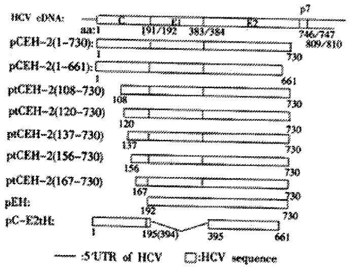
Figure 1 Schematic maps of HCV coding sequences inserted into the plasmid pTM1 and expressed under transcriptional control of the bacteriophage T7 pol promoter.
Numbers refer to amino acids of the HCV polyprotein.
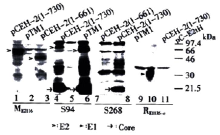
Figure 2 Detection of E2 glycoprotein species of different molecular masses.
Transient expression products were analyzed by Western blot with antibodies of distinct origin: mouse polyclonal antibody ME2116 (lane 1, 2, 3); HCV patient serum S94 (lane 4, 5, 6); HCV patient serum S268 (lane 7, 8); rabbit polyclonal antibody RE1135-C (lane 9, 10, 11). Empty vector pTM1 was used as the negative control. HCV-specific protein bands are indicated by arrowheads. The plasmids used for transfection are indicated at the top of the lanes.
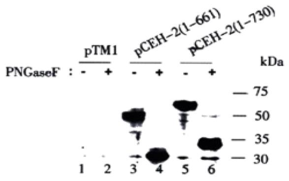
Figure 3 Deglycosylation analysis of the expressed HCV E2 proteins with PNGase F.
Transient expression products were subjected to Western blot analysis with ME2116 as the primary antibody. pTM1 transfected cells were used as control. The plasmids used for transfection are indicated at the top of the lanes. Samples incubated with deglycosylation buffer were run in parallel. +: samples digested with PNGase F, -: samples incubated with PNGase F reaction buffer.
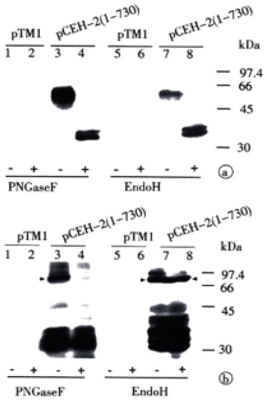
Figure 4 Different sensitivities of two glycosylated E2 species to PNGase F and Endo H.
Transient expression products of pCEH-2 (1-730) were digested with PNGase F or Endo H, respectively. The digested samples were analyzed by Western blot with ME2116 (A) and S94 (B). Empty vector pTM1 was used as negative control.
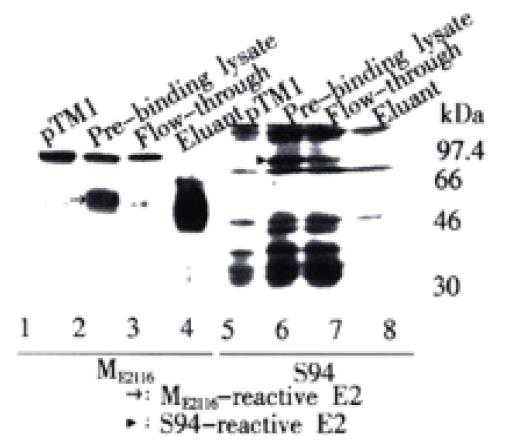
Figure 5 Different ability of differently glycosylated E2 speicies to bind to GNA-agarose.
HeLa cells infected with vTT7 and transfected with pCEH-2 (1-730) were collected, washed and lysed with lysis buffer. The cleared supernatant was then allowed to bind to GNA-agarose. The gel beads were washed and eluted with 1M α-D-mannopyranoside in lysis buffer. Pre-binding lysate, flow-through and eluant fractions were analyzed by Western blot analysis. HeLa cells infected with vTT7 and transfected with pTM1 were served as negative control. The sera used as primary antibodies are indicated at the bottom of the lanes. E2 proteins are indicated by arrowheads.
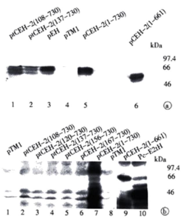
Figure 6 Requirement of the core sequence for the expression of complex-type glycosylated E2.
Expression products of differently truncated HCV structural genes were analyzed by Western blot analysis. Blots were probed with ME2116 (A) or with S94 (B). Empty vector pTM1 was used as negative control. The plasmids used for transfection are indicated at the top of the lanes.
- Citation: Zhu LX, Liu J, Li YC, Kong YY, Staib C, Sutter G, Wang Y, Li GD. Full-length core sequence dependent complex-type glycosylation of hepatitis C virus E2 glycoprotein. World J Gastroenterol 2002; 8(3): 499-504
- URL: https://www.wjgnet.com/1007-9327/full/v8/i3/499.htm
- DOI: https://dx.doi.org/10.3748/wjg.v8.i3.499


