Copyright
©The Author(s) 2002.
World J Gastroenterol. Jun 15, 2002; 8(3): 483-487
Published online Jun 15, 2002. doi: 10.3748/wjg.v8.i3.483
Published online Jun 15, 2002. doi: 10.3748/wjg.v8.i3.483
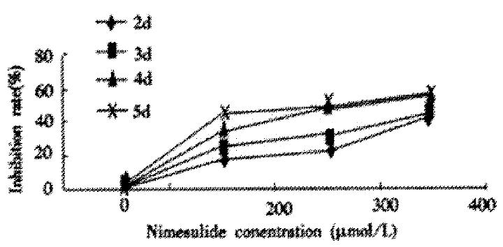
Figure 1 Inhibition rate of Nimesulide on proliferation of SMMC-7721 cells.
Cells were incubated with 200 μmol/L, 300 μmol/L, 400 μmol/L Nimesulide for 2 d, 3 d, 4 d, 5 d respectively.
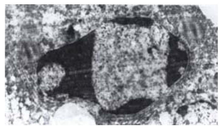
Figure 2 Transmission electron micrograph of SMMC-7721 cells treated with Nimesulide at the concentration of 300 μmol/L for 72 h.
The picture showed early change of apoptosis, the nuclear chromatin condensation.
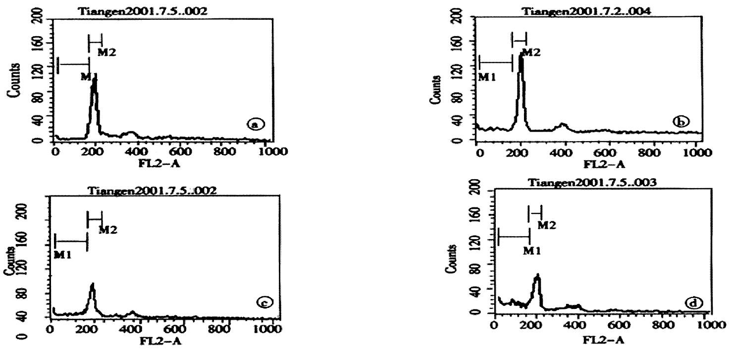
Figure 3 Cell apoptosis was determined by flow-cytometry, SMMC-7721 cells were treated with Nimesulide at various concentrations (0, 200, 300, 400 μmol/L respectively A to D).
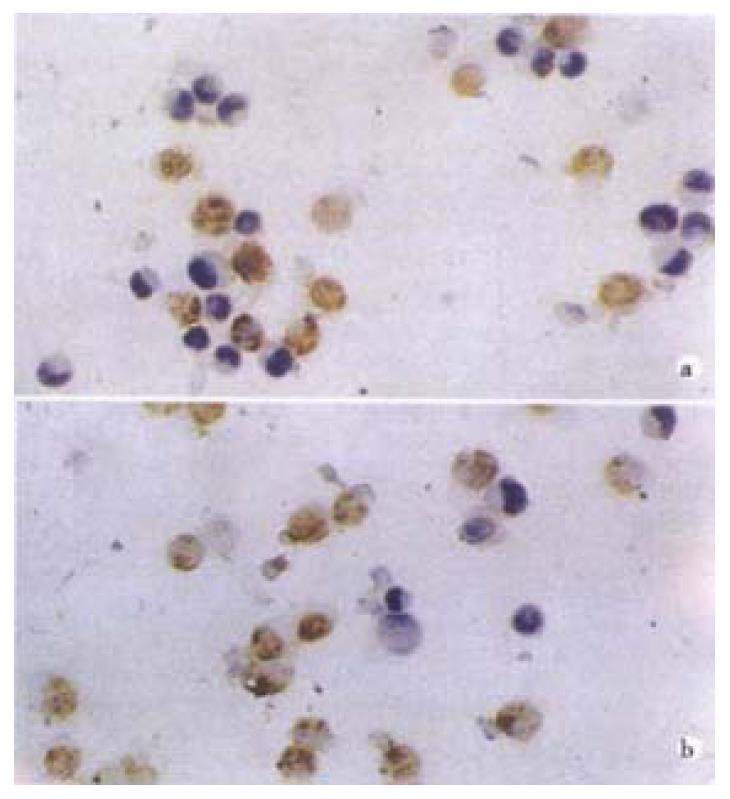
Figure 4 TUNEL stain showed SMMC-7721 cells apoptosis.
SMMC-7721 cells were treated with Nimesulide at various concentrations (300, 400 μmol/L A and B).
- Citation: Tian G, Yu JP, Luo HS, Yu BP, Yue H, Li JY, Mei Q. Effect of Nimesulide on proliferation and apoptosis of human hepatoma SMMC-7721 cells. World J Gastroenterol 2002; 8(3): 483-487
- URL: https://www.wjgnet.com/1007-9327/full/v8/i3/483.htm
- DOI: https://dx.doi.org/10.3748/wjg.v8.i3.483


