©The Author(s) 2002.
World J Gastroenterol. Apr 15, 2002; 8(2): 270-275
Published online Apr 15, 2002. doi: 10.3748/wjg.v8.i2.270
Published online Apr 15, 2002. doi: 10.3748/wjg.v8.i2.270
Figure 1 Schematic representation of the adenovirus shuttle plasmid, LTR, left inversed terminal repeats; pA, polyadenylation signal.
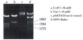
Figure 2 Map of pAdCEACD.
There was a fragment of 1.8 kb containing CEA and CD after being digested with Xba I and Hind III; As there were two corIrestriction sites, there were three fragments after treatment with EcoR I and Hind III, 0.9 kb for LTR and CEA, 1.3 kb for CD, 2.2 kb for LTR and CEA and CD.
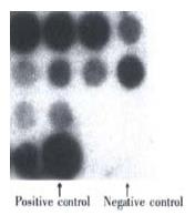
Figure 3 The identification of the recombinant adenovirus AdCEACD by dot blotting.
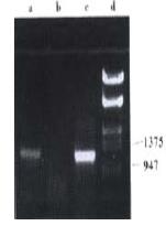
Figure 4 The identification of the recombinant adenovirus AdCEACD by PCR.
a, PCR product using AdCEACD as templet; b, PCR product using pAdE1CMV as templet; c, PCR product using pAdCMVCD as templet; d, λDNA Marker.
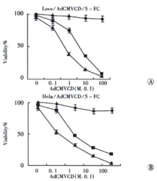
Figure 5 Sensitivity of cells infected with AdCMVCD to 5-FC.
◆: 5-FC 0; ■: 5-FC, 15 μmol•L⁻¹; ▲: 5-FC, 150 μmol•L⁻¹.
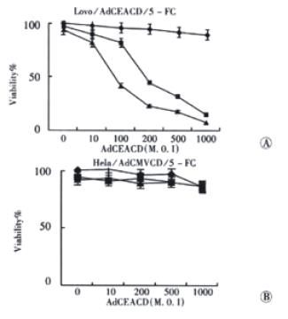
Figure 6 Sensitivity of cells infected with AdCEACD to 5-FC.
◆: 5-FC 0; ■: 5-FC, 15 μmol•L⁻¹; ▲:5-FC, 150 μmol•L⁻¹.
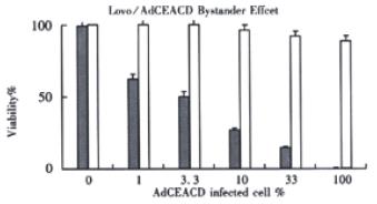
Figure 7 The bystander effect of AdCEACD/5-FC system.
□: without 5-FC; ■: 5-FC 150 μmol•L⁻¹
- Citation: Shen LZ, Wu WX, Xu DH, Zheng ZC, Liu XY, Ding Q, Hua YB, Yao K. Specific CEA-producing colorectal carcinoma cell killing with recombinant adenoviral vector containing cytosine deaminase gene. World J Gastroenterol 2002; 8(2): 270-275
- URL: https://www.wjgnet.com/1007-9327/full/v8/i2/270.htm
- DOI: https://dx.doi.org/10.3748/wjg.v8.i2.270


