Copyright
©The Author(s) 2001.
World J Gastroenterol. Dec 15, 2001; 7(6): 766-771
Published online Dec 15, 2001. doi: 10.3748/wjg.v7.i6.766
Published online Dec 15, 2001. doi: 10.3748/wjg.v7.i6.766
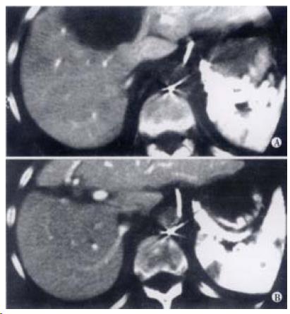
Figure 1 A: Solitary hepatic metastasis occupying segment 4, 5 and 8, managed by trisegmental resection; B: CT scan at one year showing hypertrophy of residual left lobe and segments 6/7.
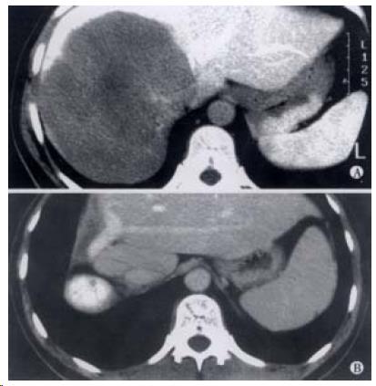
Figure 2 (A) Large solitary metastasis resected by extended right hepatectomy.
(B) CT scan showing hypertrophied left and caudate lobes with no evidence of recurrence at one year.
- Citation: Parks R, Garden O. Liver resection for cancer. World J Gastroenterol 2001; 7(6): 766-771
- URL: https://www.wjgnet.com/1007-9327/full/v7/i6/766.htm
- DOI: https://dx.doi.org/10.3748/wjg.v7.i6.766


