©The Author(s) 2001.
World J Gastroenterol. Oct 15, 2001; 7(5): 637-641
Published online Oct 15, 2001. doi: 10.3748/wjg.v7.i5.637
Published online Oct 15, 2001. doi: 10.3748/wjg.v7.i5.637
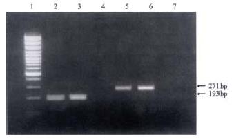
Figure 1 Target amplification fragments from HGV RNA and TTV DNA.
(1: marker; 2 and 3: HGV positive serum samples; 4: blank for HGV detection; 5 and 6: TTV positive serum samples; 7: blank for TTV detection)
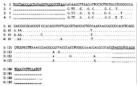
Figure 2 Homology of the nucleotide sequences from HGV RT-nested PCR products from 3 serum samples as compared with the reported sequence.
(1. The reported HGV sequence[13] ; 2-4. The sequences of HG V RT-nested PCR products from 3 serum samples. Underlined areas indicate the primer positions.)
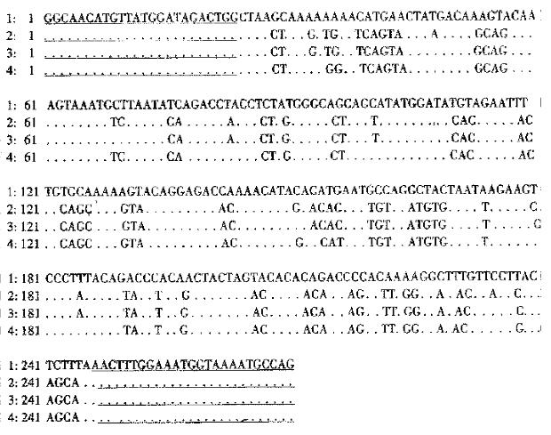
Figure 3 Homology of the nucleotide sequences from TTV semi-nested PCR products from 3 serum samples as compared with the reported sequence.
1. The reported TTV sequence[40]; 2-4. The sequences of TTV semi-nested PCR products from 3 serum samples. Underlined areas indicate the primer positions.
- Citation: Yan J, Chen LL, Luo YH, Mao YF, He M. High frequencies of HGV and TTV infections in blood donors in Hangzhou. World J Gastroenterol 2001; 7(5): 637-641
- URL: https://www.wjgnet.com/1007-9327/full/v7/i5/637.htm
- DOI: https://dx.doi.org/10.3748/wjg.v7.i5.637


