Copyright
©The Author(s) 2001.
World J Gastroenterol. Oct 15, 2001; 7(5): 630-636
Published online Oct 15, 2001. doi: 10.3748/wjg.v7.i5.630
Published online Oct 15, 2001. doi: 10.3748/wjg.v7.i5.630
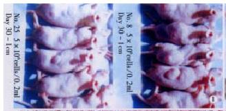
Figure 1 Subcutaneous tumor formation of the two clones.
Thirty days after sc injection of 5 × 10⁶ tumor cells for each nude mouse, the average s.c tumor diameter was (1.94 ± 0.36) cm for MHCC97-H as against (0.84 ± 0.47) cm for MHCC97-L.
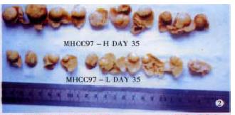
Figure 2 The liver tumor size of the two clones 5 wk after orthotopic inoculation.
The tumor geometric mean diameter (GMD) for MHCC97-H was (1.42 ± 0.11) cm as against (0.90 ± 0.26) cm for MHCC97-L.
Figure 3 Photomicroscopy of lung metastases of the two clones.
MHCC97-H (3A) produced large metastatic lesion pressing the bronchioles while MHCC97-L (3B) only formed small metastasis. HE × 100.
Figure 4 Photomicroscopy of MHCC97-L (4A) and MHCC97-H (4B) illustrating the clear differences in the number of nucleoli.
Giemsa × 400.
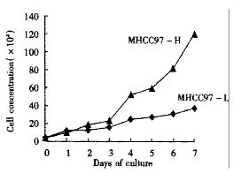
Figure 5 Cell growth curve of two clones.
4 × 10⁴ viable cells were cultured in each well of the 24 well culture plate. Cell numbers were determined for 7 consecutive days. Each time point represents the mean of duplicate cell counts.
- Citation: Li Y, Tang ZY, Ye SL, Liu YK, Chen J, Xue Q, Chen J, Gao DM, Bao WH. Establishment of cell clones with different metastatic potential from the metastatic hepatocellular carcinoma cell line MHCC97. World J Gastroenterol 2001; 7(5): 630-636
- URL: https://www.wjgnet.com/1007-9327/full/v7/i5/630.htm
- DOI: https://dx.doi.org/10.3748/wjg.v7.i5.630


