©The Author(s) 2001.
World J Gastroenterol. Jun 15, 2001; 7(3): 425-430
Published online Jun 15, 2001. doi: 10.3748/wjg.v7.i3.425
Published online Jun 15, 2001. doi: 10.3748/wjg.v7.i3.425
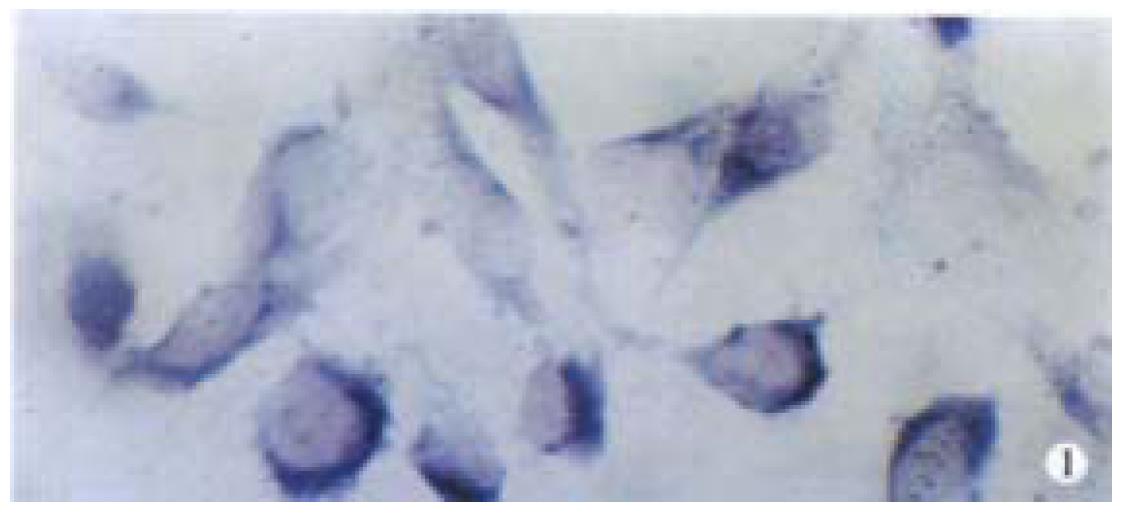
Figure 1 The expression of α1,3Fuc-TmRNA in Lovo cells shows strongly positive staining, with deep royal blue cytoplasms and visible royal blue beads.
In situ hybridization. × 400
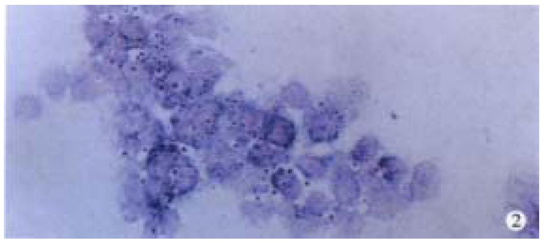
Figure 2 The expression of α1,3Fuc-TmRNA shows weakly positive staining, light royal blue with royal blue bead in cytoplasm of HT29 cells.
In situ hybridization. × 400
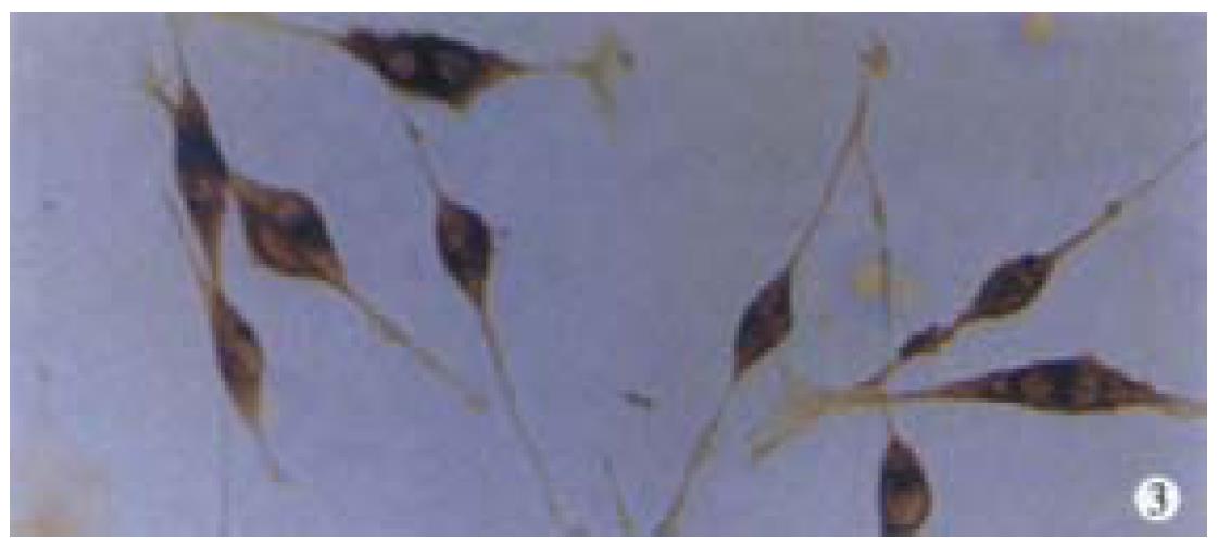
Figure 3 The expression of SLeX antigen in Lovo cells shows strongly positive staining, with deep brown in cytiplasm.
Immunohistochemistry. × 400
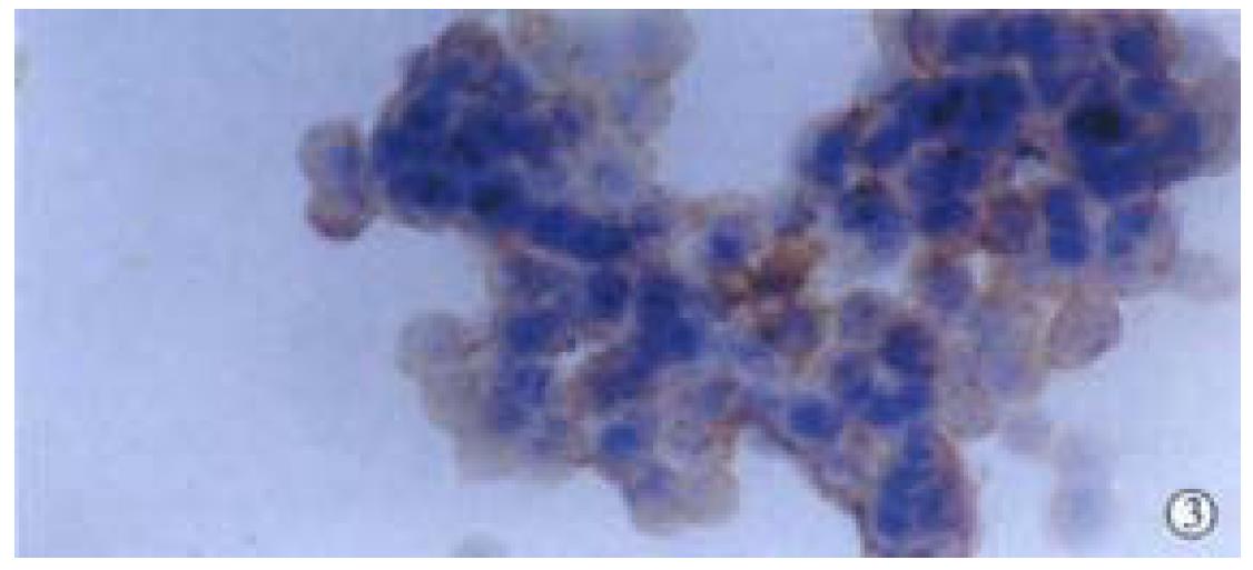
Figure 4 The expression of SLeX antigen shows positive or weakly positive staining, brown in membrane of HT29 cells.
Immunohistoc hemistry. × 400
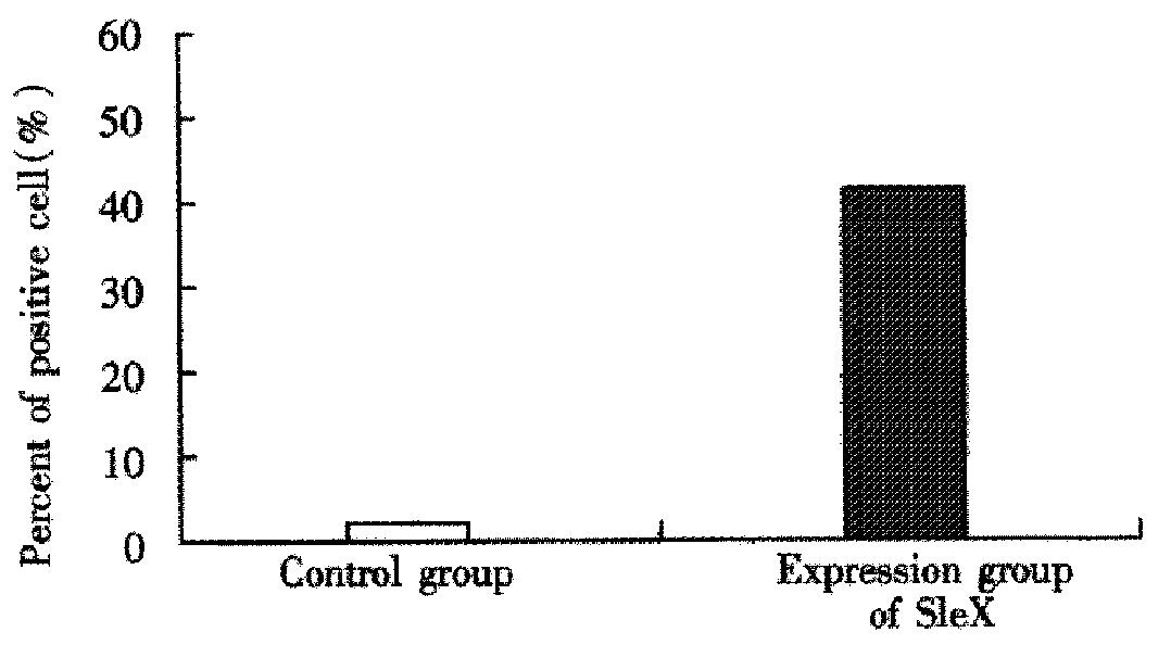
Figure 5 The detective result of Lovo cells.
flow cytometry
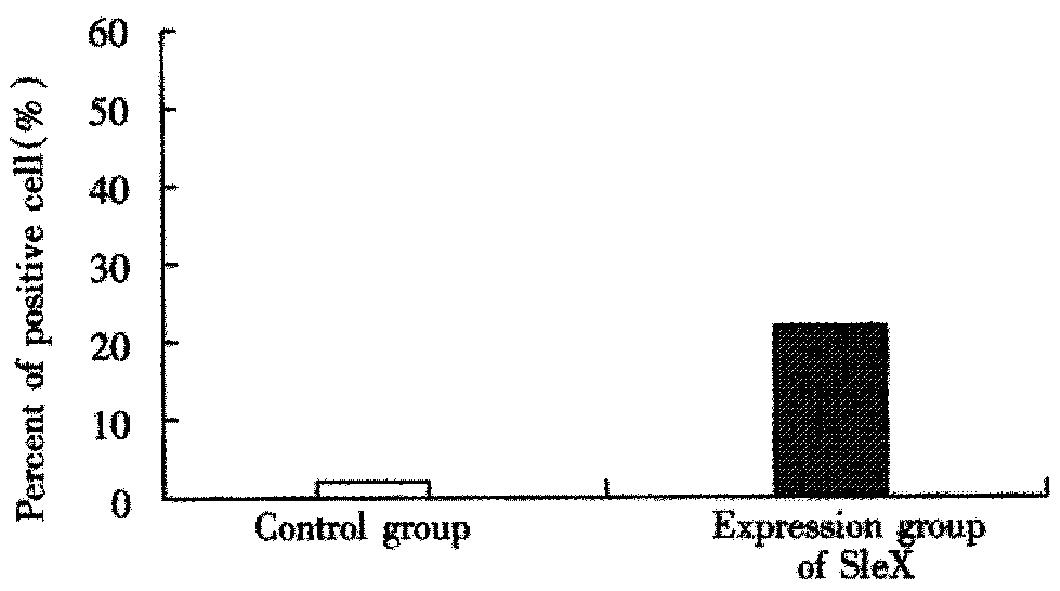
Figure 6 The detective result of HT29 cells.
flow cytometry
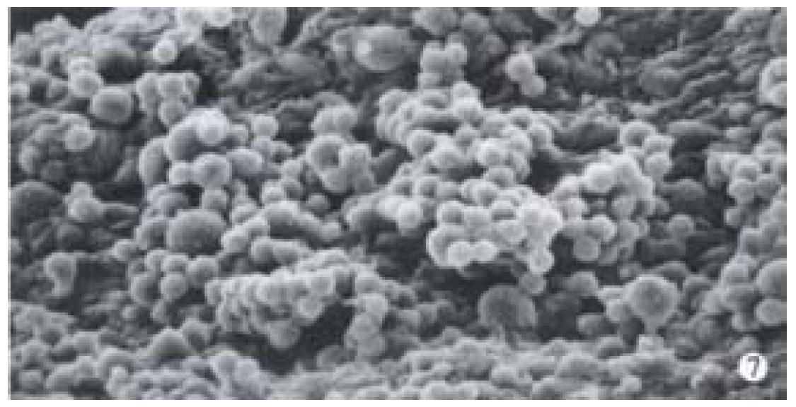
Figure 7 In control groups, a lot of Lovo cells adhered to the surface of umbilical vein endothelium, and tumor cells clustered.
SEM, × 500
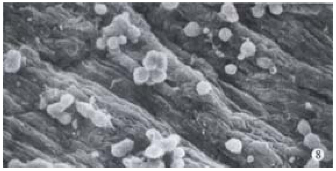
Figure 8 In control groups, The HT29 cells adhered to the surface of umbilical vein endothelium, a part of them clustered.
SEM, × 500
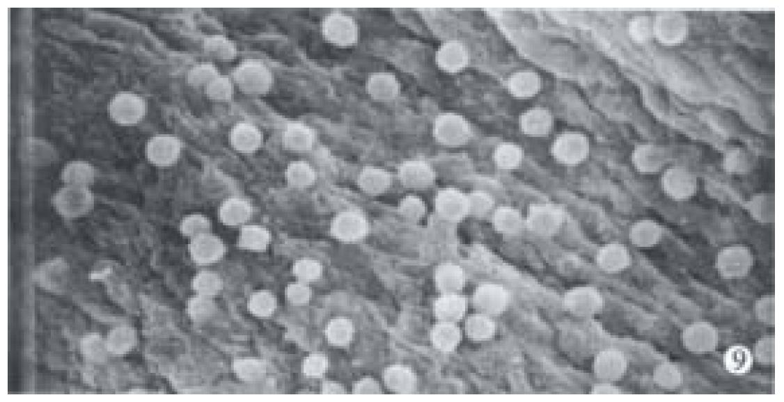
Figure 9 In experimental groups, a small amount of the Lovo cells adhered to the umbilical vein endothelium, no clustering.
SEM, × 500
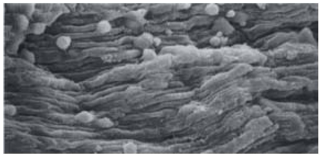
Figure 10 In experimental groups, a small amount of the HT29 cells adhered to the umbilical vein endothelium, no clustering.
SEM, × 500
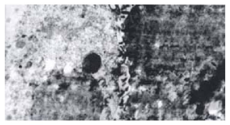
Figure 11 The Lovo cells were linked to umbilical vein endothelium via interdigitations on the cell membrane.
TEM × 20K
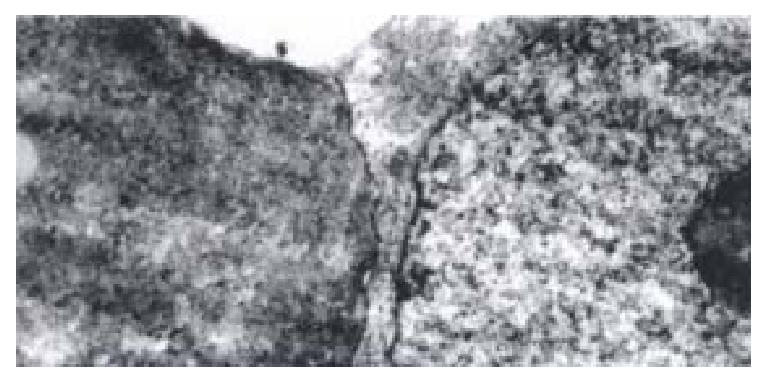
Figure 12 The HT29 cells connected endothelial cell membrane directly via the cell membrane.
TEM × 24K
- Citation: Li XW, Ding YQ, Cai JJ, Yang SQ, An LB, Qiao DF. Studies on mechanism of Sialy Lewis-X antigen in liver metastases of human colorectal carcinoma. World J Gastroenterol 2001; 7(3): 425-430
- URL: https://www.wjgnet.com/1007-9327/full/v7/i3/425.htm
- DOI: https://dx.doi.org/10.3748/wjg.v7.i3.425


