©The Author(s) 2000.
World J Gastroenterol. Apr 15, 2000; 6(2): 234-238
Published online Apr 15, 2000. doi: 10.3748/wjg.v6.i2.234
Published online Apr 15, 2000. doi: 10.3748/wjg.v6.i2.234
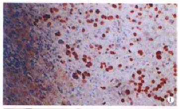
Figure 1 Strongly positive immunoperoxidase staining of the p53 protein with the anti-p53 mAb Do-7 in HCC, predominantly in nuclei.
Absence of staining in bnontumorous liver. LSAB method, × 100
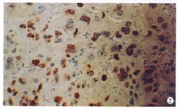
Figure 2 HCC with diffuse PCNA staining using mAb PC10.
LSAB method, × 200
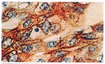
Figure 3 Cholangiocellular carcinoma showing extensive strongly membrance and plasma positivity for c-erbB-2 using poly-Ab code N.
A485. LSAB method, × 400
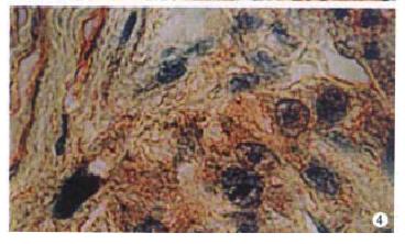
Figure 4 Diffuse H-rasp21 using mAb Ncc-Ras-001 staining in HCC and nontumorious tissues.
LSAB method, × 400
- Citation: Lin GY, Chen ZL, Lu CM, Li Y, Ping XJ, Huang R. Immunohistochemical study on p53, H-rasp21, c-erbB-2 protein and PCNA expression in HCC tissues of Han and minority ethnic patients. World J Gastroenterol 2000; 6(2): 234-238
- URL: https://www.wjgnet.com/1007-9327/full/v6/i2/234.htm
- DOI: https://dx.doi.org/10.3748/wjg.v6.i2.234


