Copyright
©The Author(s) 1999.
World J Gastroenterol. Jun 15, 1999; 5(3): 225-227
Published online Jun 15, 1999. doi: 10.3748/wjg.v5.i3.225
Published online Jun 15, 1999. doi: 10.3748/wjg.v5.i3.225
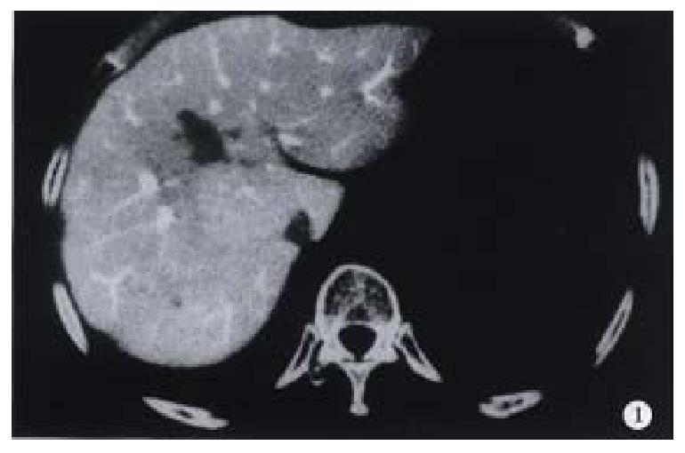
Figure 1 CTAP image obtained in a 40-year-old man with liver micro-cancer shows a punctuate perfusion defect in right lobe.
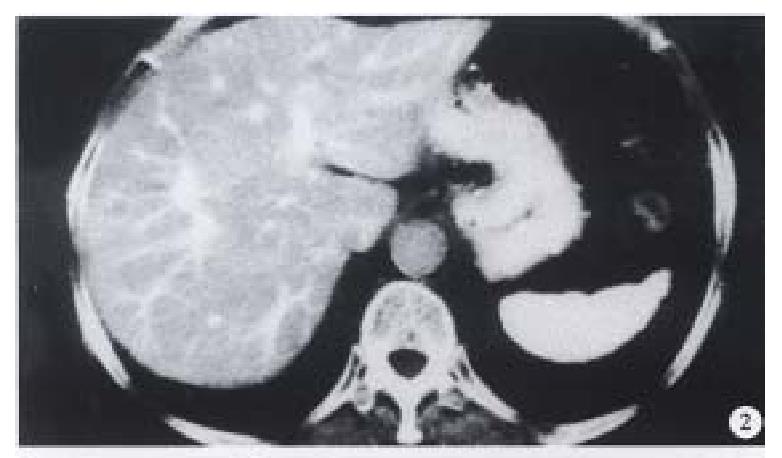
Figure 2 CTHA image obtained on the same slice to Figure 1, the micro-tumor manifested as an small round enhancement nodule.
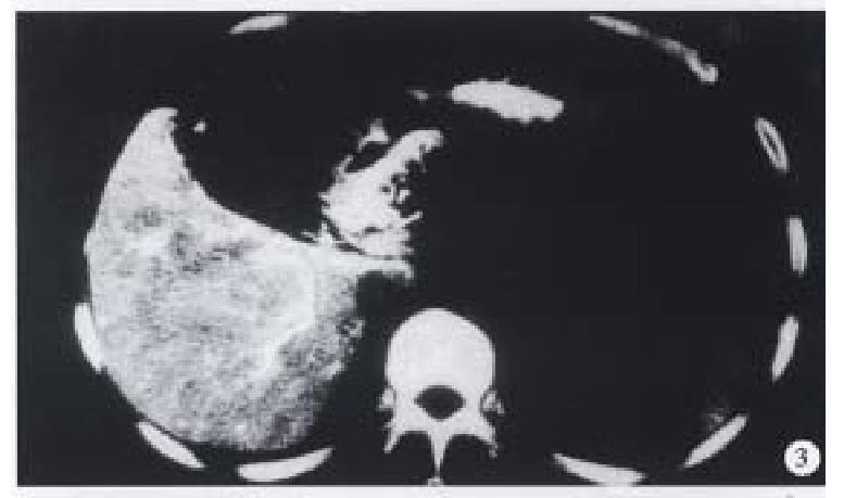
Figure 3 CTHA in a 45-year-old man shows a micro-cancer with a diameter of 0.
3 cm.
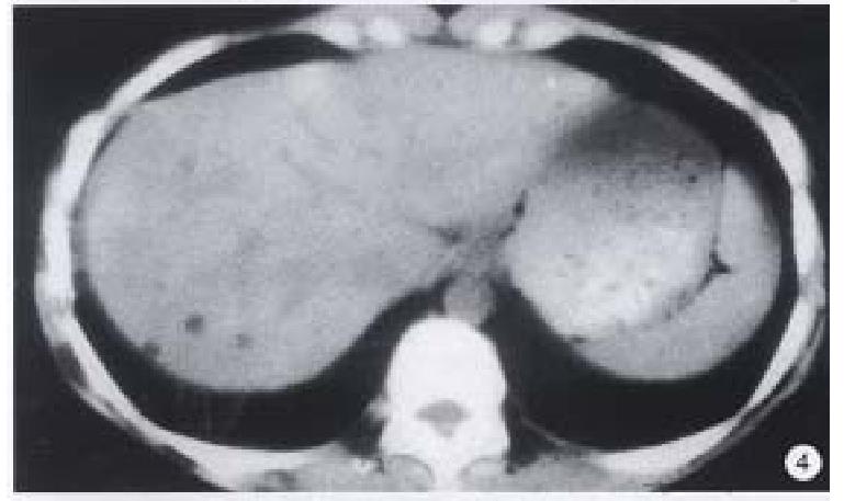
Figure 4 A punctuate lipiodol deposit focus with 0.
2 cm in diameter is shownon Lp-CT in a 28-year-old woman with elevate d serum AFP level of 860 μg/L detects which was not detected by CTAP and CTHA.
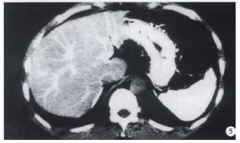
Figure 5 CTHA in a 35-year-old man shows two punct uate non-pathologic enhancement foci on the edge of right lobe.
- Citation: Li L, Wu PH, Mo YX, Lin HG, Zheng L, Li JQ, Lu LX, Ruan CM, Chen L. CT arterial portography and CT hepatic arteriography in detection of micro liver cancer. World J Gastroenterol 1999; 5(3): 225-227
- URL: https://www.wjgnet.com/1007-9327/full/v5/i3/225.htm
- DOI: https://dx.doi.org/10.3748/wjg.v5.i3.225


