©The Author(s) 1998.
World J Gastroenterol. Apr 15, 1998; 4(2): 125-127
Published online Apr 15, 1998. doi: 10.3748/wjg.v4.i2.125
Published online Apr 15, 1998. doi: 10.3748/wjg.v4.i2.125
Figure 1 Examination of codon 249 mutation using RFLP analysis.
Lane 1 and lane 2 were PCR products from cancerous tissues without and with digestion of restriction enzyme Hae III. Lane 3 and lane 4 were products from pericancerous tissues with and without digestion of restriction enzyme Hae III. Lane 5 was marker of 100 bp ladder from GIBco BRL.
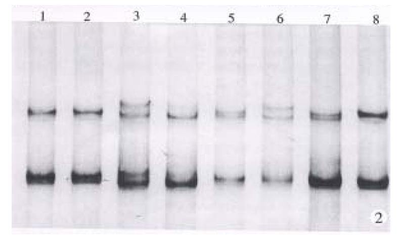
Figure 2 The PCR/SSCP detection of LOH, silver-staining.
Lane 1 and lane 2 were products from patient 1 who was homozygote. Lane 3 to 8 were products from patients 2 to 4. They were all heterozygote. Patients 2 and 4 were LOH positive. Patient 3 has mutation of one allele.
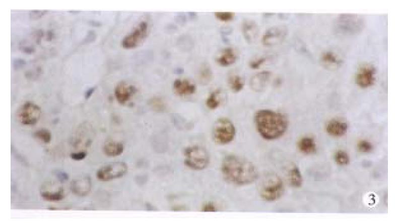
Figure 3 Expression of p53 proteins in HCC tissue using IHC (× 400).
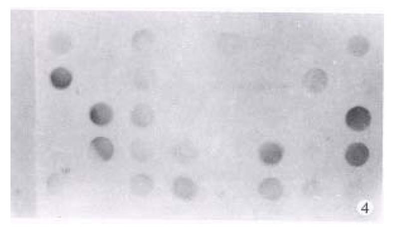
Figure 4 p53 mRNA detected using RTª²PCR/slot hybridization.
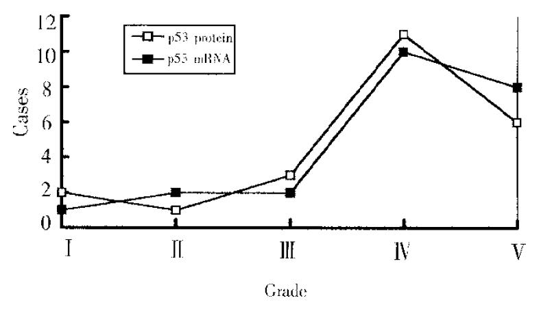
Figure 5 The relation between the expression of p53 proteins and that of p53 mRNA.
- Citation: Peng XM, Peng WW, Yao JL. Codon 249 mutations of p53 gene in development of hepatocellular carcinoma. World J Gastroenterol 1998; 4(2): 125-127
- URL: https://www.wjgnet.com/1007-9327/full/v4/i2/125.htm
- DOI: https://dx.doi.org/10.3748/wjg.v4.i2.125


