
















Copyright: ©Author(s) 2027.
World J Gastroenterol. Jan 14, 2027; 33(2): 123982
Published online Jan 14, 2027. doi: 10.3748/wjg.123982
Published online Jan 14, 2027. doi: 10.3748/wjg.123982
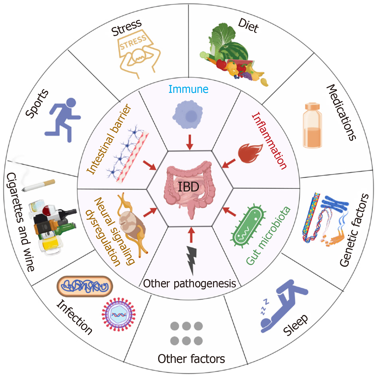
Figure 1 Risk factors for inflammatory bowel disease.
As a critical risk factor, stress interacts with genetic susceptibility and environmental triggers to promote disease via neuroendocrine-immune dysregulation, heightened gut inflammation, dysbiosis, and barrier dysfunction, collectively driving inflammatory bowel disease pathogenesis. IBD: Inflammatory bowel disease. The pattern was created with BioGDP.com[14,15].
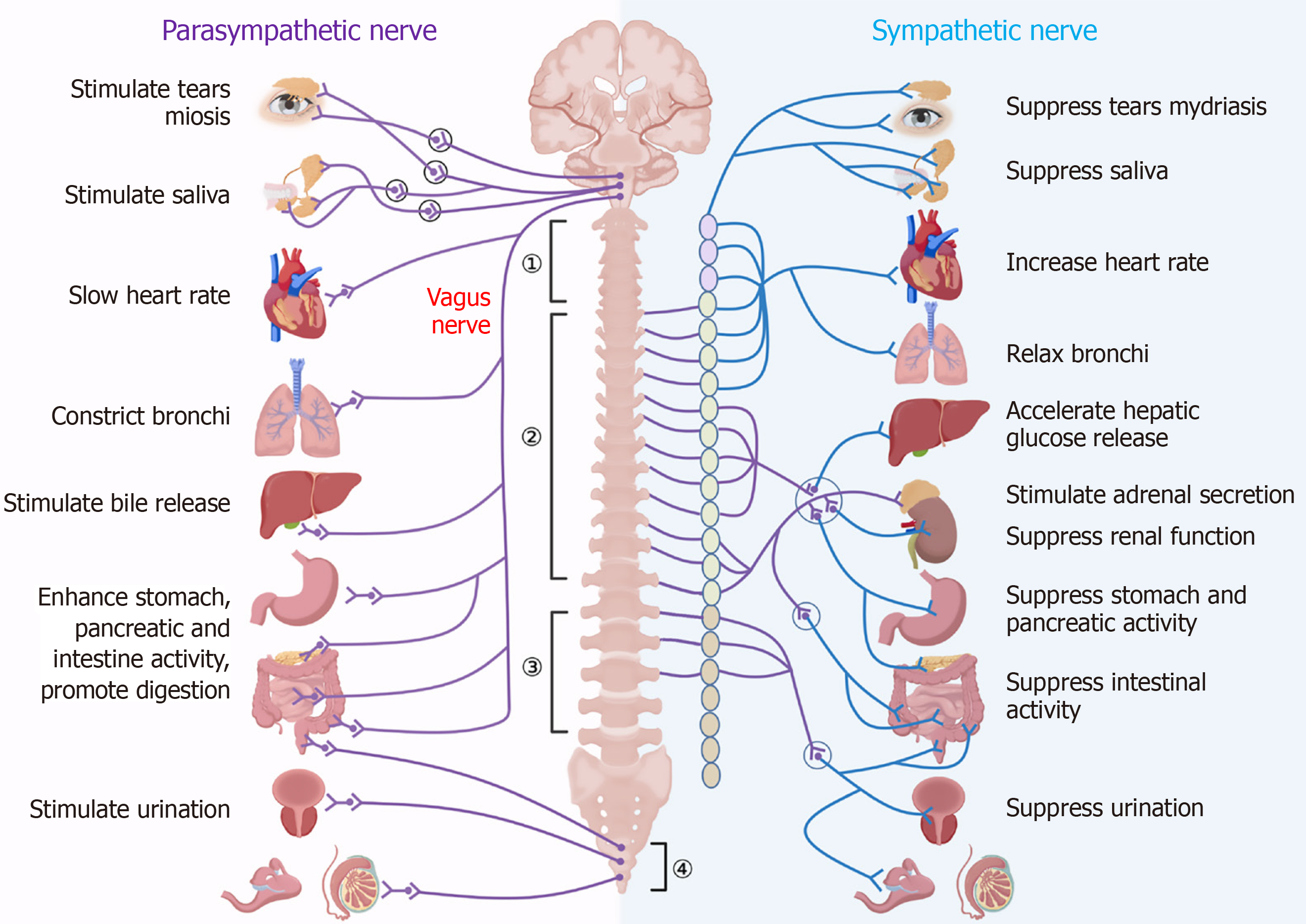
Figure 2 Bidirectional regulation of target organs by the autonomic nervous system.
(1) Cervical nerves; (2) Thoracic nerves; (3) Lumbar nerves; and (4) Sacral nerves. The autonomic nervous system regulates bodily functions through the antagonistic actions of the sympathetic and parasympathetic nerves. Activation of the sympathetic nerve leads to increased energy expenditure and organ function enhancement, resulting in elevated heart rate, accelerated respiration, increased blood pressure, bronchodilation, and pupil dilation, while concurrently inhibiting gastrointestinal motility and digestive secretion. In contrast, parasympathetic nerve activation decreases energy consumption and promotes energy storage, leading to decreased heart rate, slowed respiration, reduced blood pressure, bronchoconstriction, and pupil constriction, while also enhancing gastrointestinal motility and digestive secretion. Furthermore, these systems interact synergistically in specific physiological processes, such as sexual function, where parasympathetic dominance occurs during arousal and sympathetic dominance during orgasm. This delicate balance between antagonism and synergy allows the body to adapt efficiently to various physiological demands, including stress, digestion, and reproduction. The pattern was created with BioGDP.com[14].
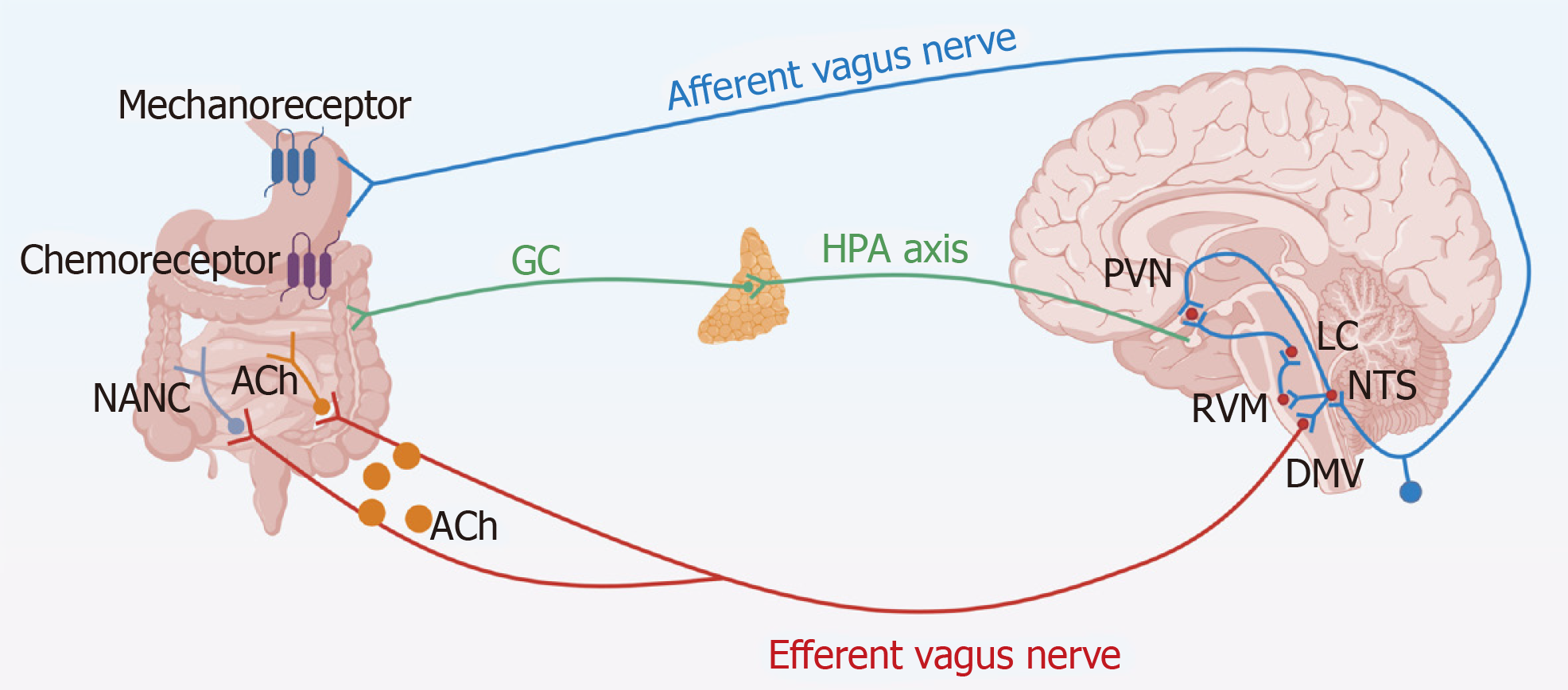
Figure 3 Bidirectional communication between vagus nerve and gastrointestinal tract.
The digestive system transmits signals through mechanoreceptors (e.g., food-related mucosal stroking, distension, and contraction) and chemoreceptors (detecting nutrients, hormones, pH, and immune stimuli). These signals travel through vagal afferent fibers to the nucleus tractus solitarius (NTS) and then to the central nervous system. The NTS processes visceral sensory information and transmits it to higher brain regions, including the paraventricular nucleus, the dorsal motor nucleus of the vagus, and the rostral ventromedial medulla. γ-aminobutyric acid, glutamate, and norepinephrine act as primary neurotransmitters in this process. The paraventricular nucleus of hypothalamus, upon the integration of signals, activates corticotropin-releasing hormone neurons to stimulate the hypothalamic-pituitary-adrenal axis, leading to the release of glucocorticoids that regulate gastrointestinal function. The rostral ventromedial medulla, influenced by NTS projections, modulates visceral pain perception through μ-opioid receptors. Simultaneously, the dorsal motor nucleus of the vagus, following signal integration, activates excitatory pathways via vagal efferent fibers to release acetylcholine or inhibitory pathways to release non-adrenergic non-cholinergic neurotransmitters like nitric oxide or vasoactive intestinal peptide in the digestive tract. Ach: Acetylcholine; NANC: Non-adrenergic non-cholinergic; GC: Glucocorticoid; HPA: Hypothalamic-pituitary-adrenal axis; PVN: Paraventricular nucleus of hypothalamus; RVM: Rostral ventromedial medulla; DMV: Dorsal motor nucleus of vagus; LC: Locus coeruleus; NTS: Nucleus tractus solitarius. The pattern was created with BioGDP.com[14].
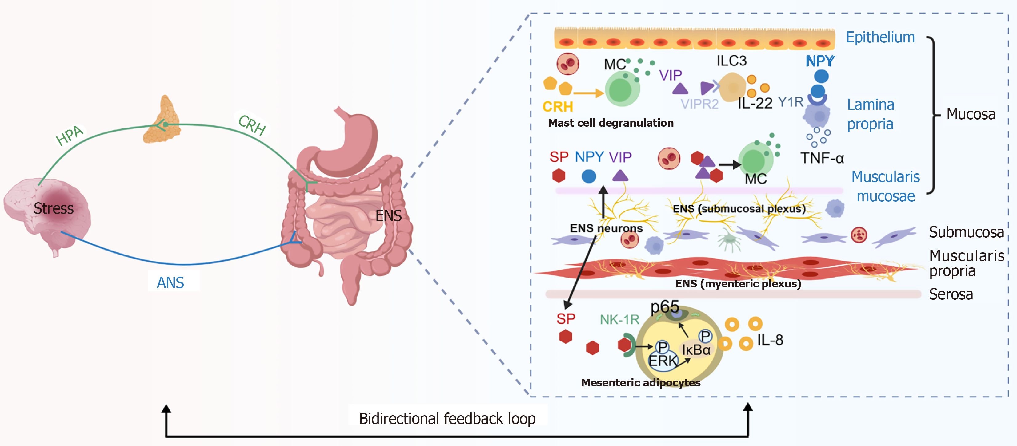
Figure 4 Stress-induced enteric nervous system signaling regulating intestinal immune responses.
Stress activates the hypothalamic-pituitary-adrenal axis and autonomic nervous system, which modulate enteric nervous system activity and neuropeptide release. Enteric nervous system-derived substance P, vasoactive intestinal peptide, and neuropeptide Y regulate immune cell activation, mast cell responses, group 3 innate lymphoid cells-mediated mucosal protection, and inflammatory signaling in mesenteric adipocytes. These neuroimmune interactions establish a bidirectional feedback loop that influences intestinal inflammation and disease progression. HPA: Hypothalamic-pituitary-adrenal axis; CRH: Corticotropin-releasing hormone; ANS: Autonomic nervous system; ENS: Enteric nervous system; MC: Mast cell; VIP: Vasoactive intestinal peptide; NPY: Neuropeptide Y; ILC3: Group 3 innate lymphoid cells; TNF-α: Tumor necrosis factor-α; SP: Substance P; IL: Interleukin. The pattern was created with BioGDP.com[14].
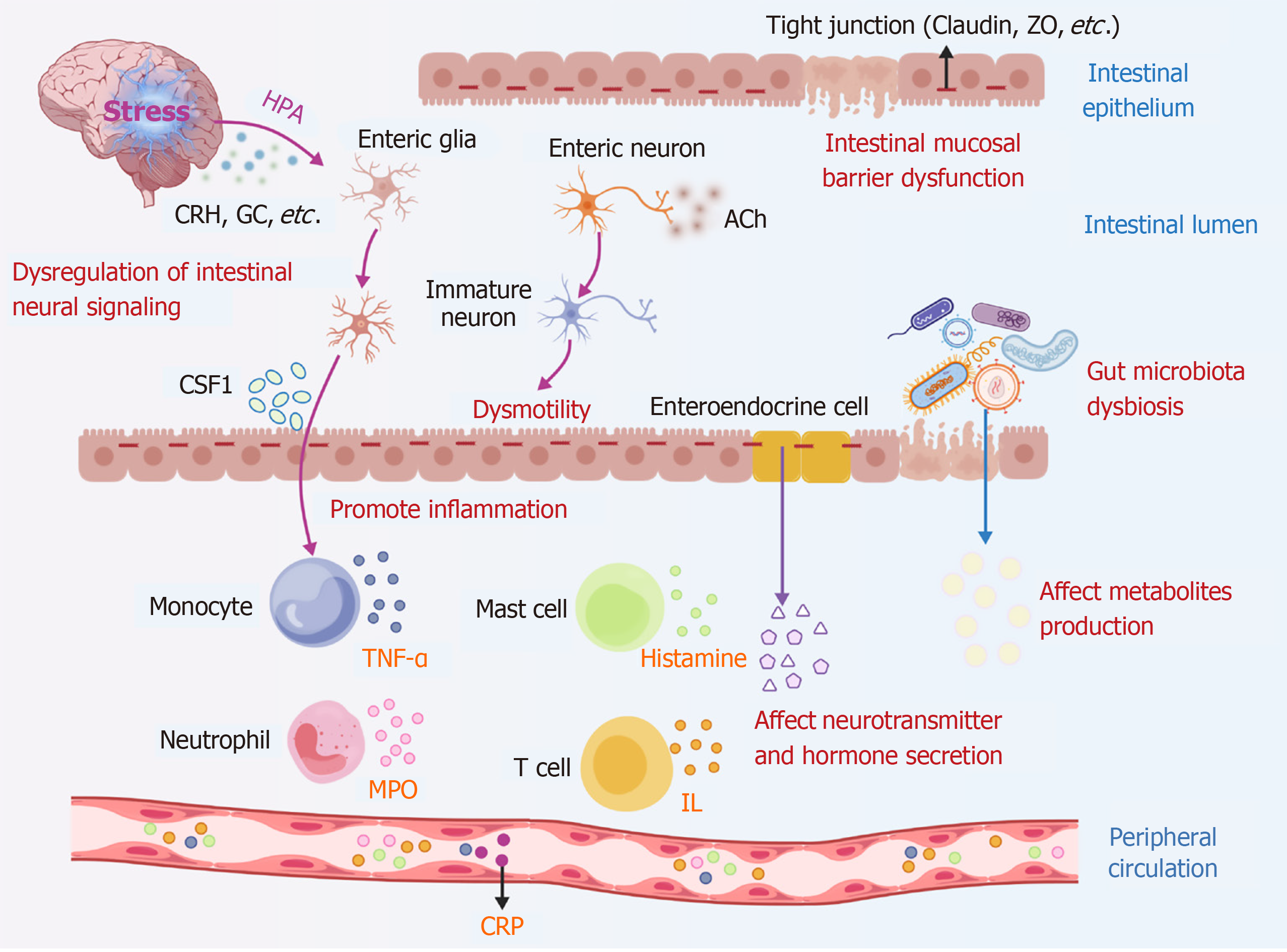
Figure 5 The impact of stress on intestinal inflammation.
Stress can lead to inflammatory bowel disease through various pathways, such as affecting the transmission of intestinal nerve information, affecting the release of neurotransmitters and gastrointestinal hormones, disrupting intestinal barrier function, causing dysmotility, participating in immune inflammatory responses, affecting intestinal microbiota balance, and the release of microbial metabolites, ultimately participating in systemic inflammatory responses. HPA: Hypothalamic-pituitary-adrenal axis; CRH: Corticotropin-releasing hormone; GC: Glucocorticoid; CSF1: Colony-stimulating factor 1; Ach: Acetylcholine; TNF-α: Tumor necrosis factor-alpha; MPO: Myeloperoxidase; IL: Interleukin; CRP: C reactive protein. The pattern was created with BioGDP.com[14].
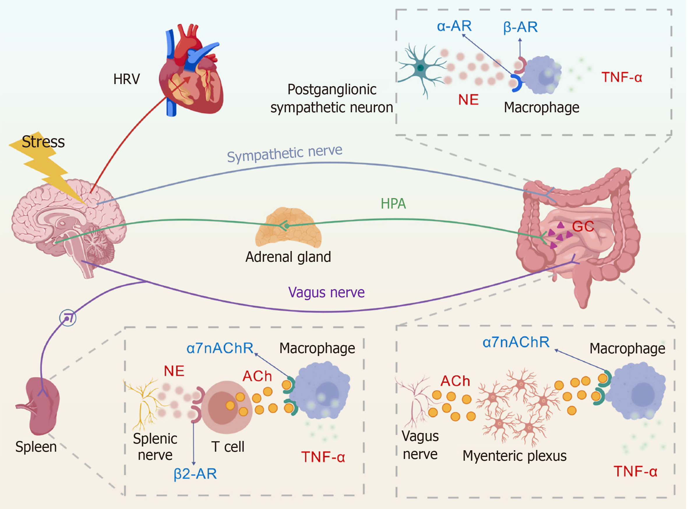
Figure 6 The role of autonomic nervous system in stress-related inflammatory bowel disease.
Stress-induced sympathetic nervous system activation increases norepinephrine (NE) release and signals through α-adrenergic receptor (AR) and β-AR. Excessive sympathetic nervous system activity can slow intestinal motility, impair the mucosal barrier, and promote inflammation. Vagus nerve stimulation may suppress inflammation through hypothalamic-pituitary-adrenal axis-dependent glucocorticoid release. The vagus nerve engages abdominal ganglia and the splenic nerve, promoting NE release from splenic nerve terminals. NE activates β2-AR on cholinergic T lymphocytes, inducing acetylcholine (ACh) release. ACh activates α 7 nicotinic ACh receptor on macrophages and inhibits tumor necrosis factor-α production. Vagal fibers also regulate the enteric nervous system through the myenteric plexus. ACh signaling can activate α 7 nicotinic ACh receptor on intestinal macrophages and reduce pro-inflammatory cytokine release. HRV: Heart rate variability; AR: Adrenergic receptor; NE: Norepinephrine; TNF-α: Tumor necrosis factor-α; HPA: Hypothalamic-pituitary-adrenal axis; GC: Glucocorticoid; ACh: Acetylcholine; α7nAChR: Α 7 nicotinic acetylcholine receptor. The pattern was created with BioGDP.com[14].
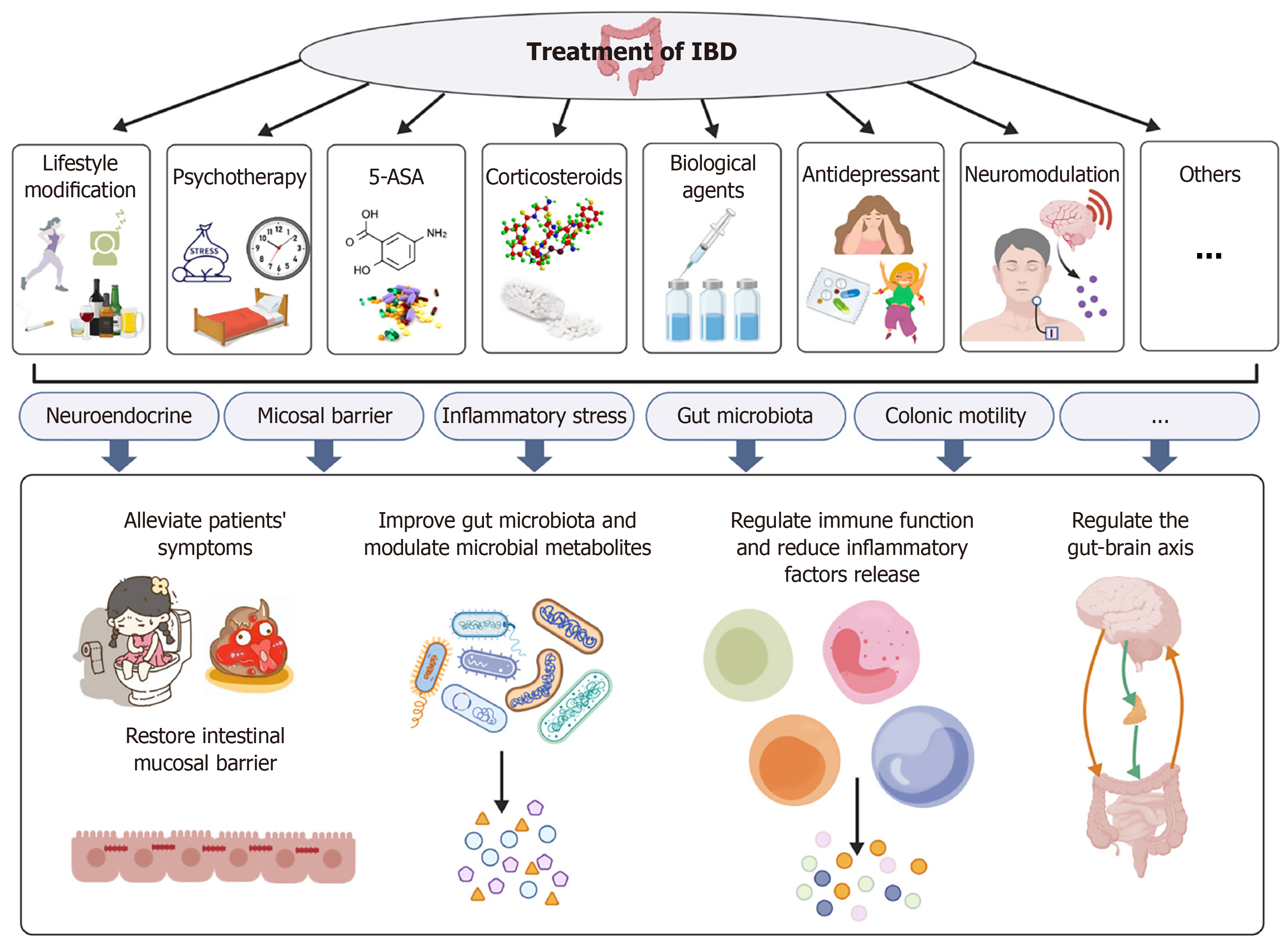
Figure 7 Treatment of inflammatory bowel disease various methods such as lifestyle changes, psychotherapy, drug therapy, and neural regulation can help treat inflammatory bowel disease by restoring the intestinal mucosal barrier, regulating the intestinal immune response, reducing the release of inflammatory factors, restoring the balance of the intestinal microbiota, and regulating the bidirectional communication between the brain and the gastrointestinal tract.
IBD: Inflammatory bowel disease. The pattern was created with BioGDP.com[14].
- Citation: Hu XX, Zhang XP, Wang CX, Yang RR, Ma LX, Wu SW, Jiang WQ, Deng PM, Zhang T, Su XL, Wei W. Autonomic nervous system as a key modulator in stress-related inflammatory bowel disease: Mechanisms and therapeutic implications. World J Gastroenterol 2027; 33(2): 123982
- URL: https://www.wjgnet.com/1007-9327/full/v33/i2/123982.htm
- DOI: https://dx.doi.org/10.3748/wjg.123982