
















Copyright: ©Author(s) 2027.
World J Gastroenterol. Jan 14, 2027; 33(2): 123795
Published online Jan 14, 2027. doi: 10.3748/wjg.123795
Published online Jan 14, 2027. doi: 10.3748/wjg.123795
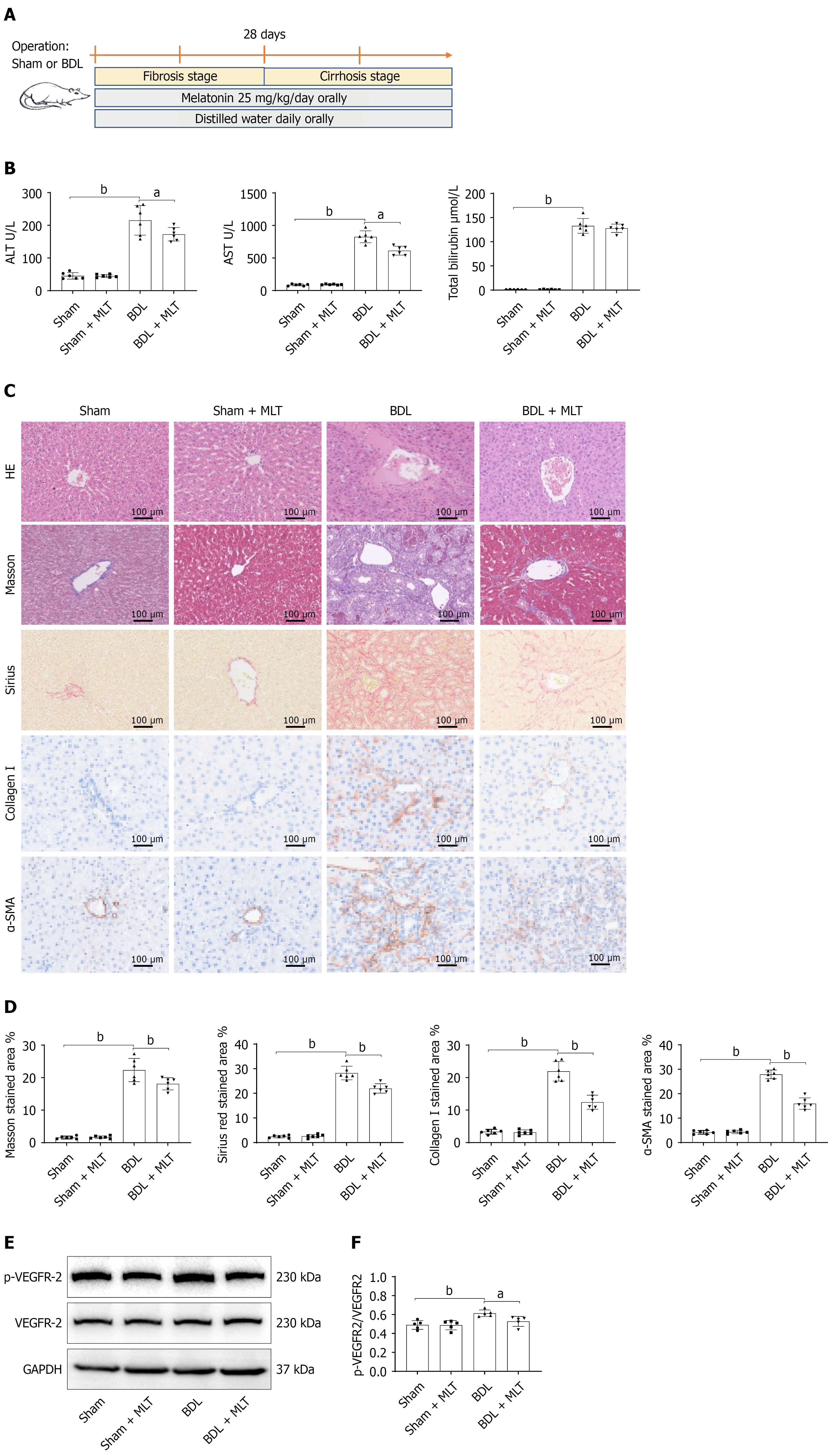
Figure 1 Melatonin alleviated bile duct ligation-induced liver cirrhosis and portal hypertension.
A: Construction of animal model of liver cirrhosis and melatonin administration; B: The levels of plasma liver injury markers alanine aminotransferase, aspartate aminotransferase and total bilirubin in rats; C: The histological staining of liver tissue sections using hematoxylin and eosin, Masson’s trichrome, and Sirius red. Immunohistochemical staining of collagen I and α-smooth muscle actin in rat livers; D: Quantitative analysis of histopathological and immunohistochemical staining in C; E: Western blotting of hepatic VEGFR-2 and its phosphorylated form; F: Quantitative analysis of VEGFR-2 phosphorylation. aP < 0.05. bP < 0.01. BDL: Bile duct ligation; MLT: Melatonin; α-SMA: Α-smooth muscle actin; HE: Hematoxylin and eosin; ALT: Alanine aminotransferase; AST: Aspartate aminotransferase.
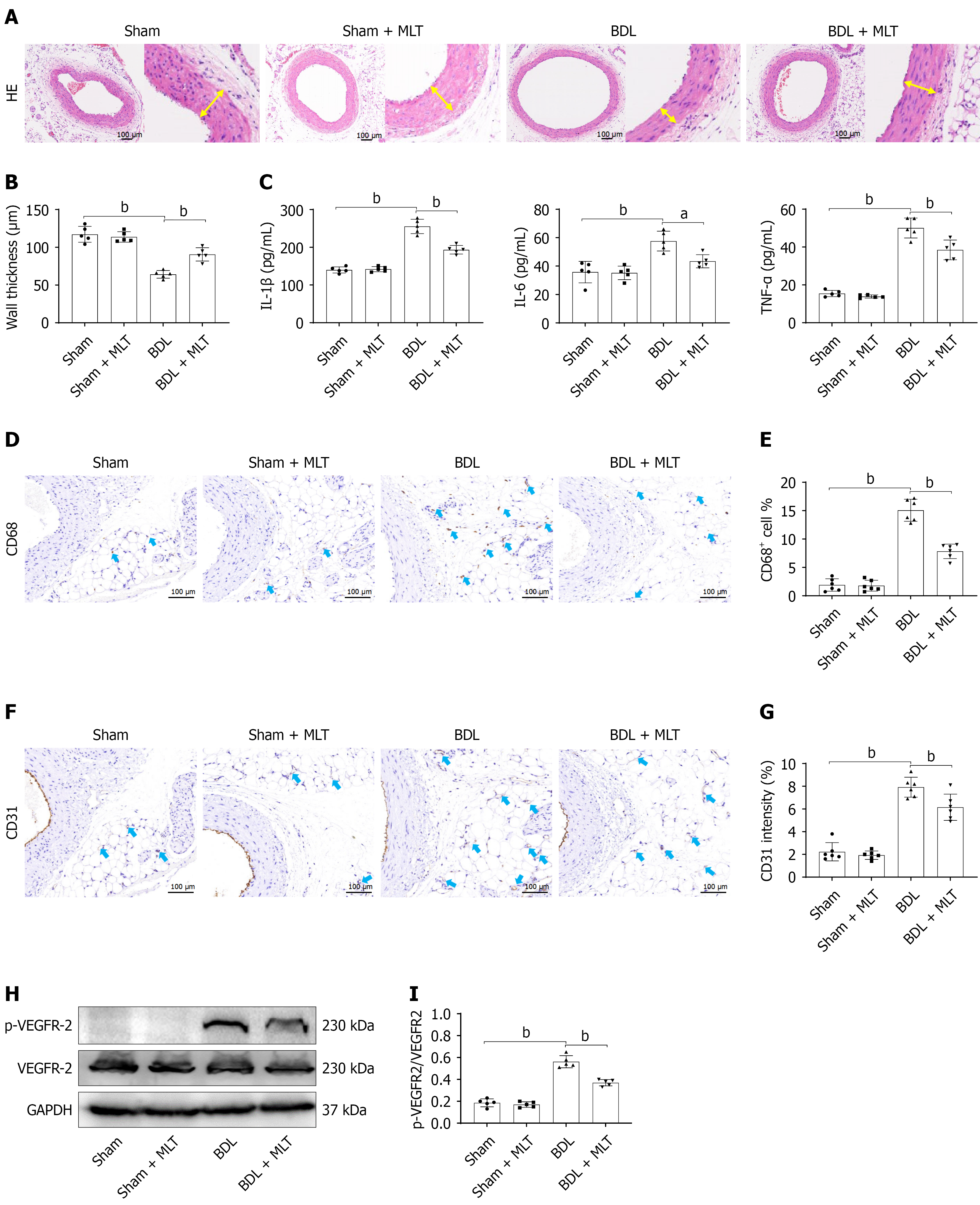
Figure 2 Melatonin alleviated bile duct ligation-induced mesenteric vascular remodeling and angiogenesis.
A: Representative hematoxylin and eosin staining images of mesenteric artery. The yellow arrows represent the thickness of vascular wall; B: Quantitative analysis of the wall thickness; C: Enzyme-linked immunosorbent assay of inflammatory cytokines in mesenteric artery tissues; D: Representative immunohistochemical images of CD68 in the mesentery; E: Quantitative analysis of CD68-positive cells; F: Representative immunohistochemical images of CD31 in the mesentery; G: Quantitative analysis of CD31 staining intensity; H: Western blotting of mesenteric VEGFR-2 and its phosphorylated form; I: Quantitative analysis of VEGFR-2 phosphorylation. aP < 0.05. bP < 0.01. BDL: Bile duct ligation; MLT: Melatonin; HE: Hematoxylin and eosin; IL: Interleukin; TNF-α: Tumor necrosis factor alpha.
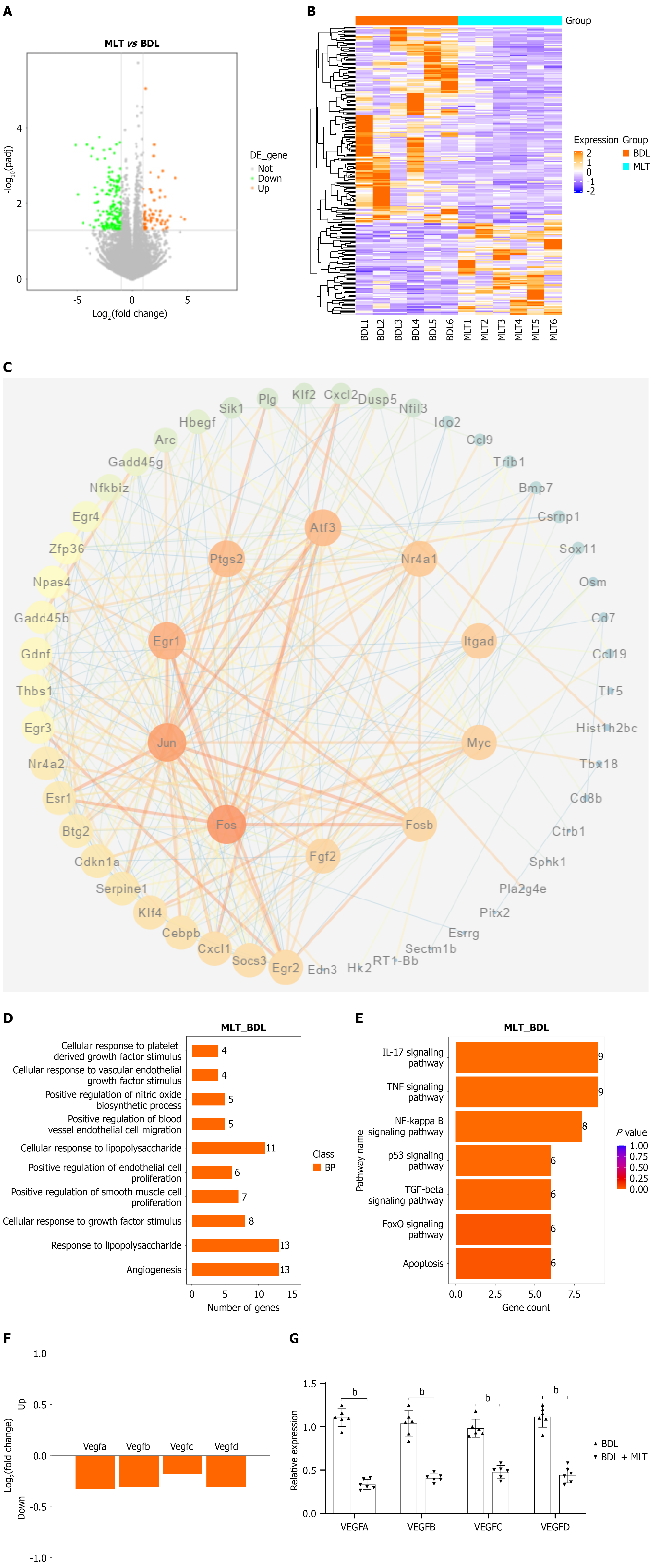
Figure 3 RNA-sequencing of mesenteric arteries from bile duct ligation rats treated with melatonin.
A: The volcano plot of differential expressed genes (DEGs) between bile duct ligation (BDL) and BDL + melatonin (MLT) groups; B: The heatmap of DEGs between BDL and BDL + MLT groups; C: The protein-protein interaction network of DEGs with 10 hub genes; D: The top 10 terms enriched by Gene Ontology enrichment analysis based on DEGs; E: The top 10 pathways enriched by Kyoto Encyclopedia of Genes and Genomes analysis based on DEGs; F: The expression profile of VEGFs between BDL and BDL + MLT groups; G: The validation of the expression levels of VEGFs by quantitative real-time polymerase chain reaction. aP < 0.05. bP < 0.01. BDL: Bile duct ligation; MLT: Melatonin.
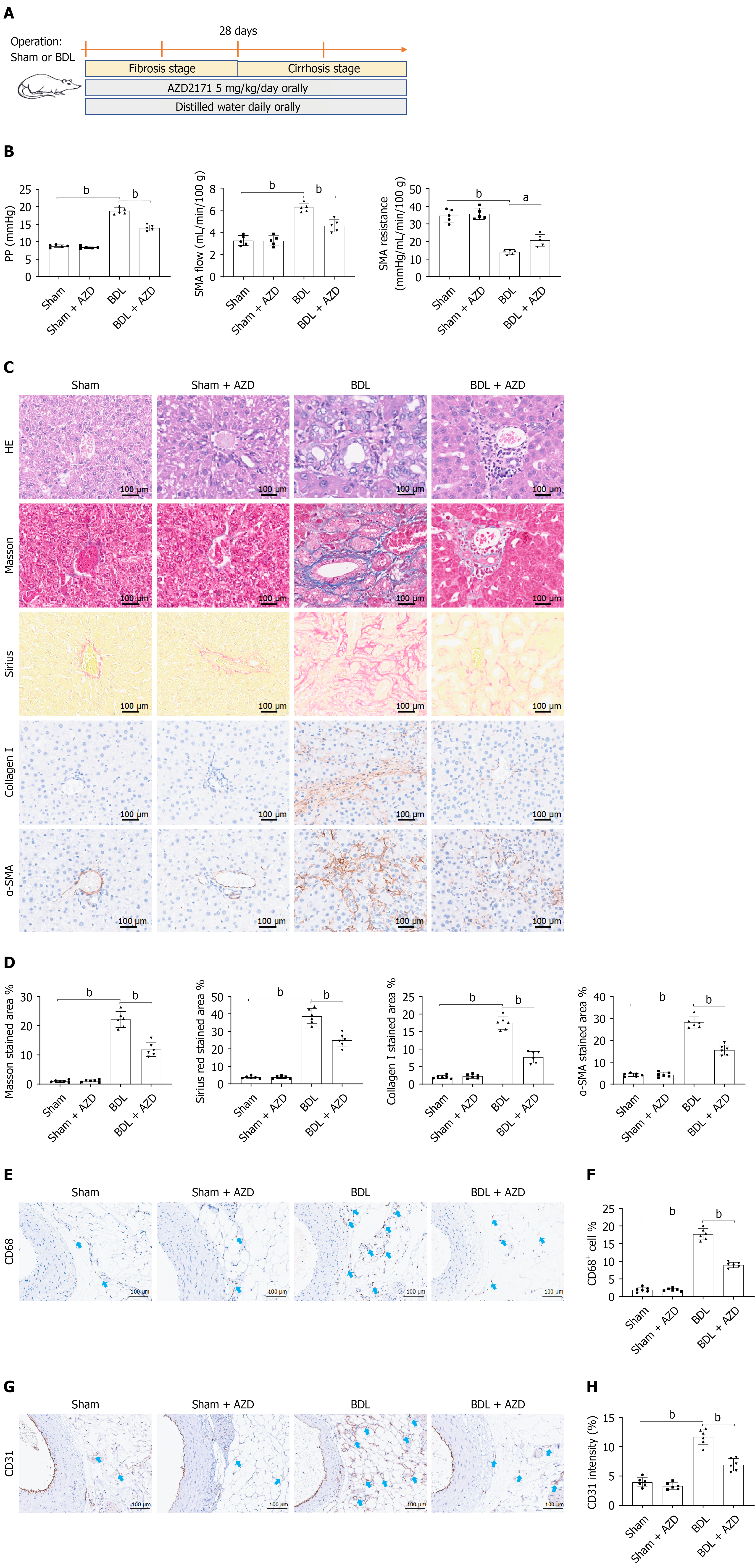
Figure 4 VEGFR-2 inhibition alleviated bile duct ligation-induced cirrhotic portal hypertension.
A: Construction of animal model of liver cirrhosis and AZD2171 administration; B: The portal hypertension hemodynamic parameters in rats from 4 groups; C: The histological staining of liver tissue sections using hematoxylin and eosin, Masson’s trichrome, and Sirius red. Immunohistochemical (IHC) staining of Collagen I and α-smooth muscle actin; in rat livers; D: Quantitative analysis of histopathological and IHC staining in C; E: Representative IHC images of CD68 in the mesentery; F: Quantitative analysis of CD68-positive cells; G: Representative IHC images of CD31 in the mesentery; H: Quantitative analysis of CD31 staining intensity. aP < 0.05. bP < 0.01. α-SMA: Α-smooth muscle actin; HE: Hematoxylin and eosin; BDL: Bile duct ligation.
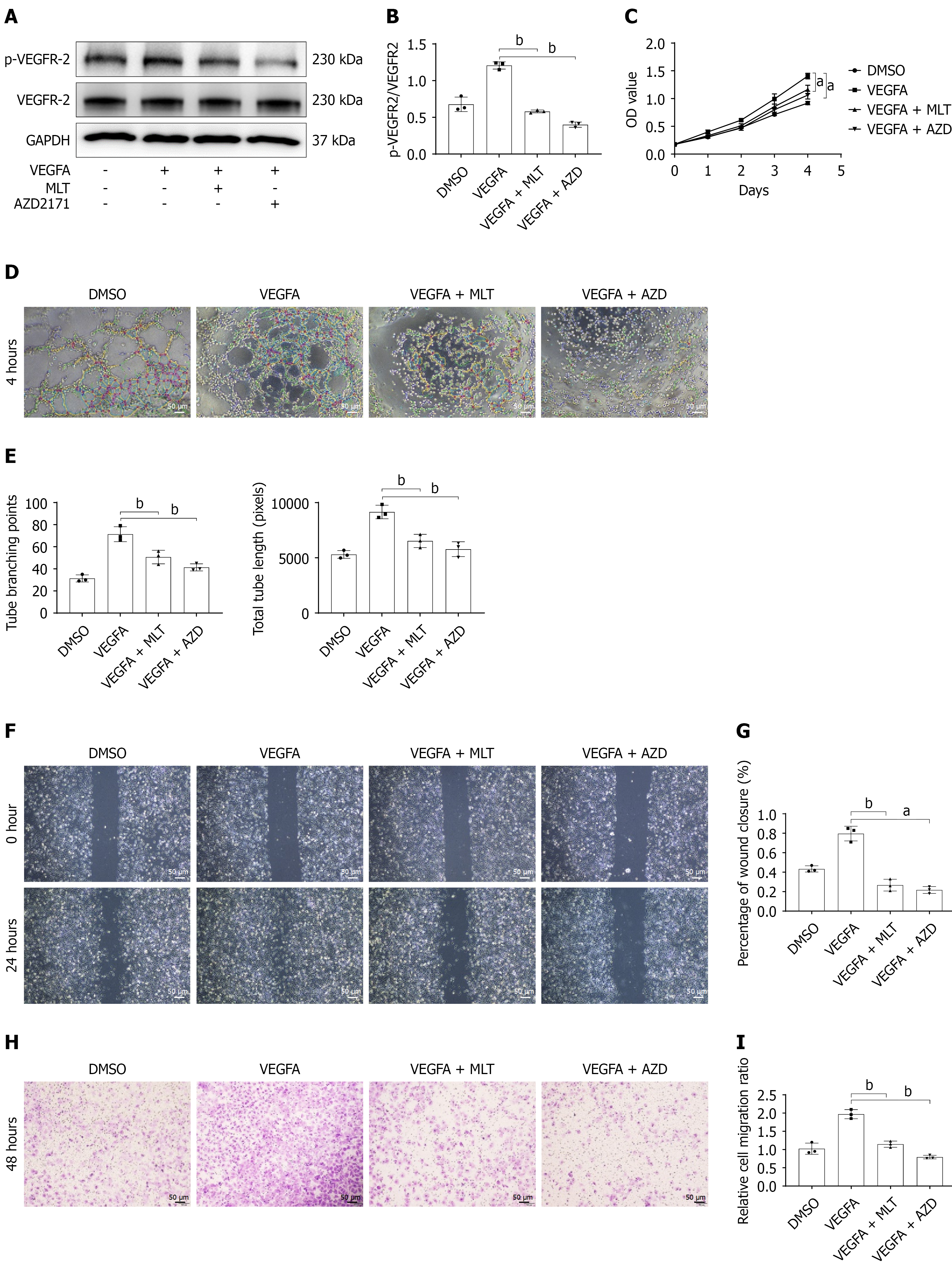
Figure 5 Melatonin and AZD2171 inhibited angiogenesis in vitro.
A: Western blotting of VEGFR-2 and its phosphorylated form in human umbilical vein endothelial cells (HUVECs) treated with VEGFA, melatonin (MLT), or AZD2171; B: Quantitative analysis of VEGFR-2 phosphorylation; C: The proliferation assay of HUVECs treated with VEGFA, MLT, or AZD2171; D: The tube formation assay of HUVECs treated with VEGFA, MLT, or AZD2171; E: Quantitative analysis of tube formation assay; F: The wound healing assay of HUVECs treated with VEGFA, MLT, or AZD2171; G: Quantitative analysis of wound healing assay; H: Transwell migration assay of HUVECs treated with VEGFA, MLT, or AZD2171; I: Quantitative analysis of Transwell migration assay. aP < 0.05. bP < 0.01. MLT: Melatonin.
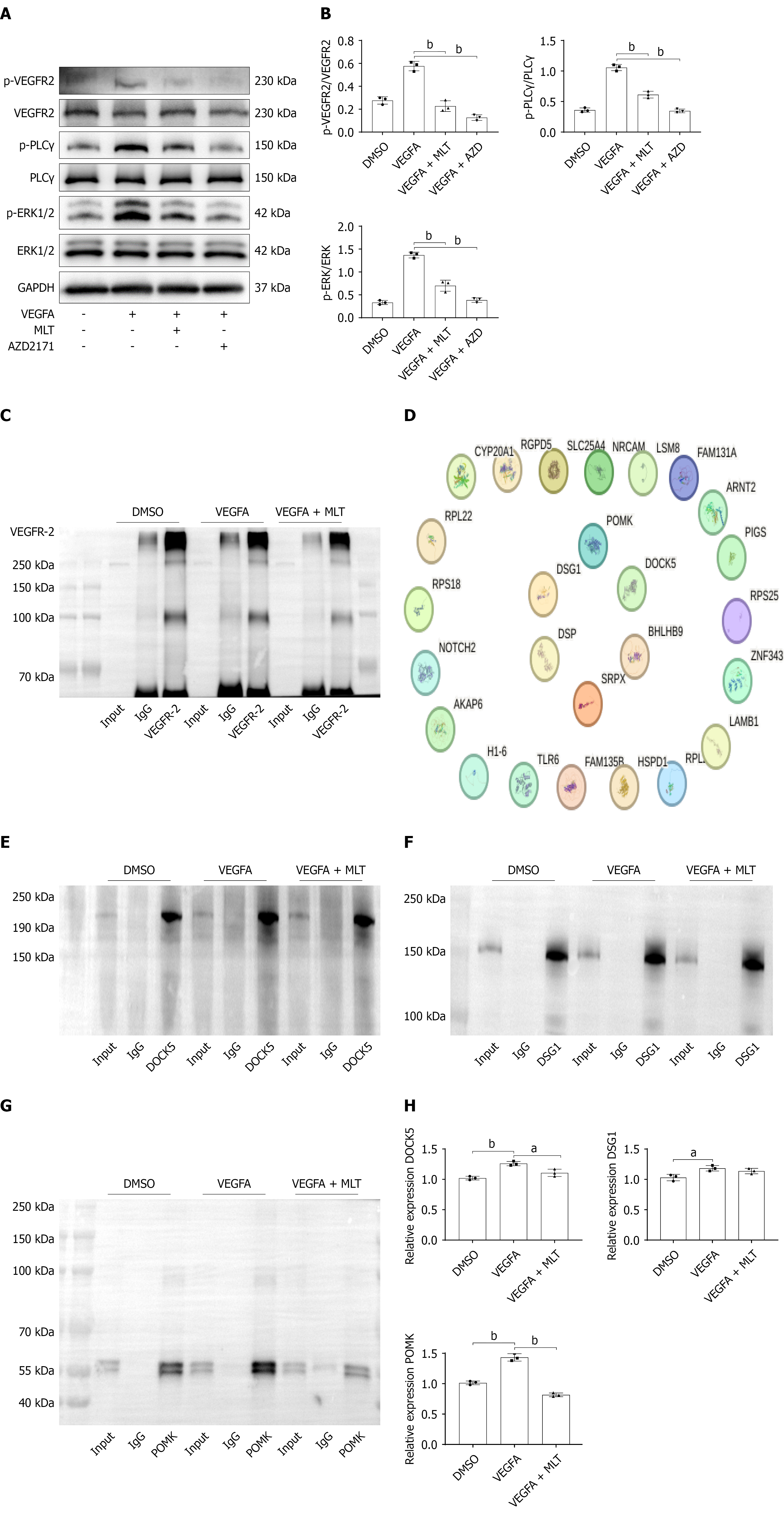
Figure 6 Melatonin suppressed VEGF signaling and interactions between VEGFR-2 and POMK.
A: Western blotting of VEGFR-2 signaling in human umbilical vein endothelial cells (HUVECs) treated with VEGFA, melatonin, or AZD2171; B: Quantitative analysis of the activation of VEGFR-2 signaling; C: Immunoprecipitation (IP) blotting of VEGFR-2 in HUVECs and subjected of IP-mass spectrometry; D: PPI network of enriched proteins by IP-mass spectrometry; E: IP blotting of DOCK5 by VEGFR-2 in HUVECs; F: IP blotting of DSG1 by VEGFR-2 in HUVECs; G: IP blotting of POMK by VEGFR-2 in HUVECs; H: Quantitative analysis of IP blotting. aP < 0.05. bP < 0.01. MLT: Melatonin.
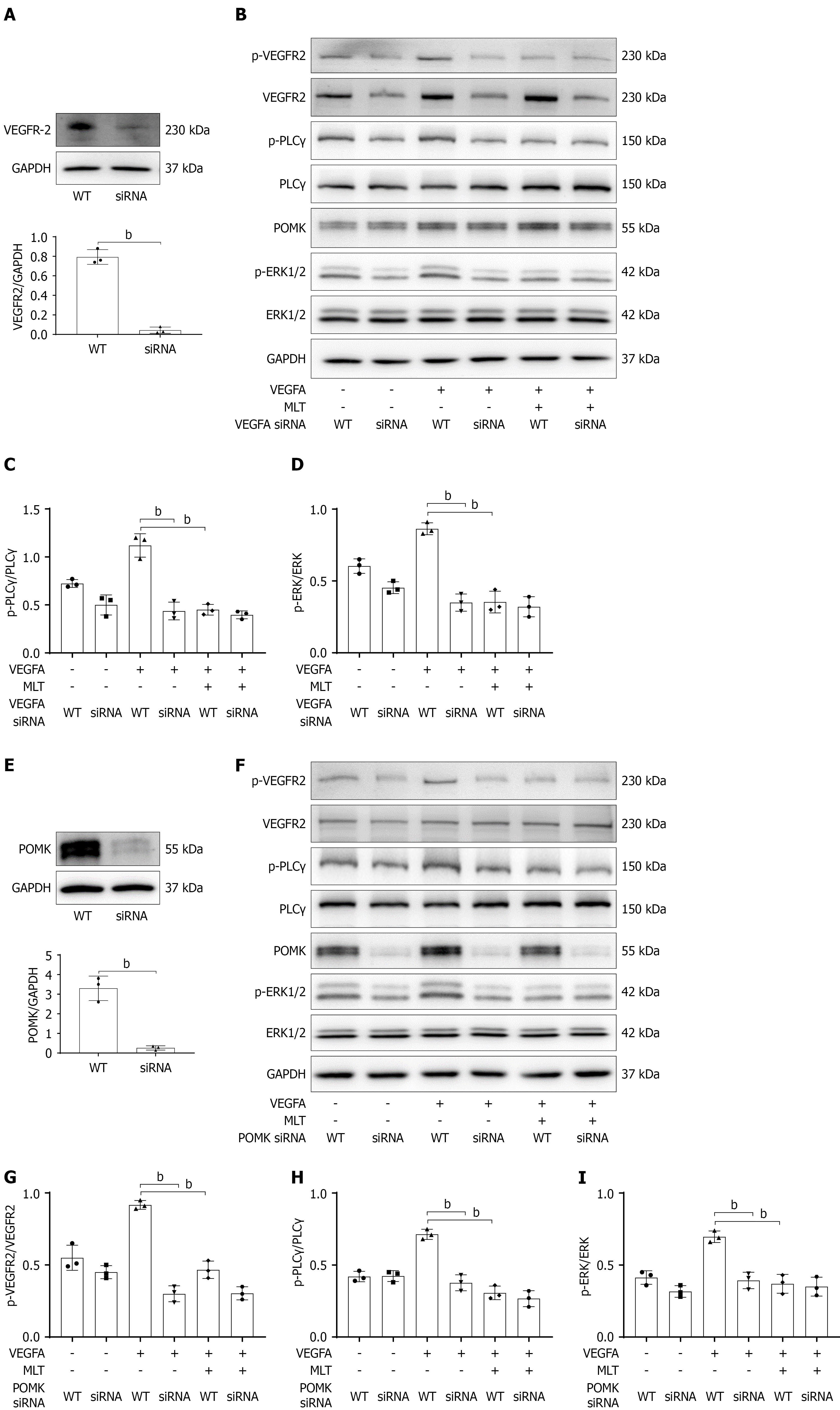
Figure 7 Melatonin suppressed VEGFR-2 signaling via a POMK-dependent manner.
A: Validation of VEGFR-2 knockdown by siRNA in human umbilical vein endothelial cells (HUVECs); B: Western blotting of VEGFR-2 signaling in HUVECs with VEGFR-2 knockdown; C and D: Quantitative analysis of PLCγ and ERK phosphorylation in B; E: Validation of POMK knockdown by siRNA in HUVECs; F: Western blotting of VEGFR-2 signaling in HUVECs with POMK knockdown; G-I: Quantitative analysis of VEGFR-2, PLCγ, and ERK phosphorylation in F. bP < 0.01. MLT: Melatonin.
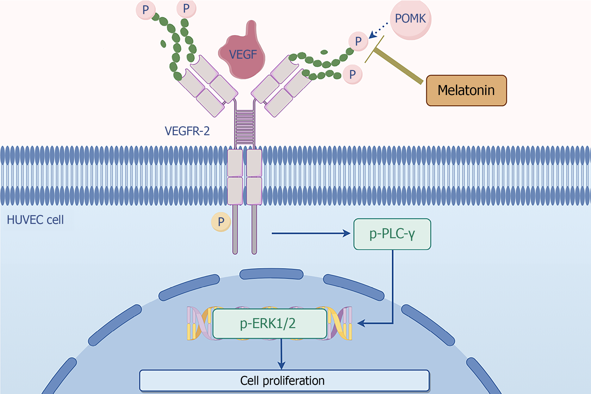
Figure 8 Proposed mechanism underlying the protective effects of melatonin against pathological angiogenesis in cirrhotic portal hypertension.
Melatonin disrupts the interaction between POMK and VEGFR-2, resulting in reduced VEGFR-2 phosphorylation and attenuation of downstream PLC-γ/ERK1/2 signaling activation. This inhibition suppresses VEGF-induced endothelial cell proliferation and angiogenesis, thereby contributing to the improvement of splanchnic hyperdynamic circulation in cirrhotic portal hypertension. HUVEC: Human umbilical vein endothelial cell.
- Citation: Wu GB, Luo GQ, Chen M, Cai KW, You XT, Han F, Zhang NZ, Luo M, Li Q, Huo HZ, Fan Q. Melatonin attenuates splanchnic hyperdynamic circulation in cirrhotic portal hypertension by disrupting POMK-VEGFR2 interaction. World J Gastroenterol 2027; 33(2): 123795
- URL: https://www.wjgnet.com/1007-9327/full/v33/i2/123795.htm
- DOI: https://dx.doi.org/10.3748/wjg.123795