
















Copyright: ©Author(s) 2027.
World J Gastroenterol. Jan 7, 2027; 33(1): 123843
Published online Jan 7, 2027. doi: 10.3748/wjg.123843
Published online Jan 7, 2027. doi: 10.3748/wjg.123843
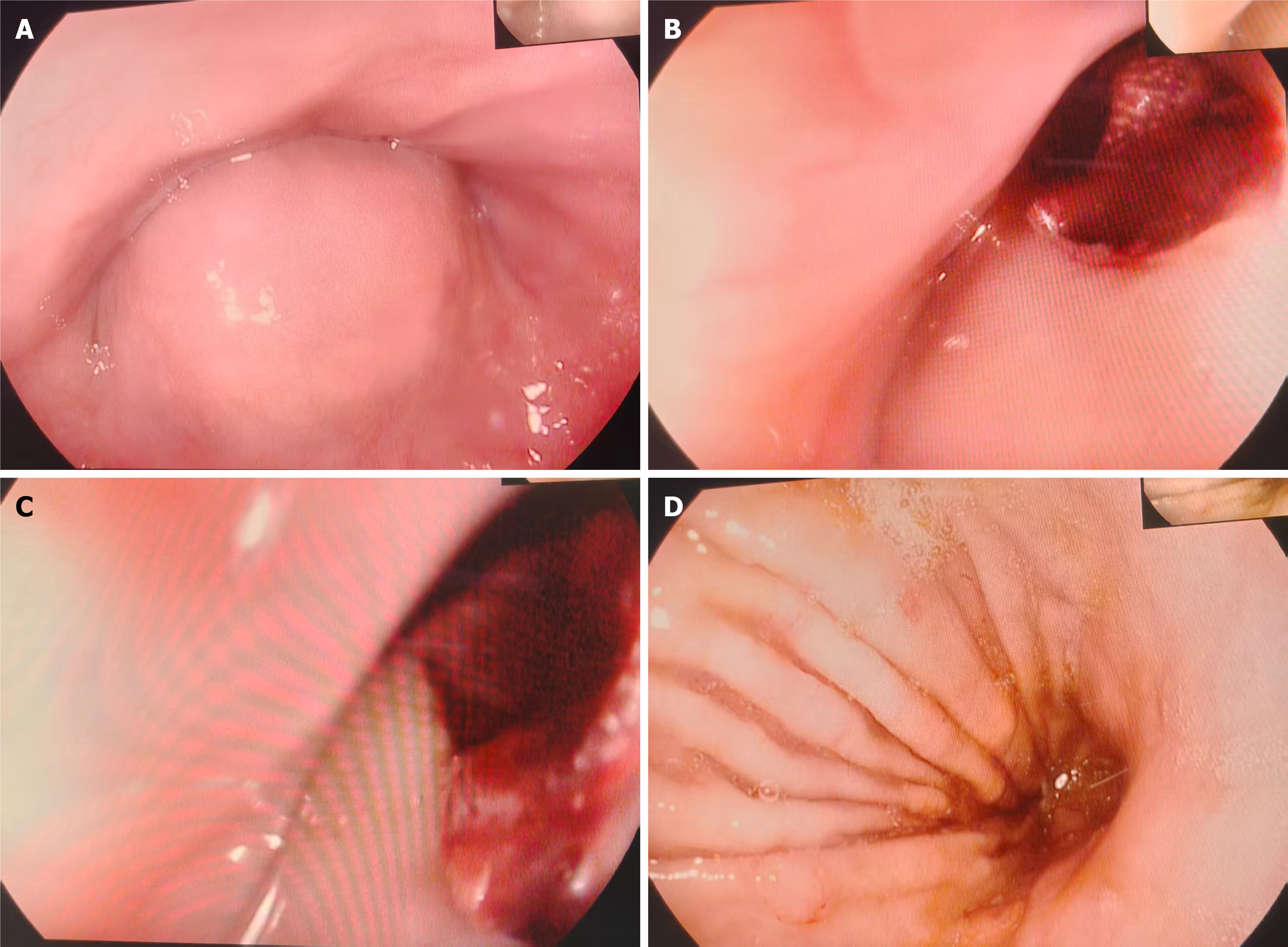
Figure 1 Upper endoscopic findings.
A: A large smooth submucosal protrusion was observed on the posterior esophageal wall, 26-32 cm from the incisors, causing partial narrowing of the esophageal lumen; B: Apical ulceration of the protruding lesion with an adherent dark-red clot; C: Close-up view of the ulcerated surface and adherent clot, suggesting the recent bleeding site; D: Endoscopic view of the gastric body showing no evidence of active intragastric bleeding.
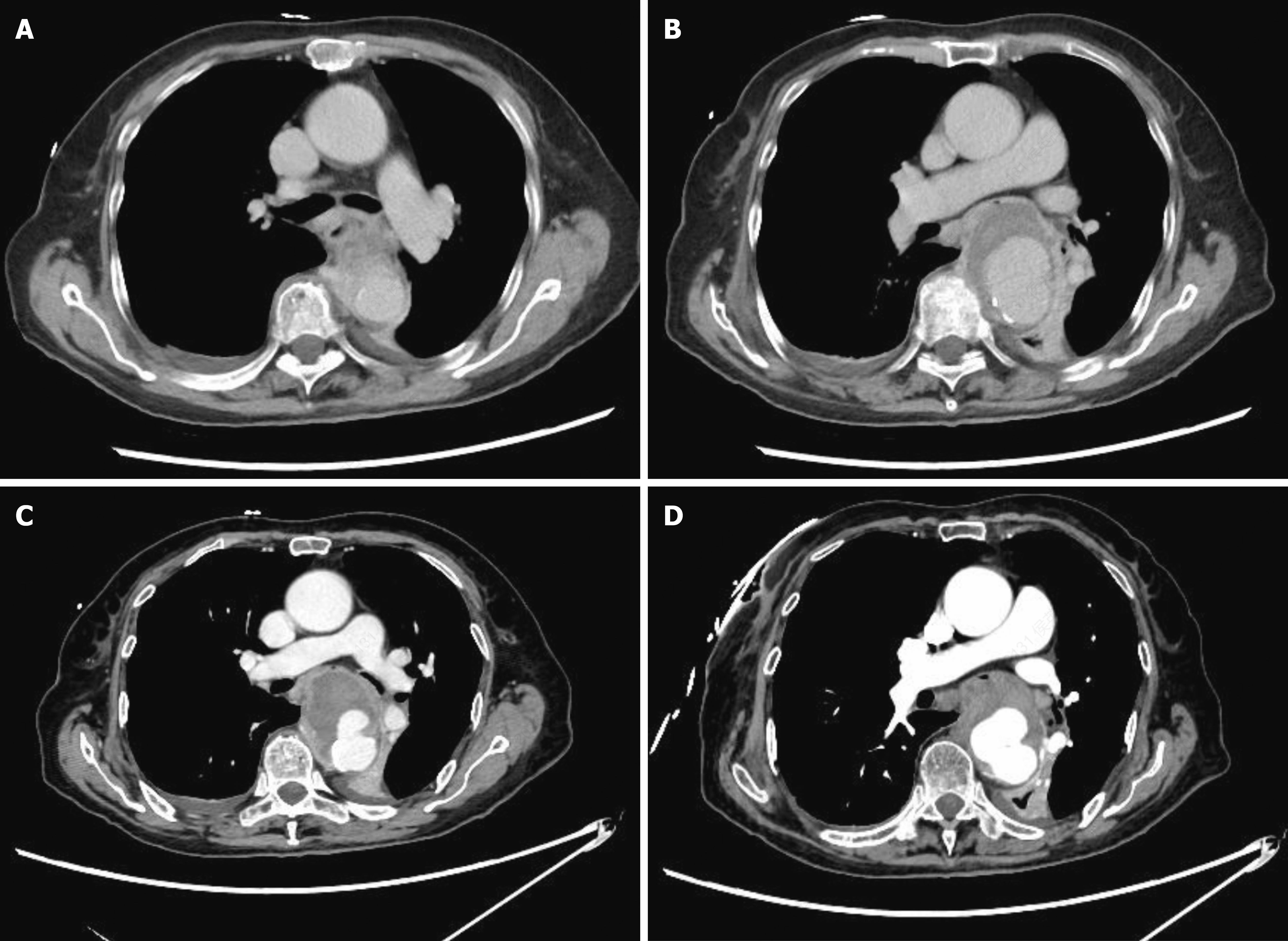
Figure 2 Axial contrast-enhanced computed tomography images of the descending thoracic aorta.
A and B: Unenhanced axial computed tomography images at different levels showing a focal saccular pseudoaneurysmal lesion arising from the descending thoracic aorta, with irregular wall calcification and peripheral soft-tissue components; C and D: Contrast-enhanced axial computed tomography images at different levels showing contrast filling of the pseudoaneurysmal sac through an approximately 1.9-cm neck communicating with the aortic lumen. The peripheral non-enhancing low-attenuation components are compatible with mural thrombus or perianeurysmal inflammatory tissue.
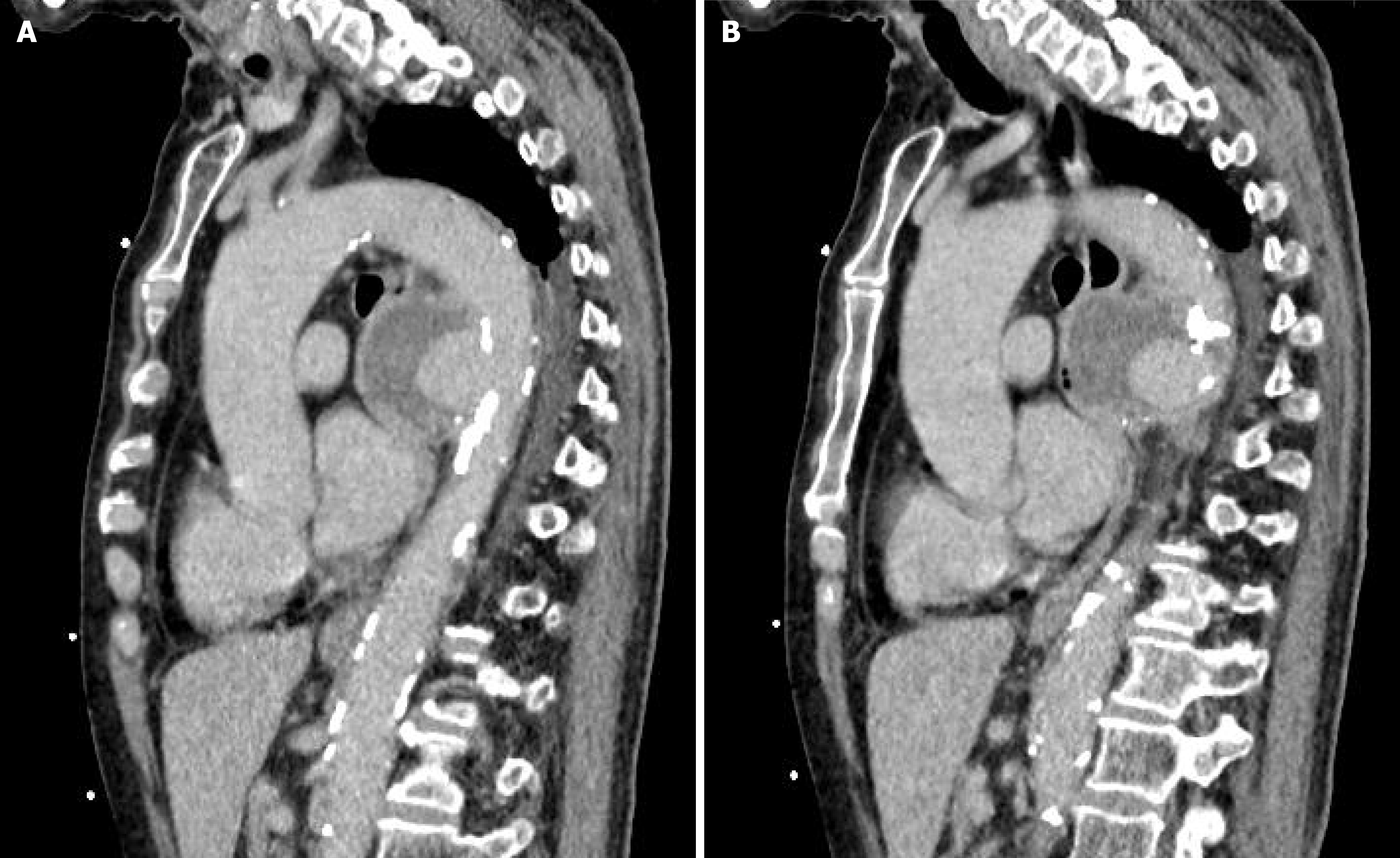
Figure 3 Sagittal computed tomography angiography of the descending thoracic aorta.
A: A sagittal image showing the saccular pseudoaneurysm with central contrast enhancement and peripheral low-attenuation components compatible with mural thrombus or perianeurysmal inflammatory tissue; B: An adjacent sagittal image showing compression and anterior displacement of the esophagus by the pseudoaneurysm.
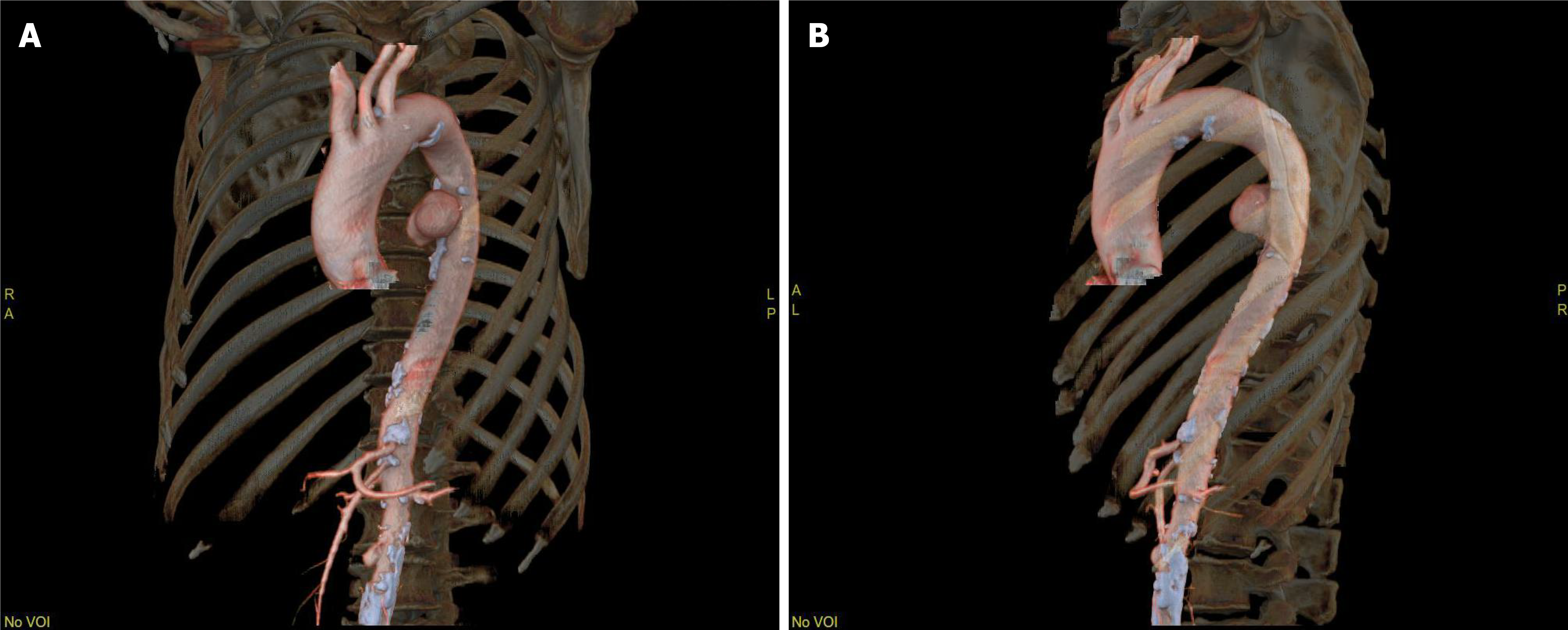
Figure 4 Three-dimensional reconstruction of computed tomography angiography images.
A: Anterior view; B: Lateral view. A localized saccular outpouching arises from the descending thoracic aorta distal to the origin of the left subclavian artery.
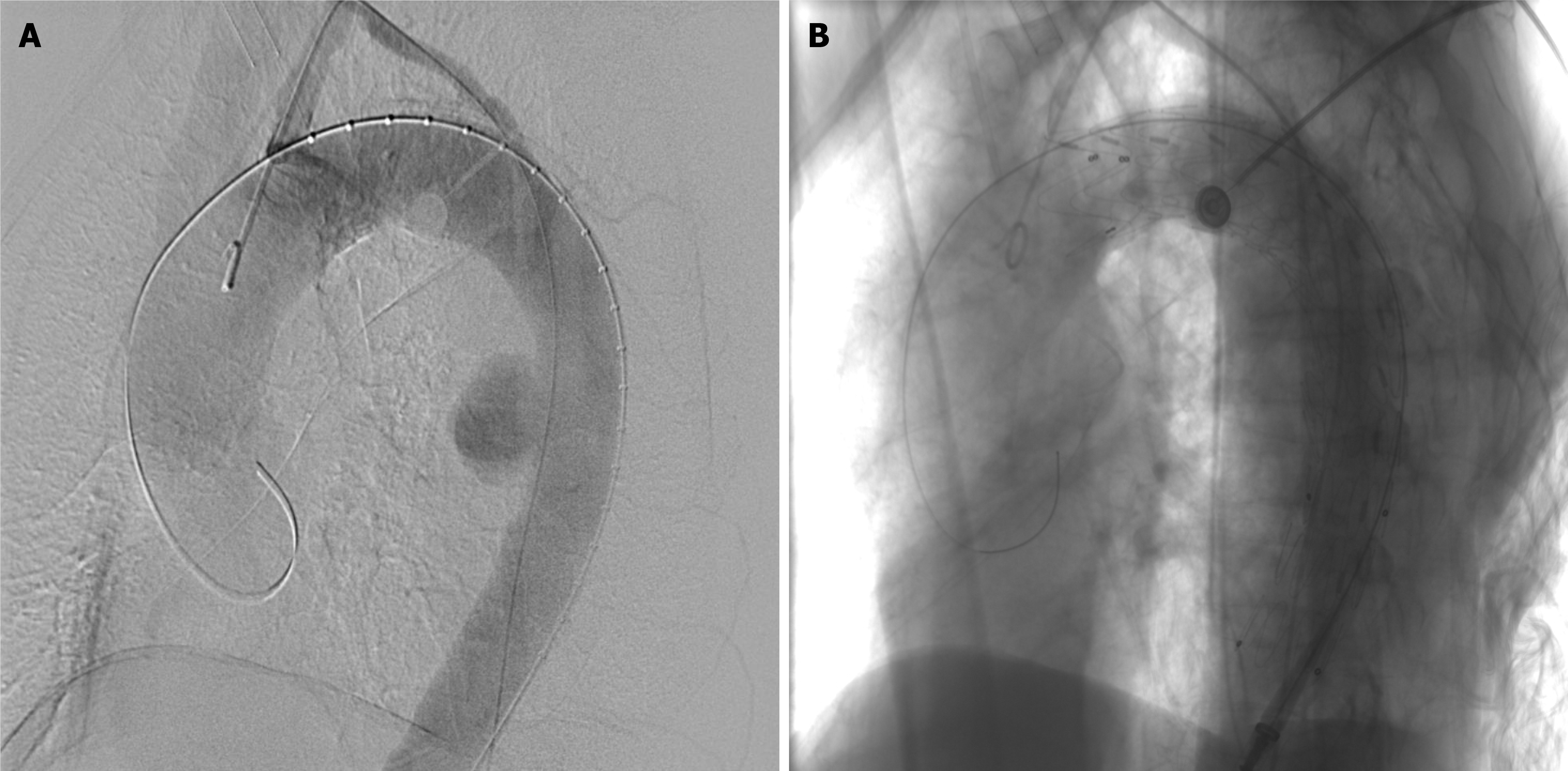
Figure 5 Intraoperative angiography during thoracic endovascular aortic repair.
A: Pre-deployment angiography showing the descending thoracic aortic pseudoaneurysm and landing zones; B: Post-deployment angiography demonstrating complete pseudoaneurysm exclusion, no endoleak, and preserved left subclavian artery perfusion.
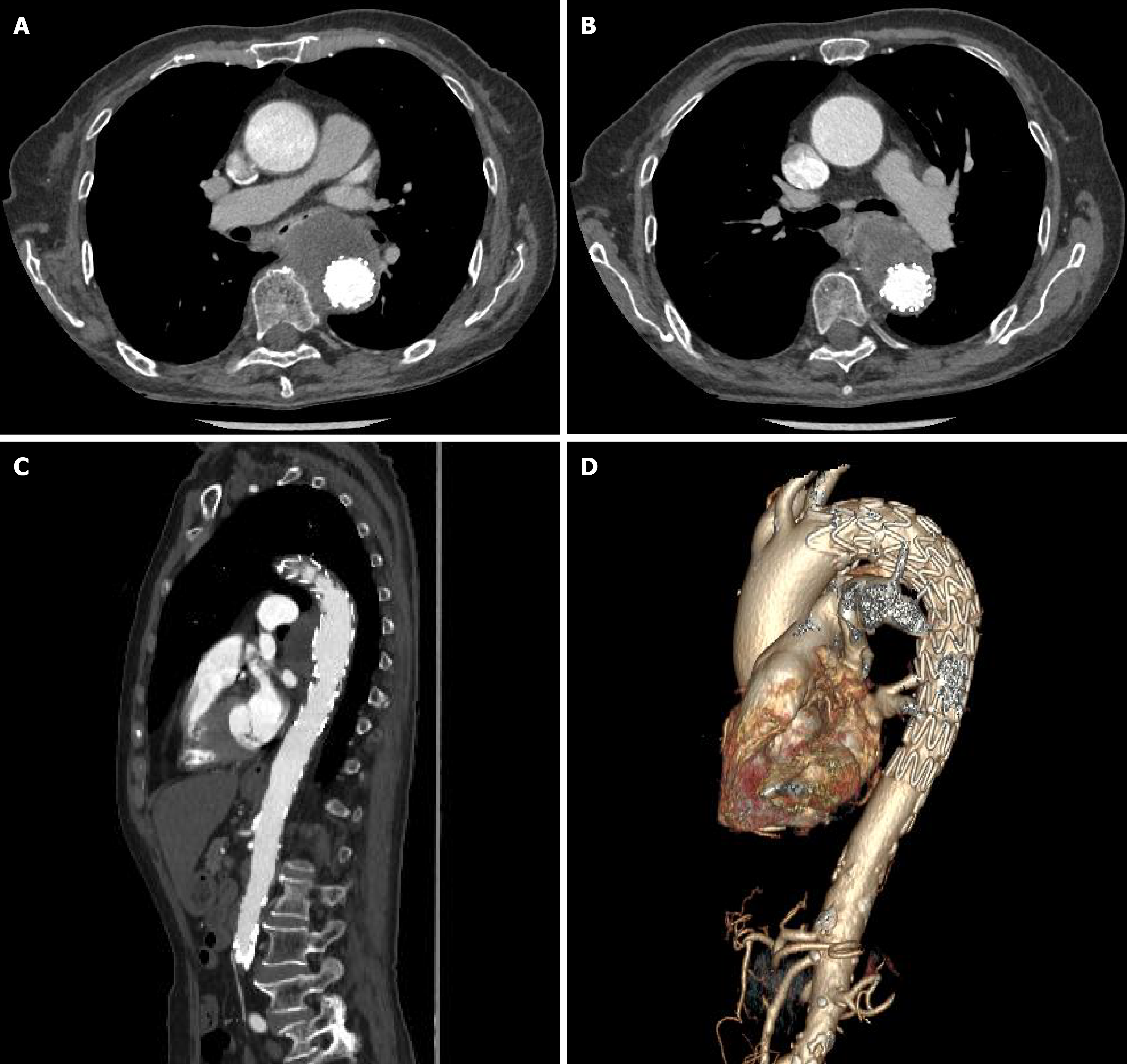
Figure 6 Follow-up computed tomography angiography performed approximately 25 days after thoracic endovascular aortic repair.
A and B: Axial; C: Sagittal; D: Three-dimensional. They show stable postoperative findings, with persistent exclusion of the descending thoracic aortic pseudoaneurysm. No endoleak, stent-related complication, or interval disease progression is identified. Axial, sagittal, and three-dimensional reconstructions collectively demonstrate maintained procedural efficacy during early follow-up.
- Citation: Xue Y, Yun XJ, Liu QY, Qi YT, Zhang LH, Jiang K, Li BD, Zhang JX. Suspected Brucella-associated descending thoracic aortic pseudoaneurysm presenting as upper gastrointestinal bleeding: A case report and review of literature. World J Gastroenterol 2027; 33(1): 123843
- URL: https://www.wjgnet.com/1007-9327/full/v33/i1/123843.htm
- DOI: https://dx.doi.org/10.3748/wjg.123843